Note: Descriptions are shown in the official language in which they were submitted.
20 1 3 1 t l
P.C. 7553
FEMALE SUSPENSION PROCEDURE
The invention relates to anchoring means and
systems for positioning anchoring means in living
tissue, particularly of use in suspending the female
urethrovesical junction, also called bladder neck.
Stress incontinence is caused by increased
abdominal pressure. One surgical method for treating
this condition involves suspension of the bladder neck
for repositioning in the correct fixed retropubic
position such that there is no voiding of the bladder
under stress and at the same time bladder outlet
obstruction is avoided. Four relatively non-invasive
surgical procedures for bladder neck suspension are
described in Hi~dley et al., Urologic Clinics of North
America, Vol. 12, No.2, p. 291 (1985). In the original
Pereyra method, a needle is passed from a suprapubic
incision to an incision in the vagina near the bladder
neck. Stainless steel suture wire is passed several
times from the bladder neck to the suprapubic incision
to suspend the bladder neck. The Cobb-Radge method
inserts the needle from below through the vaginal
incision. The Stamey procedure uses an endoscope to
prevent the surgical needle from puncturing the
bladder. Dacron vascular graft is used to anchor nylon
suture in the periurethral tissue. Finally, in the Raz
method the surgeon inserts his finger through the
vaginal incision to guide the suspension needle and
avoid penetration of the bladder by the needle. The
sutures are anchored by threading through tissue of the
vaginal wall and tissue in the suprapubic area.
201 31 1 1 -2-
A major problem encountered during surgical needle
suspension procedures such as described above is the
correct positioning of the bladder neck and the urethra
such that the position of the bladder neck with respect
to the bladder is high enough to avoid incontinence
under stress while not too high to prevent proper
bladder voiding.
The invention is an improvement over the prior art
by providing for easy adjustment of the suspending
sutures to lower or raise the bladder neck during
surgery, and for readjustment to lower or raise the
bladder neck, if necessary, after the patient has
benefited from the suspension procedure. The invention
also allows for more secure anchoring of the sutures
without extensive tissue suturing to prevent lowering
of the bladder neck over time. Furthermore, the
invention reduces the amount of tissue dissection
required to place the tissue anchors in proper
position.
A system for positioning of anchoring means used
during surgical procedures is described in U.S. Patent
4,705,040 disclosing a hollow needle containing a
retaining device attached to a filament. The retaining
device after dislodging from the needle attaches to the
interior wall of a body organ, and the filament is
pulled to draw the device against the body wall. The
filament is clamped outside the body to keep the organ
in position.
U.S. Patent 4,166,469 describes placement of a
pacemaker within a patient through a sleeve which is
positioned in the body through a needle or over a guide
wire which enters the patient within a needle and
remains after the needle is removed.
The above prior art devices are helpful in
introducing objects into the body, but do not have the
20~31 1 1
versatlllty of the systems of the lnventlon descrlbed below.
The urethroveslcal ~unctlon may be suspended by
lnsertlng sutures through an lnclslon ln the suprapublc
abdomlnal area or through an lnclslon ln the vaglnal wall,
suspendlng the urethroveslcal ~unctlon wlth the sutures, and
anchorlng the sutures at an anchorlng slte wlth an anchorlng
means such as a relatlvely rlgld hellx havlng an attachlng
means to attach the suture, or a pad adapted to be dellvered
wlthln a dellvery means to the anchorlng slte, or a fllp
anchor ln substantlal axlal allgnment wlth a placement means
and adapted to be fllpped from the axlal allgnment to an
angled posltlon wlth respect to the placement means, or an
ad~ustable tlssue anchor havlng a means for ad~ustably
attachlng the sutures. In thls context, lnclslon ls
understood to lnclude mere puncture by a needle.
The lnventlon further lncludes an anchorlng means
for anchorlng a suture ln tlssue, whlch comprlses a houslng, a
substantlally cyllndrlcal means for recelvlng a suture, sald
cyllndrlcal means contalned wlthln sald houslng, and an
ad~ustlng means in mechanlcal relatlonshlp wlth sald
cyllndrlcal means such that on ad~ustlng sald ad~ustlng means
the length of sald suture wlthin the tlssue can be regulated.
In one embodlment the ad~ustment of the anchorlng means ls by
rotatlon of the ad~ustlng means. In a second embodlment, the
cyllndrlcal means ls a rotatlng spool capable of belng rotated
through ad~ustment of the ad~ustlng means.
The lnventlon also lncludes an anchorlng means for
anchorlng a suture ln tlssue, whlch comprlses a houslng, a
_ 3 _
64680-538
20 1 3 1
-
rotating spool contalned wlthln sald houslng for recelvlng a
suture, a drlvlng gear ln reverslble engagement wlth sald
rotatlng spool such that on engagement wlth sald rotatlng
spool, sald spool may be rotated ln one dlrectlon and on
dlsengagement sald spool may be rotated ln the opposlte
dlrectlon, and an ad~ustlng means ln mechanlcal relatlonshlp
wlth sald drlvlng gear such that on ad~ustment of sald
ad~ustlng means the drlvlng gear may be reverslbly engaged
wlth sald rotatlng spool to regulate the length of sald suture
wlthln the tlssue.
Preferably, the ad~ustlng means ls adapted for
external access to ad~ust the ad~ustlng means by external
means. It ls understood that "external access" means access
from outslde the anchorlng means, and, more speclflcally, from
outslde the body, e.g. by way of puncturlng the skln or
through a small lnclslon (e.g. 2 to 4 mm) of the skln.
The system for posltlonlng an anchorlng means ln
llvlng tlssue, comprlses an lnsertlon means prlmarlly deflned
along a longltudlnal axls to flt ln a surglcal needle, the
anchorlng means havlng a substantlally axlal channel
reverslbly surroundlng sald lnsertlon means ln substantlally
axlal allgnment wlth sald longltudlnal axls of sald lnsertlon
means. The anchorlng means ln one embodlment of the lnventlon
ls adapted to be fllpped from the axlal allgnment to an angled
posltlon. Convenlently, the lnsertlon means 18 a surglcal
gulde wlre threaded through the substantlally axlal channel of
the anchorlng means.
- 4 -
64680-538
2~!31~
Generally, lntroductlon of an anchoring means lnto
the tlssue can be by lnsertlng a hollow needle carrylng the
anchorlng means attached to a suture through the skln lnto the
tlssue, releaslng the anchorlng means
- 4a -
64680-538
~ 201 31 1 ~
from the hollow needle into the tissue and withdrawing
the hollow needle.
Specific manners in which the anchoring means
maybe employed for anchoring the suture in the tissue
are described in more detail below.
Fig. 1 is a top view of a relatively rigid helix.
Fig. 2 is a side elevational view of the
relatively rigid helix of Fig. 1.
Fig. 3 is a sectional view of a flip anchor with a
placement means.
Fig. 4 is a sectional view of of an adjustable
tissue anchor placed in body tissue.
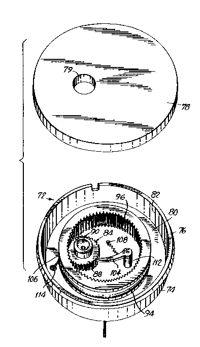
Fig. 5 is a perspective view of the externally
adjustable tissue anchor of Fig. 4.
Fig. 6 is a perspective view of externallv
adjustable tissue anchor.
Fig. 7 is a sectional view of the tissue anchor of
Fig. 6.
Figs. 1 and 2 show a relatively rigid helix 10
comprising one turn of a spiral 11 with the end of the
spiral bent toward the center and then 90 upwards
through the middle of the spiral to form shaft 12. The
shaft 12 ends in an eye 14 for attaching a suture. The
leading end 16 of the helix 10 is a sharp end to allow
for screwing of the helix lO into the body tissue.
The rigid helix may have more than onè turn of the
spiral. Conveniently, the end 16 is bend inwardly
toward the shaft 12 for protection against erosion
through adjacent critical organs such as the bladder
and vagina. The eye 14 may be replaced by other suture
attaching means such as a closed needle eye, or the
shaft 12 may be hollow to enable swaging of a suture in
the shaft in accordance with known hollow suture
needles.
201 31 1 1 -6-
Fig. 3 shows flip anchor 28 and placement means
30. The flip anchor 28 has an axial channel 32 which
snugly fits around the placement means 30. The anchor
28 includes a radial channel 34 allowing for passage of
a suture 36. The flip anchor 28 conveniently is in the
shape of a flat rectangle. The placement means 30 is a
surgical guide wire which may be placed in position by
a needle, or a needle extension. On pulling at both
~nds of the suture 36, the anchor 28 flips and becomes
positioned substantially at right angles to the means
30. In an alternative embodiment, the flip anchor may
have a means for attachment to a tool which is capable
of flipping the anchor. Once the anchor 28 is in the
flipped position, the suture 36 may be pulled from the
suprapubic end.
Fig. 4 shows an adjustable tissue anchor 38 placed
between the rectus muscle 40 and subcutaneo~s ~issue
42. Suture 44 extends between the tissue anchor 38 and
anchoring means 46 placed between periurethral fascia
48 and vaginal wall 50. The anchoring means 46 may be
properly positioned by introduction through a small
incision of the vaginal wall. Alternatively, the
anchoring means 46 may be introduced through a puncture
of the vaginal wall by a needle. Thus, the flip anchor
28 of fig. 3 may be positioned on a needle, as
described above. Alternatively, a padlike tissue
anchor may be inserted in a needle and pushed through
the needle, for instance with an obtrator. The needle
is subsequently withdrawn. The needle may have an
extension known to have a certain length equal to the
length between the desired position of the pad and the
vaginal wall. This extension helps the surgeon in
determining the distance which the needle has traveled
through the vaginal wall, and thus the distance at
which the anchoring means may be dropped.
201 31 ~ 1 -7-
Another method for positioning the anchoring means
below the urethra between the vaginal wall and the
periurethral fascia makes use of a solid needle having
a sleeve. The needle punctures the vaginal wall when
the proper position is attained, the needle is
withdrawn leaving the sleeve behind in the tissue of
the body. An anchoring means may then be inserted
through the sleeve, and the sleeve withdrawn.
Fig. 5 shows the adjustable tissue anchor 38 of
Fig. 4 in a perspective view. The tissue anchor 38
comprises substantially cylindrical anchor body 52
having snapping lip 54 to snap on cover lid 56. Anchor
bottom 58 connects anchor body 52 with inner cylinder
60. Suture 62 is wrapped around inner cylinder 60,
guided through channel 66 underneath suture-clamping
screw 64 and through outer hole 68 which in Fig. 5
extends from the top 70 of the inner cylinder 66
through the anchor bottom 58. Suture 62 is clamped to
the inner side of anchor bottom 58 by screw 64 when the
anchor 38 is placed in the body. On removal of cover
lid 56, screw 64 may be adjusted with a screwdriver.
Thus, by screwing the screw 64 upwards, suture 62 is
free for wrapping around inner cylinder 66 to shorten
the length of the suture in the body, or the suture 62
is free for unwrapping from inner cylinder 66 to
lengthen the suture in the body.
Figs. 6 and 7 show an externally adjustable tissue
anchor 72. The tissue anchor 72 comprises
substantially cylindrical anchor body 74, snapping lip
76 to snap on cover lid 78 having access hole 79,
bottom 80, tissue anchor wall 82 and rotating spool 94
on which suture 106 is wound. The spool 84 may be
rotated with the aid of driving gear 84 . Driving gear
84 is situated in opening 86 of the bottom 80. The
-8-
- 20 1 3 1
driving gear 84 comprises funnel 88 leading to hex
driving hole 90, and driving gear teeth 92. Rotating
spool 94 has inside gear teeth 96 which are capable of
engaging the driving gear teeth 92 of driving gear 84.
The rotating spool 94 is locked in place between bottom
edges 98 and 100 of the rotating spool bottom 80. Wave
spring 102 is located under the driving gear 84. When
the driving gear 84 is pushed downwards, e.g. by hand,
the wave spring 102 allows the driving gear 84 to move
down so disengaging ratcheting pawl 104. When the pawl
104 is disengaged, the rotating spool 94 can be rotated
to release suture 106 from the spool 94. Rotation of
the rotating spool 94 is with an external tool such as
an Allen wrench, capable of puncturing through the skin
into access hole 79 in cover lid 78 engaging the hex
driving hole 90. On release of the driving gear 84, the
wave spring 102 returns the driving gear 84 to its
original positicn in engagement with ratcheting pawl
104 allowing for rotation of the rotating spool 94 for
uptake of suture 106 on the spool 94. The ratcheting
pawl 104 is attached to rotating spool bottom 80
through pawl spring 106 and spring post 108 and pivots
on pawl pin 110 which is attached to pawl post 112.
The pawl post 112 in turn is attached to bottom 80 or
is part of bottom 80.
The suture 106 is spooled on rotating spool 94,
channeled through hole 114 in bottom 80, and led
through ridges or holes in extensions 116 and 118 at
the bottom 80 to the center of the anchor 72.
