Note: Descriptions are shown in the official language in which they were submitted.
:'~ c~ a~
2~:~.~.~~~F~
1
SURGICAL TREATMENT METHOD AND INSTRUMENT
BACKGROUND OF THE INVENTION
The present invention relates to surgical and
dental procedures utilizing laser radiation. The
invention relates more particularly to surgical and
dental treatment procedures and instruments utilizing
laser radiation for the removal of tooth and gum tissue.
In dental procedures, it is freguently
desirable to remove portions of tooth enamel and dentin,
and in certain cases, portions of gum tissue, in an
accurately controlled manner and there has been a
growing interest in the use of laser radiation for
performing such procedures. The use of laser radiation
is attractive because, particularly with the aid of
optical fibers, such radiation can be focused to a very
small area and is thus compatible with the dimensional
scale of dental procedures. Moreover, laser radiation
procedures can be performed without recourse to an
anesthetic.
While a number of devices of this type have
been proposed, they have not proven to be of practical
use notably because even the most effective of those
devices already proposed are useful only under limited
and very precisely defined conditions.
The enamel and dentin of a tooth include, as
one component, hydroxyapatite, which is in amorphous
form in the dentin and crystalline form in the enamel.
These portions of a tooth additionally include organic
tissues and water, but have no vascular system. Healthy
dentin is in mineralized form, while dentin which has
experienced decay is in demineralized form. Dentin has
a relatively high percentage of organic tissue, around
percent, and also a high percentage of water. These
percentages increase considerably in decayed dentin.
Tooth pulp and the gum surrounding the teeth
consist of vascularized organic tissue containing both
2
hemoglobin and water. Each of these components has a
different response to laser radiation.
Thus, it has been found, that hydroxyapatite
absorbs laser radiation in the wavelength ranges of 9 -
li , such as produced by COz lasers, and also in the
wavelength range 0.5 - 1.06 ~ , which includes the
wavelength that can be produced by a YAG laser.
The laser radiation absorption by the various
parts of a tooth at various wavelengths is influenced by
the absorption of the radiation energy by the water
component thereof. The greater the absorption by water,
the less energy is available for absorption by the other
components. Since the wavelengths of the radiation
emitted by COz lasers is absorbed to a large extent by
water, this radiation has minimal cutting effect on
enamel or dentin, and less of a cutting effect on
mineralized dentin than on demineralized dentin.
On the other hand, it has been found that
radiation at a wavelength of 1.06 is absorbed to a
lesser degree by water, and therefore has a greater
effect on mineralized tissues. Laser radiation at a
wavelength of 0.532 a is not absorbed at all by water
and can be effective on mineralized tissues if a
sufficiently high, and thus dangerous, power level is
employed.
As regards vascularized tissues, radiation at
the wavelengths emitted by CO~ lasers has an effective
cutting action because of its absorption by water,
radiation at a wavelength of 1.06 ~ does not have any
effect, and radiation at a wavelength of 0.532 ~ has a
cutting effect on soft tissues because, although not
absorbed by water, it is well absorbed by hemoglobin.
While a particular wavelength may inherently
have a cutting effect on enamel or dentin, it has been
found that the practical utilization of radiation at
such a wavelength for dental procedures is highly
dependent on the form in which the radiation is applied,
L
3
with respect to energy level, pulse duration and
repetition rate. Specifically, efforts to apply such
radiation in the form of high energy pulses of short
duration have been found to produce a highly localized
temperature increase, resulting in differential thermal
expansion which can cause mechanical damage to the tooth
as well as vascular damage to pulp tissue. Conversely,
low energy pulses of long duration cause a more
widespread heating of the tooth which results in patient
discomfort as well as pulp damage due to heating.
In the treatment of various dental. and other
medical conditions, it is frequently necessary to remove
bone, dentin, cementum or dental root material, and it
is desirable to do so without subjecting the patient to
adverse side effects.
Frequently, when performing medical procedures
within the oral cavity, the practitioner encounters
metal bodies introduced by previous dental procedures,
such bodies being constituted by metal filling material,
metal pins, and chrome posts used to secure dental
prostheses in place, and it is necessary to cut these
bodies, again without producing harmful side effects.
Also, dental practitioners frequently
encounter cysts and granulomas, which occur in the gum
adjacent the apex of a tooth, and it is necessary to
destroy, or at least substantially reduce, these
growths.
Furthermore, while a number of dental filling
materials are presently available, there is a continuing
need for material which can fill not only dental
cavities, but also cavities existing in, or created in,
bone material, and which will have a hardness comparable
to that of the natural material which it replaces and
form a strong bond with the wall of the cavity or
opening.
4
SUM~SARY OF TAE INVERTTI0~1
It is an object of the present invention to
effectively employ laser radiation in a variety of
surgical operations involving cutting, by vaporization,
of both tooth and gum tissue, as well as other
vascularized body tissue.
Another object of the invention is to
eliminate significant drawbacks of laser treatment
systems which have previously been proposed.
A further object of the invention is to
provide a single laser treatment device which can
perform a variety of operations.
Yet another object of the invention is to
perform dental treatments employing laser radiation in a
manner to minimize or completely eliminate undesirable
side effects of the treatment.
A specific object of the invention is to
employ laser radiation to cut mineralized dental tissue
without requiring high energy levels.
A further specific object of the invention is
to employ laser radiation to cut soft tissue without
requiring high energy levels.
A more specific object of the invention is to
employ laser radiation for selectively cutting bane,
dentin, cementum and dental root material, as well as
metal bodies found in the mouth, without exposing the
patient to adverse side effects, and particularly
burning of tissue adjacent the area being treated.
Another object of the invention is to provide
a novel filling material for filling cavities or
openings in both teeth and bones, and to employ laser
radiation for promoting hardening of such filling
material.
Yet another abject of the invention is to
provide an improved treatment for cysts and granulamas
in bones and in the gum.
5
According to one aspect of the invention, the
above objects are achieved by a method for filling an
opening in tooth or bone material comprising:
forming a paste composed of a liquid and a
powder containing hydroxyapatite:
filling the opening with the paste; and
irradiating the paste which fills the opening
with laser radiation in order to bond the hydrnxyapatite
to material surrounding the opening.
l0 According to another aspect of the invention,
the objects are achieved by a method of treating a cyst
or granuloma in the gum at the apex of a tooth canal, or
in bone, comprising:
opening the canal to the vicinity of the apex;
inserting an optical fiber having an output
end into the canal so that the output end is located at
the apex and
conducting a succession of pulses of radiation
through the fiber so that the radiation exits from the
output end, impinges on and opens the foramen, and then
impinges on and at least reduces the cyst or granuloma.
According to yet another aspect of the
invention, the objects are achieved by a method for
cutting bone, dentin, cementum and dental root material
in the body, comprising: generating laser radiation
having a wavelength suitable for cutting such material;
producing successive pulses of the radiation with an
energy level, pulse duration and repetition rate
selected to cut the material without causing harmful
side effects: concentrating the radiation pulses on the
material to a spot sufficiently small to cause cutting
of the material; and, simultaneously with the step of
concentrating, directing a cooling fluid onto the spot.
According to still another aspect of the
invention, the objects are achieved by a method for
cutting metal bodies in the mouth of a patient,
comprising: generating laser radiation having a
~~~~~~3~
6
wavelength suitable for cutting the metal; producing
successive pulses of the radiation with an energy level,
pulse duration and repetition rate selected to cut the
metal without causing harmful side effects;
concentrating the radiation pulses on the metal to a
spot sufficiently small to cause cutting of the metal;
and, simultaneously with the step of concentrating,
directing a cooling fluid onto the spot.
7
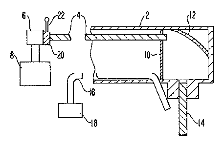
BRIEF DESCR7(FTION OF T~' DRAWING
The sole Figure is a cross-sectional view of a
preferred embodiment of an instrument for performing
laser radiation treatments according to the invention.
~fl~~~~4
DESCRIPTION OF THE PREFERRED EMBODIMENTS
The present invention is based essentially on
the discovery that laser radiation can be used to cut,
by vaporization, both tooth and gum material, as well as
other vascularized tissue, with essentially no adverse
side effects, if specific parameters are established for
the laser radiation.
According to the present invention, the
drawbacks described earlier herein can be eliminated, or
at least substantially minimized, and an effective
cutting action can be achieved, by the use of laser
radiation preferably at a wavelength of 1.06 ~ in the
form of pulses having an energy content of between 10
and 100 mJ, with a pulse duration of the order of 100 -
300 microseconds, and a repetition rate of the order of
50 Hz, and with the radiation beam concentrated at a
spot, at the treatment location, of the order of 200 -
600
A pulse duration of 100 - 300 ~C sec. has been
found to be sufficiently long to avoid subjecting the
tissue being treated to thermal shocks but sufficiently
short to enable effective control of the heating action
to be maintained.
According to the invention, laser radiation at
a wavelength of 1.06 , which can be produced by an Nd
YAG laser, can be used for cutting, or vaporizing
demineralized, i.e., decayed, enamel and dentin, without
endangering gum tissue. Laser radiation at a wavelength
of .532 ~ , which can also be produced by an Nd YAG
laser, can also be used, but this requires great care
because it has been found that radiation at this
wavelength will also cut gum tissue. Therefore,
radiation at this wavelength can be used when it is
desired to cut gum tissue.
Further, applicant has discovered that laser
radiation at the wavelength of 1.06 ~S can be made to
cut'healthy, or mineralized, dentin, and healthy enamel,
~~~~~~4
which was not heretofore considered possible, if a dark
colored region is first provided at the spot where
cutting is to begin. Specifically, it has been found
that the absorption of energy at the wavelength of 1.06
PLC by dark materials is sufficient to enable laser
radiation having a suitable energy level to create a
plasma which causes vaporization of dentin tissue.
Applicant has further discovered that once a plasma
cloud capable of vaporizing dentin has been established
at a dark colored region, the laser beam can be
displaced at a controlled speed from the dark colored
region so that the plasma cloud will remain intact and
vaporization of healthy dentin will continue.
For cutting dentin and enamel, laser radiation
at a wavelength of 1.06 should be used. Radiation at a
wavelength of 0.532 ~ has been found to be effective
only if applied at dangerously high energy levels.
Since radiation at 0.532 ~ can efficiently
cut vascularized tissue, it can be used for general
surgical procedures. In this case, the radiation pulses
should have an energy level of not greater than io mJ,
with a pulse duration of 100 - 300 p sec., and the
radiation may be focussed to a spot 200 - 600 ~ in
diameter. A pulse repetition rate of the order of 50 Hz
may be employed.
The Figure illustrates a handpiece for
supplying laser radiation in a form suitable for
performing the operations described above. A housing 2
is provided in the form normally utilized for
handpieces, which housing would be configured in a
manner known in the art for ease of manipulation. The
interior of housing 2 is provided with an optical fiber
4 having an input end coupled to a source 6 of
monochromatic light, such as an Nd YAG laser producing
radiation at a wavelength of 1.06 . Light source 6 is
connected to an operating power source 8 which supplies
10
pulses sufficient to cause light source 6 to produce
light pulses having the desired parameters.
The free end of fiber 4, in the vicinity of
the free end of housing 2, is supported by a suitable
support plate 1o to direct light radiation onto a curved
mirror 12 which deflects the radiation onto the
receiving end of a further optical fiber 14. Mirror 12
additionally performs a focusing action which can focus
the radiation emerging from fiber 4 to a point within
fiber 14, preferably in the vicinity of the outlet end
thereof. This will help to assure that the light
emerging from fiber 14 can be concentrated at a
sufficiently small spot on the tooth to be treated.
Fiber 14 preferably has a very small diameter, possibly
of the order of 250 ~.
Housing 2 additionally contains a hollow tube
16 which is connected to a source 18 of water and/or air
and which has an outlet end positioned to direct a
stream of the fluid supplied by source 18 into the
immediate vicinity of the tooth region to which laser
radiation is being applied.
In accordance with a particular novel feature
of the invention, a plate 20 which is capable of
influencing the laser radiation so as to double its
frequency is slidably mounted on source 6 and is
connected to a control handle 22 so as to be slidable,
by manipulation of handle 22, between the illustrated
position, where plate 20 is interposed in the light path
between source 6 and fiber 4, and a retracted position,
where plate 20 does not intersect the light path. With
this simple arrangement, the handpiece is given the
capability of applying either 1.06 ~ or 0.532
radiation to the area to be treated, so that only a
single laser device need be provided for the selective
performance of procedures with radiation of either
wavelength.
~01~~'~4
11
For performing endodontic treatments within a
tooth canal, fiber 14 can be given a suitable length and
diameter to be introduced into a canal in order to apply
the radiation to the canal walls for widening the canal
preparatory to filling.
According to a particular aspect of the
invention, the requisite dark spot can be formed simply
by applying a small amount of graphite, such as used in
pencils, with the aid of a small amount of glue. In
fact, it has been found possible to achieve the desired
result by applying a small quantity of glue to the point
of a sharpened pencil and then rubbing the pencil point
at the desired location.
For removal of decay, the radiation can have a
wavelength of 1.06 and be in the pulsed form described
above.
To dissipate the heat generated by the
radiation, water and/or air should be sprayed onto the
tooth in the vicinity of the spot which is being
irradiated. The rate of flow of fluid depends on the
extent to which the fluid absorbs the radiation. For
example, water absorbs radiation at 1.06 at a very low
level, but higher than radiation at 0.532
Therefore, water would be delivered at a higher rate
when the latter radiation wavelength is being employed.
When the radiation is applied to demineralized
enamel or pathological dentin, a dark spot is not
necessary and a plasma forms at the irradiation spot and
the affected material is volatilized at and around the
spot. The extent of the plasma tends to increase in a
short time and this allows for the possibility of
reducing the pulse energy to between 10 and 20 mJ.
When cutting normal tissue, the radiation
wavelength can be 1.06 ~ , which requires application of
a dark spot, and will not affect soft tissues, or 0.532
PL~u , which can cut either hard tissues, i.e., dentin
12
and enamel, or soft, vascularized tissues. Each
wavelength will be preferable for certain purposes.
Thus, the invention provides four operating
modes responsive to different needs:
1) Far cutting demineralized enamel and
pathological dentin, use is made ~f radiation at a
wavelength of 1.06 ~C , an energy level of 20 - 50 mJ,
and with the pulse parameters described earlier herein.
Labelling with a dark spot is not required.
2) For cutting n~rmal enamel and dentin, the
radiation would have the same parameters as for mode 1),
but the starting point would be labelled with a dark
spot.
3) For cutting any tissue, the same
parameters as for mode 1) would be employed, with
labelling with a dark spot where possible.
4) For cutting vascularized tissue,
including gum and other soft body tissue, laser
radiation at a wavelength of 0.532 ~ would be used,
composed of pulses having an energy level of no greater
than 10 mJ, without requiring labelling with a dark
spot.
Far dental treatments, a cooling spray will be
used whenever the operation generates a sufficient level
of heat.
The application of laser radiation in all of
the procedures to be described herebelow can be carried
out with the apparatus described above and illustrated
in the Figure.
According to the invention, a filling material
for teeth is constituted by a mixture formed from a
liquid component composed of phosphoric acid and water
and a powder component composed of a ceramic and
hydroxyapatite, with the ingredients mixed in a
proportion to form a paste having a consistency such
that the paste is workable and sufficiently self-
supporting to be applied to the opening with a spatula
13
and remain in place, and laser radiation having the
characteristics to be described below is applied to cure
and harden the mixture and bond it to the tooth. The
proportions of the mixture are not critical, however,
the following are preferred:
Liquid: Phosphoric acid 40%
water 60%
Powder: Ceramic 80%
Hydroxyapatite 20%
If the proportion of hydroxyapatite is
increased, more energy is required to harden the
mixture: if it is decreased, the strength of the
resulting bond is reduced.
The ceramic component may be composed of
corderite, silica or silicium oxide, or aluminum oxide,
for example. The powder components will have the grain
sizes normally used for dental filling materials.
The liquid and powder components should be
mixed together just prior to introduction into the
opening to be filled.
The radiation applied during this treatment
has a wavelength of 1.06 ~ and is composed of pulses
preferably having a duration of the order of 0.4 ms, a
repetition rate of the order of 50 Hz and an energy per
pulse in the range of 20-100 mJ. However, in contrast
to the various cutting operations to be described in
detail below, the beam should here be defocussed to be
at least approximately coextensive with the exposed
surface of the filling material. This can easily be
achieved by varying the spacing between the radiation
output surface of the handpiece and the tooth surface,
the area of illumination being readily visible.
The application of radiation to the filling
material will promote the growth of a crystal structure
in that material and create a strong bond between the
hydroxyapatite and the surrounding tooth material.
14
The radiation will be applied until a crystal
structure appears, this generally requiring application
of the radiation for a period of 10-30 seconds.
The above described filling material and
radiation can be used for filling breaks or gaps in bone
material.
According to another aspect of the invention,
radiation having the above-described characteristics can
be employed for treating a cyst or granuloma adjacent a
l0 tooth apex, or in bone. For this purpose, after the
canal had been opened to the foramen, a narrow optical
fiber, having a diameter of around 200~e, for example, is
threaded into the canal up to the foramen, and radiation
having the characteristics described above for cutting
soft tissue is delivered through the fiber to cut the
foramen and then eliminate the cyst or granuloma.
For this operation, use is preferably made of
radiation having a wavelength of 1.06 ~ , a pulse
duration of the order of 0.4 ms, a pulse repetition rate
of the order of 50 Hz and an energy content per pulse of
50-400 mJ:
In further accordance with the invention, it
is possible to cut, without burning, bone, root, dentin
and cementum in periods of the order of seconds by
applying radiation of the type described above together
with irrigation with a water/air mixture to control the
thermal laser beam cutting action. In this case, the
radiation wavelength is 1.06 ~ , the pulse duration is
of the order of 0.8-1.2 ms, the pulse repetition rate is
in the range of 30-50 Hz and the energy content per
pulse is 200-400 mJ. This can be done without first
forming a dark spot where the radiation is first
applied. However, application of a dark spot will
increase energy absorption and thus speed the cutting
operation. In addition, a dark spot can be applied when
it is desired to preliminarily mark or outline with a
low energy beam the place to be cut.
~0~.~~~~z~
In addition, radiation having form described
above for cutting bone can further serve to cut metal
parts in the mouth, such as metal fillings, pine, or
chrome tooth prosthesis posts. For this purpose laser
5 radiation will be created and directed to the material
to be cut in the manner described above.
In the performance of all of the cutting
operations described above, the light output surface of
the handpiece is positioned to focus the radiation to a
10 small spot, preferably having a diameter of the order of
200-600 Vie.
When dentin is cut with the aid of an optical
fiber in contact with the dentin, the end of the fiber
in contact with the dentin is subject to destruction.
15 Therefore, it is desirable to use a relatively long
fiber which will be replaced after a period of use. While
the description above refers to particular embodiments
of the present invention, it will be understood that
many modifications may be made without departing from
the spirit thereof. The accompanying claims are
intended to cover such modifications as would fall
within the true scope and spirit of the present
invention.
The presently disclosed embodiments are
therefore to be considered in all respects as
illustrative and not restrictive, the scope of the
invention being indicated by the appended claims, rather
than the foregoing description, and all changes which
come within the meaning and range of equivalency of the
claims are therefore intended to be embraced therein.