Note: Descriptions are shown in the official language in which they were submitted.
~~?~~"~"~
sNllz
_1_
A CONTINENT OsTOMATE BANDAGE
The present invention relates to bandages,
more particularly, to bandages for applications
requiring multiple bandages per day to the same
area of the skin.
New surgical procedures today far ostomy
care have in some cases eliminated the need for
external pouches. A pouch is formed internally by
placing a U-bend in the terminal portion of the
colon or intestinal track after surgical removal of
a portion of the colon. The U-bend ends in a stoma
or opening through the abdominal wall in the usual
manner but the pouch formed internally by the
U-bend is capable of storing a finate amount of
discharge. The pouch is emptied four or five times
a day by intubation. People who have undergone this
type of surgery are often referred to as~continent
ostomates or ileostomates or urostomates.
The stoma will secrete a certain amount of
mucous or discharge which is absorbed by a bandage
placed over the stoma. The bandage must be
removed and a new bandage applied each time
intubation takes place.
A common bandage approach consists of taking
a square or rectangular piece of acrylic adhesive,
placing a piece of cotton or gauze in the center
~0~~~~'~"~
_2_
SN112
and placing this over the stoma or, alternatively,
taping a piece of cotton or gauze over the stoma.
The acrylic adhesive is not particularly friendly
to skin upon removal and using this technique four
or five times a day often results in skin irrita-
tion or even excoriation. A real need exists to
provide a bandage for continent ostomates or others
such as those suffering with mucous fistulas which
bandage will not cause skin irritation, etc. when
multiple bandages are used each day to the same
area of the skin.
The present invention is directed to an
improved bandage particularly suitable for
continent ostomates or others such as those
suffering with mucous fistulas. This invention
provides a flexible, thin, hydrocolloid adhesive
layer having a plastic or non-woven fabric layer
affixed to one surface thereof and a superabsorbent
pad, smaller than the adhesive layer affixed to the
opposite adhesive surface. A porous cover pad
covers the superabs~r-bent pad and leaves a portion
of the adhesive layer uncovered to form an adhesive
border around the pads.
A protective cover covers the adhesive
layer and pads. In one embodiment, the
protective cover comprises a plastic cover with a
molded cavity having a depth and size to receive
the superabsorbent pad and cover pad. Alterna-
tively, the protective cover may be a layer of
release paper.
In one embodiment, the hydrocolloid adhesive
layer is about eight to twelve mils thick, prefer-
-3-
srr112
ably about ten mils thick, while the plastic or
non-woven fabric layer is in the range of 0.5 to 3
mils thick but preferably is a 1 mil think poly-
ethylene film. The polyethylene layer may be of an
opaque color such as flesh colored which may be
more aesthetically pleasing and serves to hide
blood stains o.r the like. Also, the polyethylene
layer may be embossed.
Preferably, the border portion of the adhesive
layer is substantially co-planar with the exposed
surface of the cover pad. This can be accomplished
by exposing the bandage covered with a layer of
release paper to a vacuum. The vacuum removes air
from around the pads and when the vacuum is removed
the flexible adhesive layer is pressed up against
the edges of the superabsorbent pad and the
overlaying cover pad.
The invention further provides for placing
a narrow strip of release paper along one edge of the
adhesive layer before applying the protective
cover or layer of release paper which extends
over the strip as well.
FIG. 1 is a top planar view of the bandage of
the present invention.
FIG. 2~ is a cross sectional view of the
bandage of FIG. 1 with protective cover and strip
of release paper taken along the lines and arrows
2-2 in FIG. 1 before applying the vacuum while FIG.
2~ is the same bandage after vacuum forming.
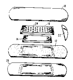
FIG 3 is an exploded isometric view of the
bandage of P°IG. 1.
~~~~'~~"l
_~,_
SN112
FIG. 4 is a top planar view of the bandage
of FIG. 1 with a layer of release paper partially
peeled away.
FIG. 5 is a cross sectional view of the
bandage of FIG. 2 with an alternate embodiment
protective cover taken along the lines and arrows
2-2 in FIG. 1 with no vacuum forming.
FIGS. 6A and 6B show the bandages of FIG. 2B
and S, respectively, in crass section applied to
the stoma of a user.
Referring now to the Figures, a bandage
designated generally 10 is shown having an
adhesive layer 12, a superabsorbent pad 14, a
porous cover pad 16 and a protective cover 18. The
adhesive layer 12 is a highly flexible, relatively
thin, occlusive hydrocolloid adhesive layer with a
backing layer 20 of polymeric material attached to
one surface thereof.
To make the bandage as flexible as
possible, it is desirable to keep the hydrocolloid
layer with backing as thin as possible. A
thickness of between 5 mils and 15 mils for the
layer 12 is suggested. In the preferred
embodiment, a thickness of between S and 12 mils
is used with 10 mils being most preferable. The
polymeric or non-woven fabric layer 20 may be
between 0.5 mils to 3 mils. Suitable non~woven
fabrics for use as layer 20 include polyester
fibers, polypropylene fibers, nylon fibers,
composite olefin fibers, and cellulose fibers.
Preferably, layer 20 is a polymeric film such as
polyethylene with 1 mil e~abossed polyethylene being
~~~~eu"~
-5-
SN112
most preferred. A suitable polyethylene film is
sold under the 'trade name Tafaflex Code XIX
available from Clopay U.S.A. The layer 12 is
formed by extruding and is laminated to layer 20.
Other polymeric backing films can be selected from
the various materials commonly employed in ostomy
and medical devices. For example, polyolefins such
as polypropylene, ethylene acrylic acids, ethylene
vinyl acetates, polyvinylchlorides, polyether
sulfones, polyether ketones, polyether urethanes,
polyurethanes, etc. can be used.
Unlike prior bandages used by continent
ostomates and others requiring like bandages, the
preferred adhesive layer 12 of the bandage 10 of
this invention is a skin friendly non-acrylic
material which is less irritating to the skin. The
overall adhesive layer 12 of this invention is
flexible and conformable to the body contours of
the user and is comfortable for the user.
The adhesive layer 12 is formulated by
blending one or more water soluble or swellable
hydrocolloids with a polyisobutylene or a mixture
of polyisobytylenes or a mixture of
polyisobutylenes and other nonacrylic elastomers.
Other materials can be included within the
adhesive formulations such as mineral oih.,
tackifiers, antioxidants, cohesive strengthening
agents, and pharmaceutically active materials such
as antiinflammatory agents, antiseptics, or
materials having skin healing or soothing
properties. Suitable occlusive adhesive
formulations are taught by Chen in U.S. Patent
3,339,546; Chen et al. in U.S. Patent 4,192,785;
Pawelchak et al. in U.S. Patent 4,393,080; Doyle
et al. in U.S. Patent 4,551,490; and by ICeyes et
-6-
SN112
al. in U.S. Patent 4,762,738. As disclosed in
these references, suitable water soluble and water
swellable hydrocolloids include sodium carboxy-
methylcellulose, pectin, gelatin, guar gum, locust
bean gum, gum karaya, and mixtures thereof.
Suitable cohesive strengthening agents include
water-insoluable cross-linked sodium caroxy-
rnethylcellulose, water-insoluble cross-linked dex-
tran, etc. Suitable non-acrylic elastomers in-
elude butyl rubber and styrene radial or block
copolymers such as styrene-butadiene-styrene
(S-B-S) and styrene-isoprenestyrene (S-I-S) block
type copolymers. Preferably, adhesive layer 12 is
an adhesive available from the Convatec division
of E.R. Squibb and Sons, Inc. used under the trade
name System III adhesive and is a blend on a weight
percentage basis of about 19% of a blend of poly-
isobutylenes (9.5% Vistanex~ LM-iii and 9.5%
Vistanex~ I~-100), about 14.5% mineral oil, and
about 6f.5% of an equal weight mixture of pectin,
gelatin. and sodium carboxymethylcellulose.
In one embodiment, porous cover pad 16 is a
1/16 inch thick square or rectangular pad made of
cellulose pulp (85 grams per square meter) and
polyolefin fibers (22 gsm) and covered by a non-
woven cover layer (20 gsm) on its top and bottom.
The non-woven cover layer is an air-laid, wet-laid
or spun-laid rayon, polyester or prefereably
polypropylene. The pad 16 is available in its
assembled state from Cellosoft Co. of Sweden and is
sold as catalogue #202.250. Alternatively, a pad
16 having a pattern of holes 24 formed through the
pad and comprising a combination of polypropylene
and tissue can be used. The pad includes a non-
woven polypropylene cover on top and bottom and is
available from IFC Non ~doven, Inc. of ,7ackson,
Florida>
~U~~;~~'~"~
_~_
srr112
Superabsorbent pad 14 is also about 1/16 inch
thick and made substantially the same as the pad 16
availabe from Cellosoft with superabsorbent powder
added thereto. A suitable superabsorbent is sold
as Salsorb 84 available from Allied Colloid. The
superabsorbent comprises about: 30% by weight of the
pad 14. "Superabsorbents" are' water insoluble
materials which are capable oa: absorbing and
retaining large amounts of water or other aqueous
fluid in comparison to their sawn weight. Dispos-
able goods manufactured using superabsorbents can
be more comfortable, less bulky, and longer lasting
than similar products made with traditional
absorbents such as cellulose fibers.
The bandage whose cross-section is depicted
in FIGS. 2A and 2~ is made by extruding a 10 rni1
layer of hydrocolloid adhesive and laminating it
with a 1 mil embossed polyethylene film on one side
and a layer of release paper on the other for
handling. The continuous web of combined hydro-
colloid adhesive, laminated backing layer and
release paper coming from the extruder moves past
several work stations to form the bandage. In a
continu~us fashion the release paper is removed
from the adhesive layer 12 to expose an adhesive
surface. At a next station, superabsorbent pads 1~4
are placed on the adhesive layer and covered by
cover pads 16. The pads 14 and 16 are pressed down
onto the adhesive layer by a platen. P~eanwhile, a
continuous narrow strip of release paper 22 is
applied to one edge of the adhesive surface. A
layer of release paper 1~ which covers the entire
width of the adhesive layer including the pads and
release paper strip 22 is applied. Alternatively,
a single widi:h of a layer of release paper can be
CA 02022577 2000-O1-19
_g_
applied over the adhesive. A score line can be
applied along one edge of the bandage so that the
release paper can be more easily removed. A
suitable release paper is a silicone treated paper
such as PolysilkT" 58003 available from HP Smith.
At the next work station, a vacuum is applied
to a region surrounding the pads on each side of
the web, i.e., to the polyethylene layer on one
side and the layer of release paper on the other.
The pads are contained within a vacuum chamber
along with a border of adhesive with an area of
polyetheylene on one side of the adhesive and an
area of release paper on top of the exposed border
area and the pads. The vacuum removes the air from
around the borders of the pads of the bandage
trapped between the adhesive layer 12 and the
release paper 18. See the regions 30 around pad 14
and pad 16 in FIG. 2A. When.the vacuum is removed
the adhesive layer 12 which is more flexible than
the release paper presses in against the edges of
the pads to eliminate most of the region 30. See
FIG. 28. The border region 32 in FIG. 2B of the
adhesive layer surrounding the pad 16 is sub-
stantially co-planar with the outwardly directed
surface 34 of the pad 16 opposite the surface in
contact with superabsorbent pad 14. A slight
bulge at the center of the pads 14 and 16 may
occur. The pads 16 assisted by the pattern of
holes 24 conforms quite well to the adhesive layer.
At the final work station, bandages are cut from
the vacuum formed portions of the web. Preferably,
each bandage is rectangular in shape with the
smaller sides having curved edges 26 and 28. Two
possibly sizes are: 4 1/4 inches by 3 inches and 3
inches by 2 5/8 inches. The superabsorbent pad 14
~p~~~~'~"~
..g_
srrllz
is 2 inches by 1 1/4 inches for the larger size and
1 1/4 inches by 1 inch for the smaller size
bandage, while the larger size porous cover pad 16
is 2 1/2 inches by 1 3/4 inches and the smaller
size is 1 3/4 inches by 1 1/2 inches. The 1/16
inch thick superabsorbent pads 14 of the size given
above are capable of absorbing 10-12 cc°s of
liquid. Of course, other size bandages and pads
are possible.
l0 The strip or tab 22 along one side of 'the
bandage facilitates removal of the release paper
18. Since the strip is itself release paper, the
strip and layer 18 are easily separated along this
edge and then the layer 18 can be stripped away.
The strip 22 is useful for holding one edge of
the bandage until it is applied and then it is
easily removed. FIG. 4 shows the release paper 18
partially peeled away to expose the strip 22.
Alternatively, as mentioned before, a single layer
of release paper could be applied over the pads and
adhesive border area and then scored or partially
cut through along one edge to assist in peeling
away the release paper.
In use, the release paper layer 18 is peeled
away starting at the overlap with strip 22. Then,
gripping strip 22, the bandage is applied to the
stoma or fistula with the pad 16 next to the stoma.
While the pad Z6 is somewhat absorbent it is also
porous and allows air and fluid to pass through.
Pad 16 functions to confine the discharge until
it is absorbed by the superabsorbent, keep the
superabsorbent from contacting the stoma at the
fistula and present a dry surface to the stoma or
fistula. The superabsorbent captures and fixes the
fluid or discharge. The non-woven layer on the pad
~~~~~s~"~~'~
-10-
SId112
16 next to the stoma or fistula remains dry. Since
the fluid is captured by the superabsorbent, the
fluid will not travel to the pad 16 from pad 14
even when the bandage is pressed.
The application of the vacuum to form the
adhesive layer 12 to the pads 14 and 16 removes
regions where fluid could collect without being
absorbed and provides a flat ;surface for
application to the skin and stoma. FIG. 6A shows
what would happen if the pad of FIG. 2A were
applied to the skin without first vacuum forming
the bandage. Region 30 is formed into reservoirs
30a and 30b about the pads where fluid might
collect. Using the vacuum formed bandage of FIG.
2B, FIG. 6B shows the reservoirs are reduced.
An alternate embodiment bandage designated
generally 40 is shown in FIG. 5. It comprises the
adhesive layer 12 with polymeric backing layer 20,
superabsorbent pad 14 with non-woven pad 16. The
protective cover 42 is a plastic part with a cavity
formed by dish portion 44. The depth and size of
the cavity is designed to accept the size and
thickness of the pads 14 and 16 where vacuum
forming of the bandage is not utilized. The
protective cover 42 may be thermoformed polyethy-
lene which is siliconized to make it releaseabie
from the adhesive border.
The bandage of this invention provides the
advantage of a skin friendly adhesive coupled with
a superabsorbent pad and a porous cover pad which
insulates th.e stoma from the superabsorbent. The
highly flexible and conformable hydrocolloid
adhesive is friendly to the periostomal skin upon
removal. Multiple bandages can be used each day.
The superabsorbent on the other hand absorbs a
-11-
SId112
relatively large quantity of liquid or discharge
from the stoma. The bandage thereby forms a
substitute for an external ostomy pouch and a good
customized design for continent ostomates or others
suffering with mucous fistulas.
When the bandage is exposed to a vacuum the
adhesive layer is substantially coplanar with the
exposed surface of the non-woven pad overlaying
the superabsorbent pad. The bandage is easily
applied to the stoma forming a low profile bandage
when applied.
