Note: Descriptions are shown in the official language in which they were submitted.
CA 02027434 1998-06-30
DIAGNOSTIC KIT FOR DIAGNOSING AND DISTINGUISHING
CHEST PAIN IN EARLY ONSET THEREOF
FIELD OF INVENTION
This invention relates to a novel diagnostic test which is used as a method of
providing an accurate simple, rapid, and portable diagnosis for assessing whether patient
chest pain is cardiac in origin and for differenti~ting between unstable angina and
myocardial infarction ("MI") at early onset of patient chest pain. In one embodiment, the
panel test will simultaneously assess the serum or plasma levels of three different
substances or markers found in serum or plasma during or after cardiac damage, I]tili7ing
an enzyme immunoassay sandwich dry chemistry format. In the preferred embodiment of
the invention, the three markers are creatine kinase (CK), myoglobin, and myosin light
chains (MLC).
BACKGROUND OF THE INVENTION
Emergency diagnosis of myocardial infarction has depended on physician acuity,
and an assessment of a patient's symptoms, such as chest pain or pressure, possibly
r~ ting down the arm and up the neck, fatigue, sense of impending doom, shortness of
breath, pallor, cold clammy skin, peripheral cyanosis or rapid thready pulse.
Most North American patients experiencing chest pain will report to a doctor or
emergency room within six (6) hours after the onset of the chest pain. It is therefore
essential that a diagnostic test be effective in the early stages of an MI.
Several cardiac tests have been used to detect MI. These tests include: ECG,
SGOT/AST, LDH, CK-MB Immunoassay and NA Latex Myoglobin Particle Enhanced
Assay. However, there is no single enzyme cardiac test which enables the emergency
department physician to identify the source of chest pain as cardiac or non-cardiac.
Further, it is only after a myocardial infarction has been confirmed may thrombolytic
therapy be initiated. The earlier such therapy is initiated, the greater likelihood of full
recovery of the patient or at least minimi7~tion of cardiac damage. It is therefore
essential for a physician to identify the pain as cardiac or non cardiac.
CA 02027434 1998-06-30
The electrocardiogram (ECG) may be used to detect an MI. However ECG is not
diagnostic until after the heart has suffered severe damage. The diagnostic specificity of
the ECG is only 51% in the initial phases of chest pain. Therefore, ECG is not suitable
for early detection of MI.
Serum glutamic oxalacetic tr~n~min~cc/aspartate transferase (SGOT/AST) is a
predominant enzyme found in high concentration in heart muscle. Serum tests to
determine levels of SGOT are used in diagnosing myocardial infarction. However,
SGOT only begins to rise about 8-10 hours following the onset of chest pain, peaks
within 24-36 hours and returns to normal after 5-7 days. SGOT is not particularly helpful
in diagnosing myocardial infarction in an emergency setting at an early stage of patient
chest pain. Also, SGOT is not specific to cardiac muscle. It is found in many tissues
including skeletal muscle, liver and kidney, being released as a result of intra muscular
injections, shock, during liver disease, and hepatic congestion, and is therefore of little
value in detecting specific cardiac tissue injury.
Lactate Dehydrogenase (LDH) is an enzyme found in high concentration in many
tissues, including heart, skeletal muscle and liver. Tests to detect the presence of LDH in
serum are used to diagnose myocardial infarction. There are five common isotypes of
which the heart contains predomin~ntly LDHl and LDH2. LDH levels begin to rise 24-36
hours after the onset of chest pain, and peak after 48-72 hours, returning to normal after
4-8 days. LDH is therefore not useful as an indicia of MI at an early stage of patient
chest pain. In addition, LDH is not specific to cardiac damage, and appears withpulmonary embolism, haemolysis, hepatic congestion, renal disease and skeletal muscle
damage. This lack of specificity also decreases the utility of LDH as a diagnostic aid.
Creatine kinase (CK) is an enzyme found in muscle tissue. CK catalyses the
conversion of creatine and adenosine triphosphate (ATP) to phosphocreatine and
adenosine diphosphate (ADP). One of several CK isoenzymes is CK-MB which is found
in cardiac tissue. CK-MB is a sensitive marker for the detection of myocardial infarction,
as it is released from damaged myocardium tissue. CK-MB thereafter is present in the
serum of an affected individual. Figure 1 illustrates the concentration of CK in the serum
CA 02027434 1998-06-30
of a patient as a function oftime. (ref. Lee T.H. et al. (1986) Ann. Intern. Med. 105, 221-233).
The CK-MB immunoassay is the standard diagnostic test for myocardial
infarction. A method describing the use of CK-MB is disclosed in U.S. Patent No.4,900,662 entitled "CK-MM Myocardial Infarction Immunoassay".
Shah, USP 4,900,662 discloses a method for deterrnining the initial elevated
concentration level of CK-MM-a, an isoform of CK-MM, and CK-MM-a and CK-MM-b
concurrently in patient serum following a myocardial infarction. Use of the method
provides an accurate estimation ofthe time ofthe infarction. The method involvesdeterrnining the combined concentration of CK-MM-a and CK-MM-b and the
concentration of CK-MM-a in serum, in order to determine the time of the acute phase of
myocardial infarction. Reagents are disclosed and comprise novel polyclonal and
monoclonal antibodies for CK-MM-a which do not bind significantly with CK-MB, CK-
MM-b or CK-MM-c, an anti-CK-MM-b antibody which does not bind significantly withCK-MB, CK-MM-a or CK-MM-c, an anti-CK-MM-a+b antibody which binds with CK-
MM-a and CK-MM-b but does not bind significantly with CK-MB or CK-MM-c, labelledderivatives of these antibodies, insoluble supports to which these antibodies are adhered,
and kits cont:~ining one or more of these reagents. Enzyme labelled and radiolabelled CK
reagents are particularly useful.
There are difficulties with the use of CK-MB alone as a diagnostic marker. First,
serum levels of CK-MB do not peak until 12 hours after the onset of myocardial
infarction, making early emergency diagnosis and treatment diff1cult.
Secondly, the CK-MB test must be conducted in a laboratory by trained laboratorytechnicians. In non-urban locations, it may not be feasible to have the test conducted and
the results interpreted expeditiously, resulting in increased delay in diagnosis and hence
increased costs to the health care system in terms of hospitalization costs of a patient
awaiting diagnosis.
Thirdly, CK-MB has been located in normal skeletal muscle tissue, consequently
rendering the test less cardiac specific, and the diagnosis less certain.
CA 02027434 1998-06-30
;
Myoglobin is another protein located near the skeletal or myocardial cell
membrane. It is expelled from the cell as soon as the cell membrane becomes abnormally
permeable, for example, during myocardial ischemia, a reversible state. Myoglobin is
detectable in the serum within 1.5 hours of the onset of chest pain. The medical research
5 community believes that myoglobin is released by myocardial necrosis, and it is therefore
a useful early marker of myocardial injury. Figure 2 illustrates the concentration of
myoglobin in the serum as a function oftime. (ref. Grenadier E. et al. (1981) Am. Heart J.
105, 408-416; Seguin J. et al. (1988) J. Thorac. Cardiovasc. Surg. 95, 294-297).
In determining the origin of chest pain, an acute myocardial infarction can be
excluded if no elevation of serum myoglobin is detected within 2 - 3 hours after the onset
of pam.
An NA Latex Myoglobin Particle Enhanced Assay is a commercially available
assay kit for the detection of myoglobin. The assay is based on the reaction between
antigens present in human body fluids and antimyoglobin antibodies covalently coupled
to polystyrene particles. The sample, N Myoglobin Reagent, a solution for the
elimin~tion of nonspecific reactions and N Reaction Buffer are pipetted automatically
into a cuvette. Light scattering is measured by a nephelometric procedure after 12
minutes of incubation time and the myoglobin concentration is calculated from a
calibration curve.
Myoglobin may also be assayed using a radioimmunoassay but there is no
enzyme-linked immunosorbent assay (ELISA) format yet available.
There are difficulties with the use of myoglobin alone as a diagnostic marker.
Myoglobin does not indicate a particular type of myocardial injury, such as myocardial
infarction. Myoglobin can also be present during such diverse conditions as shock, renal
disease, rhabdomyolysis, and myopathies. Additionally, myoglobin concentrations in
serum and plasma generally depend on age and sex and vary over a wide range in normal
healthy humans. Serum concentrations up to 90 llg/L are generally regarded as the upper
limit of the reference range for healthy people. Therefore, what may be a normal level for
CA 02027434 1998-06-30
.
one individual may be indicative of a serious problem in another individual, making
diagnosis somewhat less accurate than would be desirable.
Myosin light chains (MLC) are integral parts of the myosin myofibril, but their
S functional role is still unclear. MLCs exist in slow, fast, atrial, and ventricular muscles.
It is known that MLCs are highly sensitive for myocardial ischemia. MLCs appear in the
serum rapidly, and their levels remain elevated for up to 10 days following myocardial
necrosis. Figure 3 illustrates the concentration of MLC in patient serum as a function of
time. (ref. Wang J. et al. (1989) Clin. Chimica. Acta 181, 325-336; Jackowski G.,
Symmes J. C. et al. (1989) Circulation Suppl. 11 80,355.) MLC also has prognostic
value in deterrnining the success of thrombolytic therapy. Higher levels of MLC, indicate
a worse prognosis, and also corresponds to a larger infarction. Falling levels over several
days indicate a tendency towards patient recovery, whereas a spiking or stadico pattern
indicates a tendency towards infarction and a need for intervention.
There are two principal types of MLC known as MLC1 and MLC2, which exist as
a soluble pool in the myocardial cell cytoplasm and also integral with the myosin
myofibril. In the ventricular muscle, MLC2, and perhaps MLCl, is identical with the
isotype expressed in slow skeletal muscle. MLCl is elevated in 80-85% of the patients
with cardiac pain. MLCl is a very sensitive indicator of unstable angina and coronary
heart disease.
Other cardiac markers, low molecular weight cardiac proteins (LMWCP) may be
used as cardiac markers. Examples of such cardiac markers include components of the
contractile apparatus, namely, troponin, troponin-T, troponin-I and troponin C,
mitochondrial enzymes, such as triose P isomerase, low molecular weight polypeptides
which are readily released from the heart, and sarcolemmal membrane proteins or protein
fragments which may be released early after the onset of ischemia, in particular, a 15kd
sarcolemma protein and a 100kd complex glycoprotein which are cardiac specific.
The cardiac isotype troponin-I inhibits the interaction between actin and myosinmolecules during rest periods between conkactions of the heart muscle. Troponin-I
appears in serum of patient within 4-6 hours after MI and remains elevated for 7 - 8 days.
CA 02027434 1998-06-30
- 6 -
Figure 4 illustrates the concentration of troponin-I as a function of time. (ref. Cllmmin~
B., Auckland M.L. and Cllmmin~ P. (1987) Am. Heart J. 113, 1333-1344.) It is cardiac
specific and has a greater sensitivity than other markers in detecting cardiac versus
skeletal muscle injury.
Troponin-T is part of the troponin-tropomyosin complex of the thin filament and
serves as a link between the tropomyosin backbone and the troponin-I troponin C
complex. Troponin-T is a basic protein and has isotypes in cardiac and fast and slow
skeletal muscles. It appears in serum within 3 hours and remains elevated for at least 10
days following MI. Figure 5 illustrates the concentration of troponin-T as a function of
time. (ref. Katus H.A. et al. (1989) J. Mol. Cell Cardiol. 21, 1349-1353.) Troponin-T
follows a biphasic release pattern. It is cardiac specific and very sensitive for MI.
Myosin heavy chains (MHC), and tropomyosin, are heavier molecular weight
proteins which may also be used as cardiac markers. MHC is part of the major contractile
protein of muscle. Fragments of MHC can be released from the ventricule into serum
after myocardial cell necrosis and subsequent irreversible membrane injury. Although
MHC fragments do not appear quickly in the serum following myocardial cell necrosis,
MHCs remain elevated for at least 10 days following MI, and peak levels of MHC are
observed 4 days after MI. Figure 6 illustrates the concentration of MHC as a function of
time. (ref. Leger J.O.C. et al. (1985) Eur. J. of Clin. Invet. 15, 422-429, Seguin J.R. et al.
(1989) J Thorac. Cardiovasc. Surg. 98, 397-401.) The area under the MHC release curve
correlates very well with the extent of myocardial cell damage. However, MHC levels
are of little clinical value during the acute phase of MI.
Tropomyosin is a dimer formed from two polypeptides which are part of the
regulatory system in muscle contraction. Tropomyosin is detectable in serum
approximately 7-8 hours after myocardial infarction, and like CK-MB, is very sensitive
for myocardial infarction. Figure 7 illustrates the concentration of tropomyosin as a
function oftime. (ref. Cllmmin.~ P. et al. (1981) Clin. Sci. 60, 251-259) However,
tropomyosin is not cardiac specific since it is elevated in conditions of skeletal muscle
trauma.
CA 02027434 1998-06-30
There are limitations for each of the current standard diagnostic methods for
myocardial infarction. None provide a highly sensitive, specific, rapid, and simple
diagnostic test which may be conducted soon after the onset of chest pain, for example, in
an ambulance or doctor's office.
The present invention combines and measures three different markers of cardiac
damage present in the blood or serum of a patient in early onset of chest pain in order to
provide an improved method of diagnosis of myocardial infarction for use in the early
stages of unstable angina or MI. It is contemplated that additional markers may be used
as well.
SUMMMARY OF THE INVENTION
The disadvantages of the prior art may be overcome by providing an accurate,
rapid, and portable diagnostic test to be used in emergency settings to detect the presence
of three (or more) markers of cardiac damage in a patient's serum. The test results will
determine whether the patient is suffering from unstable angina or whether a myocardial
infarction has taken place. Early detection of MI enables thrombolytic therapy to be
commenced at an early stage. Cardiac damage will therefore be minimi7:~d, and the
patient's chance of survival will be increased. The results of the test will distinguish
between unstable angina and myocardial infarction, even up to several days following the
onset of pain. In a preferred embodiment, the panel test will utilize an enzyme
immunoassay sandwich dry chemistry format. Serial temporal measurements with thepanel will offer prognostic information to the physician as to the extent of muscle damage
and the success of thrombolytic intervention. In the preferred embodiment of theinvention, the three markers are creatine kinase (CK), myoglobin, and myosin light chains
(MLC).
According to one aspect of the invention, there is provided a kit for rapidly
diagnosing the cause of chest pain in the early onset thereof. The kit comprises three
antibodies specific for each of three different markers of cardiac damage present in blood,
plasma or serum, the antibodies being selected so that one binds to an ischemic marker,
another binds to an ischemic marker released only as a result of myocardial infarction and
CA 02027434 1998-06-30
a third binds to a cardiac specific ischemic marker or an ischemic marker released only as
a result of myocardial infarction, wherein at least one of the ischemic markers released
only as a result of myocardial infarction is cardiac specific. It is contemplated that there
may be more than three such antibodies and markers. The combined response of reagents
indicates the diagnostic condition of the patient. According to a further aspect of the
invention, the kit further comprises means for detecting binding of each antibody with its
marker. In a preferred embodiment, one antibody binds to myoglobin. In a furtherpreferred embodiment, one antibody binds to CK-MB. In a yet further preferred
embodiment, one antibody binds to troponin I.
In accordance with another aspect of the invention, there is provided a kit for
rapidly diagnosing the cause of chest pain of a patient in the early onset thereof which
comprises three antibodies specific for three markers, where the first antibody specifically
binds myoglobin, the second antibody specifically binds CK-MB and the third antibody
specifically binds to a protein selected from the group con~ ting of myosin light chain,
troponin I and troponin T. Again, it is contemplated that there may be more than three
such antibodies and markers.
The antibodies may be labelled and the means for detecting binding of each
antibody can be a labelled detector reagent.
In accordance with another aspect of the invention, there is provided a method for
rapidly diagnosing the cause of chest pain of a patient in the early onset thereof. The
method comprises (a) simultaneously detecting the presence of increased levels of three
different markers of cardiac damage present in a blood, serum or plasma sample from a
patient after onset of chest pain by (i) contacting the sample with three antibodies wherein
a first antibody binds to an ischemic marker, a second antibody binds to an ischemic
marker released only as a result of myocardial infarction, and a third antibody binds to a
cardiac specific ischemic marker or an ischemic marker released only as a result of
myocardial infarction, wherein at least one of the ischemic markers released only as a
result of myocardial infarction is cardiac specific, each of the antibodies being specific to
a different one of the three markers and (ii) detecting binding of the antibodies to the
markers, whereby the presence of increased levels of a marker in the sample is indicated
CA 02027434 1998-06-30
by binding of the marker with the respective antibody and (b) correlating the detected
presence or absence of increased levels of each of the three markers, thereby to determine
the cause of chest pain. In a further method embodiment, a first antibody specifically
binds myoglobin, a second antibody specifically binds CK-MB and a third antibodyS specifically binds to a protein selected from the group con~ ting of myosin light chain,
troponin I and troponin T. Again, it is contemplated that there may be more than three
such antibodies and markers. The detection of increased levels of markers can bedetected by homogeneous immunoassay or heterogeneous immunoassay.
As will be apparent from this specification, the word "simultaneous" or
"simultaneously" as it applies to the present invention does not necessarily mean only "at
the same time".
In drawings which illustrate the embodiments of the invention,
Figure 1 is a graph illustrating the level of CK in serum as a function of
time;
Figure 2 is a graph illustrating the level of myoglobin in serum as a function
of time;
Figure 3 is a graph illustrating the level of MLC in serum as a function of
time;
Figure 4 is a graph illustrating the level of troponin-I in serum as a function of time;
Figure S is a graph illustrating the level of troponin-T in serum as a function of time;
Figure 6 is a graph illustrating the level of MHC in serum as a function of
time;
CA 02027434 1998-06-30
- 10-
Figure 7 is a graph illustrating the level of tropomyosin in serum as a
function of time;
Figure 8 is a plan view of the preferred embodiment;
Figure 9 is an exploded perspective view of the embodiment of Figure 8;
Figure 10 is an oblique view of the membrane of the embodiment of Figure 8;
and
Figure 1 1 is an oblique view of a second embodiment of the membrane.
DETAILED DESCRIPTION OF THE INVENTION
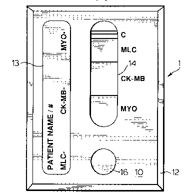
The invention is generally illustrated in Figure 8. In the pl~rel.ed embodiment the
invention is in a panel format identified as 1.
The panel format to be used is known and is commercially available. The panel
format is similar to a format currently being used in association with pregnancy testing
and is commercially available under the trade-mark BIOSIGN.
The panel consists of a polypropylene card having a front panel 10 and a back
panel 12. Front panel 10 has a display window 14, one for each cardiac marker and a
sample window 16, as illustrated in Fig. 8. Underneath front panel 10 is an exposed dry
chemistry membrane 18 which is affixed to the back of front panel 10 by suitable means.
Back panel 12 is provided with a lip 20 which extends around the perimeter of back panel
12 for receiving front panel 10 in a snap fit thereby sealing the membrane 18 between the
front and back panel.
While the front and back panel have been described as being snapped together,
there are numerous other suitable methods of joining the two together which would be
a~p~lll to a person skilled in the art.
CA 02027434 1998-06-30
Front panel 10 may also be provided with an area 13 upon which the patient's
name or identification may be written. Also space may be available to write the results of
the test.
With reference to figure 10, membrane 18 is the carrier of the monoclonal or
polyclonal antibodies. In the preferred embodiment, the flow of blood or serum is from
one end to the other end as shown by the arrow. End 22 is aligned with sample window
16. An immobilized captured antibody 24 is layered against or bonded to an antibody-
enzyme conjugate 26 which is directed against a different epitope on the antigen than that
which is recognized by the antibody 24. Antibody 24 is complementary to the myoglobin
protein. Similarly, antibody 28 is layered with a corresponding reagent 30. Antibody 28
is complementary to CK-MB. Likewise, antibody 32 is layered with a reagent 34.
Antibody 32 is complementary with the myosin light chain. Antibody 36 is one which is
complementary to any protein found in normal serum or blood. Antibody 36 is layered
with reagent 38.
The monoclonal and polyclonal antibodies can be prepared by using conventional
procedures with any m~mm~l used for polyclonal antibody production.
In the preferred embodiment, a labelled detector reagent is used. The antibody
detector reagent is labelled or chemically bonded to a distinctive moiety which can be
observed or measured to verify or quantify the presence of an antibody in the serum or
blood or on the dry chemistry membrane. Ligands and groups which can be conjugated
to the antibodies of this invention for use as a diagnostic tool include elements,
compounds or biological materials which have physical or chemical characteristics which
can be used to distinguish the antibodies to which they are bonded from other antibodies.
At least two antibodies of the type monoclonal or polyclonal per cardiac marker
are required. The antibodies are aff1nity purified against their specific cardiac
immunogen and then further purified by cross-adsorption against a non-related species to
elimin~te non-specific immunoglobulins.
CA 02027434 1998-06-30
In use, the diagnostician, for example a physician, ambulance attendant or nurse,
adds three drops or less than 100 ~lL ofthe patient's serum or blood to the sample
window 16. The sample will migrate along the membrane 18 by capillary action and will
successively come into contact with the antibody and reagent pairs 24 and 26, 28 and 30,
32and34and36and38.
The specific cardiac marker if present in the sample binds to the antibody
immobilized on the membrane. The corresponding detector reagent will also react and is
visualized by a change in colour of the detector reagent. The colour change is
proportional to the concentration of the marker in the sample. Therefore if the test kit is
used in timed intervals the increase or decrease in marker concentration can also be
determined and used as a diagnostic tool. The results of the test should be completed
within 3 - 5 minutes.
In the plere.led embodiment, a blue band will show for each cardiac marker
which is present in the sample. The intensity ofthe band is qll~ntifi~ble using a
reflectometer, which relates the colour intensity to the concentration level of a particular
marker. The reflectometer may contain a microprocessor, so that the quantified result for
each cardiac marker being tested in the panel may be produced and printed out as a
concentration of each marker along with the patient's name or identification.
The test preferably is sensitive to marker concentrations from .5ng/ml to 25ng/ml
using 3 drops or less than 1 OO~lL of serum or plasma with a within run and between run
precision coefficient of variation of less than 15%.
The cardiac markers utilized in the test will depend on the properties of those
markers. In the preferred embodiment, there will be a panel having myoglobin, MLC,
and CK-MB, as illustrated in Figure 8.
Myoglobin is released very early from the myocardial cell, is not cardiac specific,
has a very high sensitivity for myocardial infarction and necrosis and is not released by
anoxic injury in the absence of necrosis. MLC is cardiac specific, and permits
differentiation of cardiac from non cardiac pain, and is released early but not as early as
CA 02027434 1998-06-30
myoglobin. CK-MB differentiates angina from myocardial infarction, but is not
detectable until approximately six hours after the onset of chest pain and therefore is not
of use alone as an emergency diagnostic test.
Referring to figures 1, 2 and 3 and if the three cardiac markers to be used are CK-
MB, myoglobin, and MLC, the following int~ lion of the results would provide a
diagnosis.
If the panel shows positive for MLC and negative for myoglobin and CK-MB, it
would indicate that the patient's chest pain is cardiac and that the source is unstable
angina.
If myoglobin and MLC are positive and CK-MB is negative it would indicate an
early evolving myocardial infarction and intervention therapy could be initiated.
If all three are positive, it would indicate a myocardial infarction.
If MLC and CK-MB are positive and myoglobin is negative, it would indicate a
myocardial infarction.
If myoglobin and CK-MB are positive and MLC is negative, the patient could
have skeletal muscle trauma (a false positive) or be in the midst of a myocardial
infarction.
The test could not distinguish between a false positive and a "small" myocardialinfarction in this case, as the MLC release curve has slight dips at several intervals and
the patient may have a small subendocardial infarction and be tested at the time of a
"dip". When the infarct is small, the "dip" is down to almost normal levels, and therefore
the patient would test negative for MLC. Positive diagnosis would rely on the presence
of CK-MB.
CA 02027434 1998-06-30
- 14-
In the event that the patient is having a large myocardial infarction, the "dip" in
MLC levels will not be so large as to be the same as normal levels, and therefore, MLC
will remain detectable.
In other embodiments, the test panel may utilize different combinations of
antibodies in the same format, such that different cardiac markers are assessed. In order
to ensure that the panel will detect cardiac tissue damage at an early stage of patient chest
pain, it is necessary to utilize at least one antibody corresponding to a marker which is
present in large quantities at an early stage of cardiac damage, such as CK, myosin light
chains or myoglobin. Low molecular weight cardiac proteins having the characteristics
and properties of CK, myosin light chains or myoglobin may also be used in the kit.
Suitable proteins and enzymes may be selected from the following: troponin,
troponin-I, troponin C, troponin-T and sarcolemmal membrane proteins, triose P
isomerase or any heavy molecular weight cardiac proteins having the characteristics and
properties of creatine kinase, myoglobin or myosin light chains.
Other proteins such as tropomyosin, and myosin heavy chains may also be added
to the kit. The kit would then be able to detect MI if the patient arrives for diagnosis
many hours after onset of chest pain where the patient is in the later stages of MI.
In a second embodiment, membrane 18 may have a layer of captured antibody 124
and a corresponding reagent 126. Similarly for each other marker to be detected, a
corresponding pair of antibodies and reagents are provided, i.e. 128 and 130, 132 and 134
and control pair 136 and 138. In use, the sample is dropped onto each pair and the results
are read in the same manner as described above.
The dry chemistry membrane 118 can be supported by absorbent material 120.
Absorbent material 120 will enhance the draw of the serum through the membrane.
A further embodiment for the test kit is to use a blood sample tube which is
commonly used to draw blood samples from patients. The inside wall of the tube could
act as a carrier for the monoclonal and polyclonal antibodies and reagents. After the
CA 02027434 1998-06-30
- 15 -
sample is drawn from the patient, the user simply shakes the tube so that the antibody
reacts with the blood. Colour changes as described above will take place if the cardiac
protein is present in the blood.
Although the disclosure describes and illustrates p~f~ d embodiments of the
invention, it is to be understood that the invention is not limited to these particular
embodiments. Many variations and modifications will now occur to those skilled in the
art. For a definition of the invention, reference is to be made to the appended claims.
MCTET2#3531333