Note: Descriptions are shown in the official language in which they were submitted.
~3~
PATENT
INCISE SYSTEM
BACKGROUND OF THE INVENTION
The present invention relates to an incise material
suitable for use in surgical procedures. ~ore specifically,
the incise material is a two layer structure which has a
removable top layer to facilitate su~uring of an incision upon
completion of a surgical procedure.
Many of today~s surgical procedures involve the use of
an incise material. An incise material is usually a clear
polymeric film with an adhesive on one side which is in turn
covered with a release paper. Two suppliers of incise
material are the Minnesota Mining and ManufactUring Company
and T. J. Smith and Nephew Ltd. Examples of incise material
can be found, by way of example only, in U.S. Patent Nos.
4,310,509; 4,323,5S7; 4,45~,845; RE. 31,886 and RE. 31,887.
Most typically incise material is used in connection with
towels or surgical drapes to maintain the surgical area as
clean and sterile as possible to help reduce the risk of
postoperative infection. Once the surgical area of the
patient has been scrubbed and treated with a bacteriostat, the
surgical site is squared-off by the use of sterile towels or
a surgical drape which has a fenestration of a size which ls
larger than the expected size of the incision. An incise
material is then used to cover all or a portion of the
patient's skin left exposed by the towels or the fenestration
in the surgical drape or mainsheet. Some surgeons prefer to
use incise materials which themselves have fenestrations which
are only slightly larger than the incision area. Other
surgaons prefer to use incise materials which completely cover
the incision area with the incision being made directly
through the incise material. In either case, to apply the
incise the releasable backing is removed and the adhesive side
of the material is applied directly to the skin of the
,
---` 203~
patient. one purpose in using the incise material is to help
reduce the migration of germs and bacteria to the inclsion
site. This is because, despite the cleansing o~ the skin, the
pores still contain additional germs and bacteria which can
migrate to ~he surface as the skin is moved and worked during
the course o~ the surgical proce~ure. By covering the skin,
it has been found that this migration can be reduced.
When an incise is used which completely covers the
incision area the surgeon will cut right through the incise
material attached to the skin. In this manner the amount of
exposed skin surrounding the incision is minimized. A problem
arises, however, when it comes time to close the incision.
At this point the incise material directly surrounding the
incision must be peeled back so as to expose sufficient skin
for the suturing procedure. This process of peeling the
incise back is, at the very least, a time consuming and
annoying process. Because of this problem, some surgeons
elect to use the above-described fenestrated incise material
to eliminate the need for peeling back the incise material.
This, however, is often a compromise decision since, in using
a fenestrated incise material, more skin is exposed about the
incision site thereby increasing the risk of bacteria and
germs making their way to the incision and causing infection.
It is therefore an object of the present invention to
provide an incise system which will allow the surgeon to cut
directly through the incise material while at the same time
permitting easy exposure of the skin about the incision when
it comes time to close the incision~ This and other objects
of the present invention will become more apparent upon a
further review of the following specification, drawings and
claims.
- , ,
:
'
~3~8P~
BRIEF DESCRIPTION OF THE DRAWINGS
Fi~ure 1 is a top plan view o~ an incise system according
to the present invention.
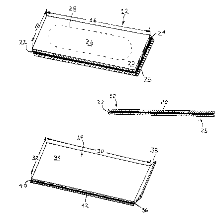
Figure 2A is a perspective ~iew of the first or bottom
layer of an incise system according to the present invention.
Figure 2B is a side view of the material shown in Figure
2A.
Figure 2C is a perspective view of the second or top
layer of an incise system according to the present invèntion.
Figure 2D is a side view of the material shown in Figure
2C.
Figure 2E is a side view of an incise system according
to the present invention.
Figure 3 is a top plan view o~ an incise system according
to the present invention attached to a surgical drape with the
second layer of incise being separated from the first layer.
Figure 4 is a cross-sectional view of Figure 3 taken
along line 4-4.
Figure 5 is a side view of the first and second layers
of incise with separate release liners according to the
present invention.
Figure 6 is a side view of a first and second layer of
incise when joined along a common edge according to the
present invention.
- 3 -
' :
, :~
., ~, '~
~3~
Figure 7 is another perspective view of an incise system
according to the present invention.
DESCRIPTION OF THE PREFERRED EMBODIMENT
Referring to Figures 1 through 7 there is shown an incise
- system 10 according to the present invention. The incise
system 10 is comprised of a first layer 12 and a second layer
14 of incise. Such incise is readily available and well known
to those having ordinary skill in the art. Examples of an
incise including adhesive suitable for the present invention
include Opsite~ Incise 63110; 63210; 63310 and 63410 from T.
J. Smith and Nephew Inc., 2000 South Beltline Blvd., Columbia,
South Carolina, U.S.A. 29205. The incise system 10 including
the first layer 12 and second layer 14 may be manu~actured and
sold as a separate unit or it may be sold as part of a
surgical pack in which case the system 10 may be unattached
or pre-attached to a surgical drape or mainsheet 11 having a
primary fenestration 13, a top edge 8 and a bottom edge 9
joined by a pair of opposed side edges 15 and 17. The
mainsheet 11 further includes a top surface 19 and a bottom
surface 21. See Figure 3. In any event, the first layer 12
should be of a size sufficient to cover all sides of the
fenestration 13 in the surgical drape 11.
Referring to Figures 2A and 2B, the first layer 12 is a
continuous layer of incise having a first length 16 and a
first width 18. The first layer 12 has a top surface 20 and
a bottom surface 22. Affixed to the bottom surface 22 is a
first adhesive layer 24, the composition of which is well
known to those having ordinary skill in the art. Attached to
the first adhesive layer 24 is a release paper 25, the
composition of which is also well known to those having
ordinary skill in the art. AAditionally, the first layer 12
has a separate removable fenestrated area 26 located within
and connected to the first layer 12 by a line of perforations
-- 4 --
. .,~ , .
, . . , ~
203~7
28 which will permit the fenestrated area 26 to be separated
fro~ the remainder of the first layer 12 when sufficient force
is exerted on the perforations 28. The fenestration area 26
should generally be at least an inch longer at either end than
the intended length of the incision. The width of the
fenestration area 26 should also generally be at least an inch
wider on either side of the incision. This will assure that
their is sufficient skin exposed to permit suturing.
Referring to Figures 2C and 2D, the second layer of
incise 14 is a continuous sheet having a second length 30 and
a second width 32 which are generally coextensive with the
first length 16 and first width 18 of the first layer 12.
Layer 14 also has a top surface 34 and a bottom surface 36.
As shown in Figures 1 and 7, first layer 12 is slightly larger
than the second layer 14, however, the first layer 12 also may
be smaller than or equal in size to the second layer 14 so
long as the second layer 14 covers the perforated fenestration
area 26 of the first layer 12. To facilitate removal of the
second layer 14 from the first layer 12, the second layer 14
may be provided with one or more pull tabs 38 located along
the edges of the second layer 14. In its simplest form the
pull tab 38 may be a section of the second layer 14 folded
over and adhered to itself via an adhesive. Alternatively,
the pull tab 38 may be a separate element such as an adhesive-
coated paper with instructions or other indicia locatedthereon and affixed to one of the ends of the second layer 14.
The tab 38 should be of sufficient size to permit adequate
grasping and pulling of the tab 38 when releasing the second
layer 14 from the first layer 12. Generally a height of one
inch or more should suffice.
To releasably adhere the first layer 12 and the second
layer 14 together, a second adhesive layer 40 similar to the
previously mentioned adhesive layer 2~ is positioned between
the two layers 12 and 14. This second adhesive layer 40 may
be applied to the top surface 20 of the ~irst layer 12 or to
'~ g3 3 ~
the bottom surface 36 of the second layer 14. In Figure 2D
the adhesive layer 40 is shown attached to the bottom surface
36 of the second layer 14. Generally, the two layers 12 and
14 are adhered to one another during the manufacturing of the
present invention so that the two layers may be applied
together to the patient's skin thereby lessening the chance
of wrinkling. See Figure 2E. Alternatively, however, as
shown in Figure 2D a release liner 42 may be attached to one
side of the axposed second adhesive layer 40 to keep the two
layers 12 and 14 separated until attachment of the two layers
is desired. In such a configuration, the first and second
layers 12 and 14 may be completely separated as shown in
Figure 5. On the other hand, the two layers 12 and 14 may be
joined along a common edge 44 as shown in Figure 6. In
addition, this common edge 44 may be attached to the top
surface 19 of the mainsheet 11 adjacent the fenestration 13.
Most commonly this edge 44 is opposite the pull tab 38.
Furthermore, the release paper 42 should not extend completely
across the juxtaposed bottom surface 36 of second layer 14 and
the top surface 20 of the bottom layer 12. Instead, the
release paper 42 should extend from the end of the layers 12
and 14 adjacent the pull tab 38 to a point adjacent the joined
common edge 44. In this manner the adhesive 40 can be used
to adhere and hinge the two layers 12 and 14 together. See
Figure 6.
As stated at the outset, the incise system 10 of the
present invention may be either unattached to the drape (as
shown in Figures 1, 5, 6 and 7) or preattached (as shown in
Figures 3 and 4). When the incise system is attached to the
drape 11, it should be large enough to cover the fenestration
13 in the drape 11 as shown in Figures 3 and 4. Also, in most
cases the release paper 25 will be attached ~o the underside
of the drape 11 as shown in cross-section in Figure 4 via the
adhesive layer 24.
.
:
Having thus described several embodiments of the incise
system 10, the application and removal of the system will be
explained .
When the incise system 10 of the present invention is
preattached to a surgical drape as is the case in Figures 3
and 4, the drape ll is applied to the patient in the same
manner as would normally be prescribed for the specific type
of drape. To apply the incise, the release paper 25 is
removed thereby exposing the adhesive 24 for adhesion to the
patient's skin (not shown) in the intended area of the
surgical procedure. The only added precaution is that the
fenestrated area 26 be properly aligned so that the incision
50 is made within the confines of the fenestrated area 26.
once the drape 11 and incise system 10 are in place, an
incision 50 can be made through layer 12, the fenestrated area
2 6 of layer 14 and the patient's skin within the area defined
by fenestrated area 26. Both the skin and incise can then be
pulled back and clamped to facilitate the surgical procedure.
To use the incise system shown in Figure 5 which employs
two release papers 25 and 42, the two layers are applied in
sequence with the first layer 12 being first applied to the
skin of the patient (not shown) followed by the application
of layer 14. As with the first embodiment, once the system
has been applied, an incision is made in the skin through the
two layers of incise 12 and 14 in the area of the perforated
fenestration area 26 of layer 12. Both the skin and the
incise can then be pulled back and clamped to facilitate the
surgical procedure.
To use the incise system 10 shown in Figures 2E and 7,
the release paper 25 is removed from the bottom 22 of the
system 10 thereby exposing the adhesive 24 on the bottom of
the first layer 12. The adhesive side is then applied to the
skin (not shown) of the patient and an incision is made
through the skin and the area of the second layer 14 which
directly overlies the fenestration area 26 in the first layer
-
.
~3~
12. Both the skin and the incise can then be pulled back and
clamped to facilitate the surgical procedure.
To use the incise system 10 shown in Figure 6, the
release paper 25 is remo~ed from the bottom 22 of the system
10 thereby exposing the adhesive 24 on the bottom of the first
layer 12. The adhesive 24 is applied to the patient's skin
such that the fenestrated area 26 directly overlies the
intended site of the incision. Next, the release liner 42 is
removed from the second layer of incise 14 thereby exposing
the adhesive 40 for adhesion of the second layer 14 to the top
of the first layer 12. As with the other embodiments, once
the system 10 has been applied, an incision is made through
the skin (not shown) and the two layers of incise 12 and 14
in the area of the perforated fenestration area 26 of layer
lS 12. Both the skin and the incise can then be pulled back and
clamped to facilitate the surgical procedure.
Once the procedure has progressed to the point of
closure, the top or second layer 14 is removed by grasping and
pulling the tab 38 in the direction of arrows 36 as shown in
Figure 3. As the second layer is pulled/peeled back, the
perforated fenestration area 26 of first layer 12 is removed
along with the second layer 14 thereby providing an area of
exposed s~in about the incision to permit suturlng without
interference from the incise material. To ensure the
fenestrated area 26 of the first layer 12 is peeled off along
with the second layer 14, the adhesive peel strength of the
second layer 14 should be great enough to overcome the force
of the adhesive 24 holding the fenestrated area 26 to the
patient's skin and the force needed to separate the perforated
fe}lestration area 26 from the remainder of the first layer 12.
Consequently, the peel strength o~ the second layer 14 (second
peel strength) should be greater than that of the first layer
12 (first peel s~rength). One standard that can be used to
measure such peel forces/strengths is PSTC-l (November 1970
revised version of the September 1955 test) developed by the
. -: , :, '
.
. - . . : .
.~. ~, ` , ' .
r3;
Pressure Sensative Tape Council, 1201 Waukegan Road, Glenview,
Illinois 60025. Also note that to make separation of the
perforated fenestration area 26 from the remainder of first
layer 12 easier, the number of perforations 28 can be
increased. Note too that in removing the second layer 14 from
the first layer 12 it may be necessary to hold down the first
layer 12 to assist in keeping the lower layer 12 from
separating prematurely from the skin of the patient. Finally,
having completed the suturing, the first layer 12 of incise
may be removed from the patient, most commonly in conjunction
with the removal of the main sheet 11.
Having thus described ~he invention in detail it should
be appreciated ~hat various other modifications can be made
without departing from the spirit and scope of the appended
claims. For example, the fenestrated area Z6 instead of being
located entirely within the incise may extend over to one
edge of the incise to create a "U-shaped" or other cut-out
portion for limb surgery.
.. .
' ' ' ' :
.,
,
:
