Note: Descriptions are shown in the official language in which they were submitted.
-1- 203;~S95
3 CARDIOTOMY RES~SRVOIR
Field of the Invention
6 The present invention pertains to cardiotomy
7 reservoir structures. Cardiotomy reservoirs are
8 currently used in major surgical procedures, such as open
9 heart surgery, for receiving blood from a cardiotomy
~ sucker and other sources, defoaming the blood, filtering
11 out debris and returning it to the patient.
12
13 Description of Prior Art
14 U.S. 3,507,395 discloses a cardiotomy reservoir
which includes a plate in the path of incoming blood for
16 spreading the blood into a thin sheet before it is passed
l7 through filter material. The thin sheet of blood
18 facilitates removal of air bubbles therefrom.
19 In U .S. 3,768, 653 there is disclosed a
filtering cardiotomy reservoir which discharges blood
21 into a device in a flat stream tangentially against a
22 tubular sidewall of the device in a manner said to cause
23 deposition of surgical debris on the sidewall.
24 In U.S. 3,891,416 and 3,993,461 there are
disclosed cardiotomy reservoirs said to be improvements
26 over that of U.S. 3,507,395. Blood entering from a
27 bottom inlet flows into a central receiving chamber from
28 which it passes to a cylindrical filter through
29 perforations in the chamber wall. Blood may also enter
the receiving chamber from the top of the device through
31 a resupply port. No provision is made to prevent
32 splashing or turbulence as a result of the blood entering
33 from the top.
34
36
~03~i;95
-2-
1 In U.S. 4,157,965 there is described another
2 cardiotomy reservoir in which blood entering from the top
3 of the device falls onto the apex of an upwardly
4 extending cone, flowing downward and outward in a sheet-
like manner from the apex toward a filter means. This
6 structure is said to reduce the formation of air emboli
7 and to reduce blood cell damage which results from
8 splashing and turbulence.
9 In U.S. 4,208,193 there is described a
cardiotomy reservoir in which blood entering at the top
11 of the device is passed immediately into a central
12 chamber filled with a defoaming sponge material. The
13 blood flows outwardly from the central chamber through
14 perforations in the chamber wall and then through
lS additional filter and defoaming elements. A similar
16 device is shown in U.S. 4,243,531.
17 In U.S. 4,737,139 there is described a
18 reservoir in which blood from surgical and venous sites
19 are separately treated and then combined. The cardiotomy
blood enters at the top of the device and flows through a
21 funnel onto the apex of an upward pointing cone and then
22 downwardly and outwardly along the outside of the cone
23 toward a surrounding defoaming and filtering element.
24 Four vertically extending flow directing fins quarter the
upper portion of the inverted cone.
26 Additional cardiotomy reservoir structures are
27 described in the background sections of the above
28 mentioned patents and in U.S. patents 4,705,497,
29 4,568,367 and 4,743,371.
31
32
33
34
36
66742-342
Z032595
Summary of the Invention
The present invention is a cardiotomy reservoir which
is distinguished by a novel structure which provides for a smooth
blood flow with low turbulence into the device in a manner
different from that disclosed or suggested in the prior art.
The cardiotomy reservoir of the invention employs a
novel inner wall member which supports a blood treatment means
on the outside thereof. The inner wall member is tapered
inwardly to provide a smooth blood flow along an imperforate
arcuate portion which extends from the top to the bottom thereof
into a blood receiving chamber defined by the wall member.
Apertures in another portion of the wall member allow fluid
communication between the receiving chamber and the blood treat-
ment means. Blood flow directing means direct blood entering
the inlet port of the device downward toward the bottom of the
receiving chamber along the arcuate imperforate portion of the
inner wall so that the inflowing blood does not splash and has
low turbulence.
According to a broad aspect of the invention there is
provided a cardiotomy reservoir device comprising a housing
enclosure having top and bottom portions, at least one cardiotomy
blood inlet port in the top portion of the housing for feeding
blood from a surgical site into the interior of the housing, a
blood outlet port in the bottom portion of the housing, a blood
receiving chamber having top and bottom portions within the
housing below the inlet port for receiving blood from said inlet
port, said rec~iving chamber defined by a receiving chamber wall
-3a-
66742-342
20325!95
member having at least one aperture therethrough on a first
portion thereof, blood treatment means including blood defoaming
means surrounding said receiving chamber wall member, a blood
collecticn chamber between an inner wall of the housing and the
blood treating element for collecting treated blood, said
collection chamber communicating with the outlet port and
constructed and arranged to facilitate blood drainage through
the outlet port, the receiving chamber wall member further having
a second portion having a downward and inward tapered shape and
providing a continuous imperforate surface from said cardiotomy
blood inlet port to the bottom portion of said receiving chamber,
and first blood flow direction means for directing blood from
said cardiotomy blood inlet port downward along said second
portion continuous imperforate surface toward the bottom portion
of said receiving chamber.
Brief Description of the Drawlngs
-
Fig. 1 is a side elevational view of the preferred
device of the invention.
Fig. 2 is a top plan view of the device of Fig. 1.
Fig. 3 is a top plan view of the inner receiving
chamber wall member.
Fig. 4 is a view taken at line 4-4 of Fig. 3.
Fig. 5 is a bottom plan view of the subject Figs. 3
and 4.
Fig. 6 is a side elevational view of the device of the
invention with parts cut awav to show the interior assembly
thereof.
2C)3;~:5~
-4-
1 Fig. 6a is a greatly enlarged fragmentary
2 detail view of a part of Fig. 6.
4 Detailed Descri~tion of the Preferred Embodiment
While this invention may be embodied in many
6 different forms, there are shown in the drawings and
7 described in detail herein specific preferred embodiments
8 of the invention. The present disclosure is an
9 exemplification of the principles of the invention and is
not intended to limit the invention to the particular
11 embodiments illustrated.
12 The cardiotomy reservoir of this invention
13 comprises a rigid exterior housing generally designated
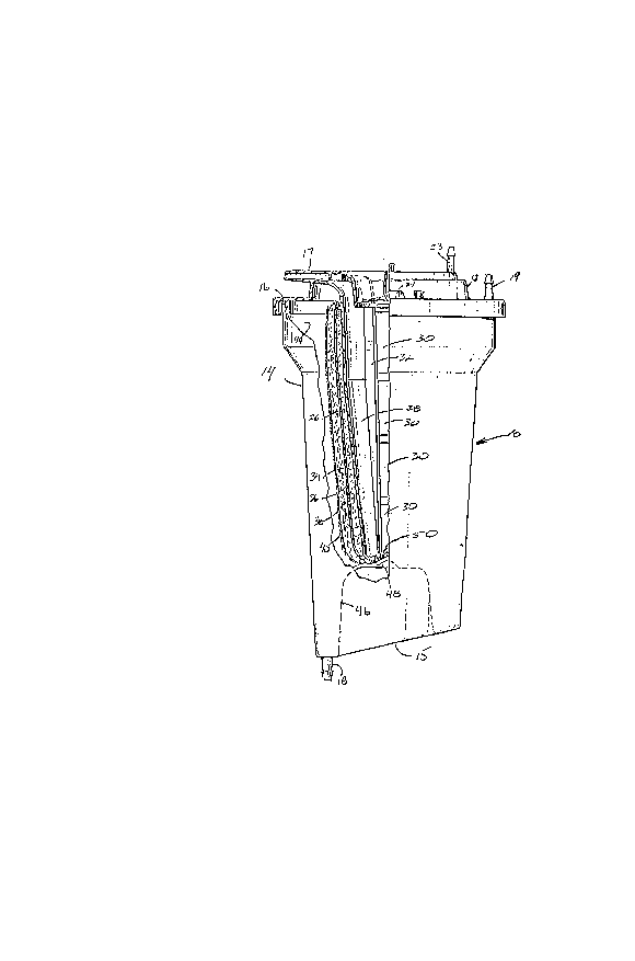
14 by the numeral lO. The specific shape of the housing is
not critical, but it is preferably a generally
16 cylindrical structure as shown in the Figs. 1 and 6.
17 Suitably the housing is made of a rigid moldable plastic
18 material such as polycarbonate, polyacrylate or polyester
19 thermoplastic which is preferably clear to enable
observation of blood levels within the device. Suitable
21 plastics are the polycarbonate resins sold by General
22 Electric Co. under the LEXAN trademark and by Mobay
23 Corporation under the MAKROLON trademark.
24 Housing 10 comprises a top 13, generally
cylindrical sidewall 14 and a bottom 15 integrally molded
26 with sidewall 14. Desirably the sidewall 14 is tapered
27 inwardly from top to bottom to assure drainage. The top
28 13 is suitably a separately formed cover member. A
29 circumferential gasket 16 provides a fluid tight seal
between the top 13 and the upper edge of sidewall 14.
31 Alternatively the top 13 may be sealingly bonded to the
32 sidewall 14 by adhesive, heat or ultrasonic bonding
33 techniques. Fluid communication into and out of the
34 device is provided by at least one cardiotomy inlet port
17 at the top of the device and an outlet port 18 in the
36
~0~25i~
1 bottom portion thereof. Generally it is desire~ that a
2 plurality of cardiotomy inlet ~orts 17 be provided so
3 that blood may be received from several locations at the
4 surgical site.
Also located at the top of housing 10 is a vent
6 port 19. Optionally the device is further provided with
7 luer ports 20 and 21 ~or introducing medicines or the
8 like, a quick priming port 22 and one or more chest
9 drainage ports 23. A dual action pressure release valve
24 may be included to assure that the internal pressure
11 of the device stays within predetermined values.
12 Normally all the parts in the device will be
13 covered by removable caps, not shown, until the port is
14 to be accessed.
10cated within the housing 10 is an inner wall
16 member 26 which defines a central blood receiving chamber
17 within the device. Receiving chamber wall member 26 has
18 a generally conical or horn-like shape which slopes
19 downwardly and inwardly from the top to a closed bottom.
Wall member 26 includes a first arcuate portion 28 which
21 provides a continuous imperforate surface from just below
22 the inlet port opening to the bottom of the device so
23 that cardiotomy blood entering the device will flow
24 smoothly into the receiving chamber along the surface of
portion 28 without splashing and with minimal turbulence.
26 Another arcuate portion 29 of member 26 includes at le,ast
27 one aperture or window 30 to allow blood to exit the
28 receiving chamber. Vanes 32 and 33 separate imperforate
29 arcuate wall portion 28 from the windowed portion 2~ and
provide blood direction means for directing blood
31 entering the housing onto the imperforate arcuate portion
32 28 as the blood flows into the receiving chamber.
33 Surrounding the outside of the receiving
34 chamber wall 26 is a blood treatment means which
comprises at least a blood defoaming element. Preferably
36
~:~32~95
--6-
1 the blood treatment means comprises a first defoaming
2 element 34, a depth filter element 36, a second defoaming
3 element 38 and a sock-like element 40. The sock-like
4 element 40 is a mesh or knit fabric which holds the
defoaming and filtering elements in place against the
6 outside of wall member 26. Suitably the sock-like
7 element is held in place by means of a tiestrap 42
8 surrounding the top of the sock and engaging receiving
9 chamber wall member 26 above a ridge 27 which extends
circumferentially around the outside of the receiving
11 chamber wall above the highest windows 30.
12 Defoaming elements 34 and 38 are preferably
13 formed of a thermally reticulated pol~urethane foam.
14 Typically defoaming elements 34 and 38 will have an
average pore size of about 20 pores per inch. Desirably
16 both are coated with a suitable defoaming agent. The
17 depth filter element may be a 20 micron felt filter.
18 Between the sock 40 and the inner surface 44 of
19 sidewall 14 is a space which provides a collection
chamber for treated blood. Outlet port 18 opens into the
21 bottom of this collection chamber.
22 Suitably the bottom 15 of the housing is
23 tapered downwardly toward outlet port 18 to facilitate
24 drainage from the blood receiving chamber to the outlet
port. Desirably a central portion 46, shown in phantom
26 in Fig. 6, of bottom 15 is indented and .includes a
27 central hump 48 which serves as a location means for the
28 bottom of receiving chamber wall member 26. Structures
29 46 and 48 reduce volume at the bottom of the blood
reservoir and, therefore, further facilitate drainage
31 toward the exit port.
32 A corresponding hump 50 in the bottom of
33 receiving chamber wall 26 mates with bottom hump 48 of
34 the housing to provide positive location of the receiving
36
2032~i~5
-7-
1 chamber wall. Hump 50 also reduces dead volume at the
2 bottom of the blood receiving chamber below the lowest
3 window 30.
4 If the inventive device is intended to be used
solely in surgical operations, windowed portion 29 of the
6 receiving chamber wall 26 may encompass the entire
7 arcuate surface of wall 26 on the outside of vanes 32 and
8 33. However, in the preferred structure shown in the
9 figures, wall member 26 also includes a second
imperforate arcuate portion 52 extending from the top to
11 the bottom of the receiving chamber wall. Second
12 imperforate portion 52 is disposed below the chest
13 drainage ports 23. A second set of vanes, 54 and 55,
14 direct blood received from the chest drainage ports down
along the imperforate portion 52 as blood flows into the
16 central receiving chamber. This structure allows the
17 device to continue to be used after surgery for treating
18 blood which is collected from a chest drain and returned
19 to the body. Suitably portion 52 is disposed opposite
portion 28 so that wall member 26 includes two apertured
21 portions 29 between the respective sets of vanes 32, 54
22 and 33, 55. Imperforate portion 52 may comprise a
23 smaller arcuate portion of wall 26 than portion 28 since
24 the chest drainage tubes will usually be fewer in number
and provide a lower blood flow rate than the cardiotomy
26 blood inlat ports.
27 In operation blood from a surgical site is fed
28 into the cardiotomy inlet ports 17 and is directed in a
29 sheet-like flow along imperforate portion 24 of receiving
chamber wall 26 into the central receiving chamber of the
31 device. The blood then passes via apertures 30 through
32 the respective defoaming and filtering elements 34, 36,
33 38 and 40 into the treated blood collection chamber from
34
36
-8- ~32~!~5
1 which it exits via outlet port 18. The blood may then be
2 fed back into the body, usually after having been further
3 oxygenated and/or cooled.
4 After surgery the cardiotomy ports 17 are
sealed by cutting and clamping the connecting tubing or
6 by removing the connecting tubing and recapping the ports
7 17. If the patient requires chest drainage, the open end
8 of a drain tube sewn into the patient may be connected to
9 a chest drainage port 23 so that the drainage may be
filtered and returned to the patient. The return may be
11 accomplished with an I.V. pump programmed to match the
12 rate of bleeding or by periodic gravity drainage into the
13 patient in a batch process. The cardiotomy ports and
14 chest drainage ports are desirably separately provided in
the device since the connecting tubing for the two
16 functions conventionally have differently sized
17 connecting ports. If medicines or the like are desired
18 to be added to the blood they may be added in the
19 receiving chamber via luer ports 20 or in the treated
blood collection chamber via luer ports 21.
21 This completes the description of the preferred
22 and alternate embodiments of the invention. Those
23 skilled in the art may recognize other equivalents to the
24 specific embodiment described herein which equivalents
are intended to be encompassed by the claims attached
26 hereto.
27
28
29
31
32
33
34
36