Note: Descriptions are shown in the official language in which they were submitted.
BACKGROUND OF T~IE INVENTION
Field of the Invention
This invention relates to ligament anchor systems and devices
for use in arthroscopic surgical procedures involving securing
an and of a ligament, such as the anterior cruciate ligament,
stint, or the li~e, under tension within a bone mass.
Prior Art
In certain ligament replacement surgical procedures,
particularly arthroscopic knee surgery, invo~ving a cruciate
ligament replacement, it is usual to form a tunnel through
bone masses on both sides of a joint for installing a ligament
therein. Such installation has generally involved fitting the
ligament through the prepared tunnel and attaching its ends
onto the bone cortex surfaces as with staples, or the like,
for maintaining the ligament under tension across the joint.
Exampies of arrangements for attaching ligament ends within a
bone mass are shown in a United Kingdom patent, No. G.B.
2,084,468A; and a patent of the present inventors, U.S. Patent
No. 4,772,286. A U.S. patent application of the present
inventors, Serial No. 235,194, entitled "Channel ~igament
Clamp and System", shows a device for securing a ligament end
onto a bone mass. Additionally, another earlier patent of the
present inventors, U.S. Patent No. 4,870,957, entitled
"Ligament Anchor System", shows a stud for mounting a ligament
end within a ligament tunnel that involves a threaded sleeve
or footing that is turned into a tapped endosteal bone.
Another U.S. patent application of the present inventors,
Serial No. 352,153, entitled "Interference Screw, System and
Process", provides an interference screw and system for
turning it into a ligament tunnel, alongside a ligament end
therein.
All of the above-cited devices and systems involve
hardware and systems for connection of a ligament to the
endosteal portion of the distal femur, and the "Ligament
Anchor System" application involves a separate stud and
footing for mounting a ligament end to bone. Whereas, the
present invention employs a single stud only that is ~or
`
insertion to closely fit in a ligament tunnel and to lock
into the cortex surface, and is arranged for mounting a
ligament end to a rear end thereof. The stud o~ the present
invention is for sliding along the prepared ligament ~unnel.
The stud ~orward end sections are split by a slot, allowing
the section to be squeezed together as the stud is urged into
the tunnel. The stud forward end, as it emerges from a cortex
end of the ligament tunnel, flexes outwardly, a hook end
section of the stud to extend beyond ~he tunnel edge. Which
hook edge thereby binds into the surrounding bone mass or over
the outer cortex when the stud is pulled back into the
ligament tunnel, securely and permanently mountin~ that stud
end to that bone cortex.
SU~!IARY OF THE INVENTION
It i5 a principal object o~ the present invention to
provide an endosteal fixation system for mounting a ligament
in a bone tunnel.
Another object of the present invention is to provide an
endosteal fixation stud that is for closely fitting through a
straight bone tunnel that is formed at an angle to the bone
cortex surface, the stud to include a hook end that exits the
tunnel cortex end and flexes outwardly thereat, the hook end
edge to flex over the tunnel cortex edge, locking thereto and
prohibiting withdrawal of that stud back through the bone
tunnel.
Another object of the present invention is to provide an
endosteal fixation stud with an arrangement for mounting it to
a ligament end, the stud and ligament for fitting in a
prepared ligament boné tunnel, the stud to travel therealong,
a stud hook end on exiting which tunnel to flex over the bone
cortex tunnel edge, prohibiting withdrawal of that stud and
ligament back through the tunnel.
Still another object of the present invention is to
provide an endosteal fixation stud that is formed from a
suitable material for human implantation that will exhibit
resilient ~uali~ies.
Still another object of the present invention is to
provide an endosteal fixation stud whereto can be attached a
ligament or the like, which stud is for locking into the end
of a bone tunnel formed through khe femur and is preferably
re-absorbable.
The present invention is in an endosteal fixation stud and
system for permanently mounting a ligament end, or the like,
within a bone tunnel. The stud is a cylindrical sectivn
having a slightly smaller diameter than does a bone tunnel
wherein a ligament end is to be mounted. Which bone tunnel is
formed at an angle to the plane of the bone cortex tunnel end
whereby a hook end of the stud will flex over, as to lock onto
the edge of that bone cortex sur~ace. Which angle is
optimally forty-five (~5) degrees but may be in a range of
angles between twenty-five (25) degrees and sixty-five (65)
degrees, within the scope of this disclosure.
The endosteal fixation stud consists of a cylindrical body
that preferably includes a bridge arrangement at its rear most
end for attaching a ligament graft, or the like, mounted
between parallel legs thereof. The nose of the stud
cylindrical body is preferably ~lared outwardly and rearwardly
at approximately a sixty (60) degree angle to the vertical
forming an arcuate segment as a hook end. An outer edge of
which hook end is to extend beyond the bone tunnal edge for
binding into or onto the bone cortex. To allow the stud with
outwardly extending hook end to slide along the ligament
tunnel, a longitudinal 610t iS formed in that stud forward end
to approximately a mid-point thereof. The slot divides the
stud end into segments, that, when comprPssed as when the stud
is fitted through the bone tunnel, will flex together, and
will spring apart on exiting the tunnel end. To provide this
flexure, the s~ud is preferably formed from a resilient
material that is sùitable for human implantation, such as a
resilient metal or a plastic like DelrinTM, polyethene or re-
absorbable material.
Additionally, a bridge, or the like, that includes an arm
or spaced arms is provided as a stud end for use in attaching
a ligament end. Further, a threaded suture, wire or rod can
be used with the invention for turning into a tapped hole that
is formed in the stud forward end, the suture, wire or rod for
pulling the stud thrsugh a bone tunnel. Whereafter, the
suture, wire or rod is removed by turning it out of that stud
end tapped end hole.
In practice, for securing an end o~ a ligament, or like
graft, either biological or prosthetic, utilizing the
endosteal fixation stud of the present invention, a tunnel is
formed through a bone or bones, for receiving the ligament.
The bone tunnel is to receive the stud and, provide a proper
binding surface therefore. Accordingly, it is preferably
angled ~rom the plane of bone cortex surface at approximately
a forty-five (45) degree angle, plus or minus twenty (20)
degrees to conform to the angle of the stud hook end. So
arranged, a ligament end is attached at the bridge rear end of
the stud cylindrical body. The stud is to travel through the
bone tunnel and exit the bone cortex surface. On exiting, the
stud hook end will flex or spring outwardly, an edge thereof
extending beyond to rest on the
tunnel edge. With tension then applied through the ligament,
that anchor stud hook edge will seat into the bone cortex,
prohibiting ligament withdrawal back through the bone tunnel.
In one installation procedure of an anterior cruciate
ligament, a first endosteal fixation stud mounting a anterior
cruciate is urged through a bone tunnel, the stud hook edge
emerging from a femoral cortex end to bind into the bone at
the tunnel edge. This passage can involve passing the suture,
wire or rod, threadjed end first, through the femoral cortex
tunnel end to the tibial cortex end. Thereat, the stud is
turned onto that wire, or rod end. The suture, rod or wire is
then drawn back through the ligament tunnel, with the
connected stud and ligament drawn therewith until the stud
hook end extends beyond and is pulled back to engage the bone
tunnel femoral cortex ~endO The ligament free end is then
placed under tension àt the tibial cortex end as by attaching
it with a standard staple, or the like, to the cortex surface.
Alternatively, the ~e~dosteal femoral fixation stud can be
fitted through an art~hroscopic port into the patient's knee
and pushed with an appropriate instrument outwardly through
the femoral bone tunnel section from within the intra
articular joint, and wi~h the free ligament end fitted from
the intra articular joint through the tibial bone tunnel
sectionO
To release a stud hook end, the stud forward end sections
can be collapsed together, until the hook end edge aligns with
the ligament tunnel wall. The stud can thereafter be pushed
back into the ligament tunnel, allowing for its removal.
DESCRIPTION OE' THE DRAWINGS
In the drawings that illustrate that which is presently
regarded as the best mode for carrying out the invention:
Fig. 1 is a side elevation perspective view taken from a
r~ar end of an endosteal fixation stud of the present
invention;
Fig. 2 is a side elevation perspective view of the
endosteal fixation stud of Fig. 1 taken from a forward end and
showing a ligament mounted between opposing parallel flanges
of a stud rear end;
Fig. 3 is a side elevation sectional view taken along the
line 3-3 of Fig. l;
Fig. 4 is a side elevation view of a longitudinal cross-
section of a distal femur wherein a ligament tunnel has been
formed, and counter-sunk to accommodate a endosteal fixation
stud of the present invention fitted therein that includes a
ligament mounted thereto: and
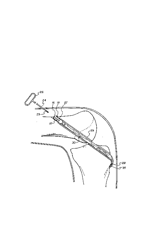
Fig. 5 is a side elevation sectional view of a patient's
knee wherein a ligament tunnel has been formed in an anterior
cruciate ligament replacement surgical procedure, showing the
ligament of Fig. 4 connected at its one end to the endosteal
fixation stud, with the ligament free end extending beyond and
bent onto the tibial cortex whereat it is secured by a staple.
DETAILED DESCRIPTION
Fig. 1 shows a side elevation perspective view taken from
a rear end of the present invention in an endosteal fixation
stud 10 of the present invention, hereinafter referred to as
stud. Stud 10, as shown best in Figs. 1 through 3, includes a
cylindrical body 11 that include~ body 12 as a rear end. The
bridge 12 includes s~aced apart parallel arms 13 that extend
parallel to one another and rearwardly from the face of the
,
cylindrical body. A stud forward or front end 14 is shown in
Figs. 2 and 3 as rounded at 15. Which stud front end includes
an arcuate section that is formed into a hook 16, as shown in
Figs. 1, 2 and 3.
The hook 16 is formed as an outwardly and rearwardly
projecting extension o~ a section of ~he stud cylindrical body
front end 14, extending rearwardly from the rounded forward
end 15. The hook 16, as shown best in Fig. 3, is preferably
angled at approximately forty-five (45) degrees rearwardly
from the cylindrical body 11 surface, which angle is
illustrated as arrow A, and is preferably the angle of a bone
tunnel to a bone cortex. ~ forward face of the hook 16 is
shown formed at approximately a sixty (60) degree angle from
the vertical plane of the stud front end 14, illustrated as
arrow B. In practice, the hook is preferably formed as a
section of less than one hundred eighty (180) degrees of arc,
and has essentially parallel opposite edges 16a, with a hook
edge or lip 18, and is an extension of the stud forward or
front end.
As will be discussed in greater detail hereinbelow, the
stud 10 is intended to fit into and travel along a bone tunnel
that is formed through a bone mass, shown herein as the distal
femur, exiting the anterolateral cortex. Which bone tunnel is
counter-sunk to just accommodate the stud cylindrical body
forward end at the anterolateral cortex exit, which
anterolateral cortex end is of lesser cross-section than the
cross-section of the stud forward end with the stud hook 16
extended. Accordingly, to allow for collapse of the stud hook
16, to where the stud will slide therethrough the femoral
tunnel section, a slot 17, as shown in Figs. 1 through 3, is
formed longitudinally into the stud cylindrical body. This
slot splits that cylindrical body to approximately the mid-
point thereof. So àrranged, the opposite slot edges arespaced apart equidis~ ntly along the slot, the stud sections
capable of being collapsed together.
The slot 17 is~ to allow the sections of the stud
cylindrical body 11, at the hook end, to be squeezed together.
The hook edge or lip 18 is thereby recessed to where it can be
fitted into ths ligament tunnel. At the anterolateral cortex
end of which tunnel, the cylindrical body sections to flex or
return to their uncompressed state, extending the hook edge 18
over the tunnel edge. That hook edge 18 will thereby bind
into a section of the bone surrounding that tunnel end when
the stud is pulled back into the bone tunnel. To provide
which flexure the stud 10 is formed from a resilient
material, such as DELRINTM plastic material, or the like, but
can also be formed of an appropriate metal, as required.
Shown in Fig. 2, the bridge 12 is formed as the rear end
of the cylindrical body and is stepped outwardly as the
parallel spaced apart arms 13. Which arms 13 are to receive a
ligament end 31 fitted therebetween. Shown best in Fig. 2,
the ligament end 31 is preferably secured between the stud
arms 13 as with screws 34, that are fitted through aligned
holes 33 that are formed through the stud arms. ~hich screws
preferably pass through both the ligament end 31 and a bone
plug 32 that is arranged as a stiffener with that ligament
end. The bone plug 32 is shown as provided to wedge the
ligament end between which arms 13, as well as for receiving
the screws 34 turned therein.
Alternative to bridge 12, the stud 10 rear end can be
provided with a screw extending rearwardly therefrom, not
shown, from turning into the ligament 30 end, or can involve
an eyelet end, not shown, for receiving a ligament and/or
stint, not shown, threaded therethrough.
The stud forward end 14, as shown best in ~ig. 2,
preferably includes longitudinal parallel arcuate grooves 21
that are formed in the center of the slot 17 opposing
surfaces. As shown best in Fig. 3, a center longitudinal hole
22 is formed into the end of stud slot 17 that is tapped with
threads 23. Which hole 22 aligns with, as an end of, the
arcuate grooves 21. The hole 22, as shown in broken lines in
Fig. 5, is to receive a thre.aded end 25 of a suture, wire or
rod. In Fig. 5, a wire or rod 2~ is shown that preferably
includes a handle 26 formed on the opposite end thereof to
threaded end 25, which handle is for manually guiding and
turning the wire or rod threaded end 25 into the threads 23 of
the stud longitudina] hole 22. So connectedl an operator can
move the stud 10 as by pulling or pushing on handle 26 along
the bone tunnel. In such travel, the stud cylindrical body
sections are compressed into slot 17, khe arcuate grooves 21
to collapse towards the wire or rod 24.
Figs. 4 and 5 illustrate an example of a practice of a
process of the present invention for replacing a patient's
anterior cruciate ligament 30. Which procedure can be adapted
to provide a bone/tendon/bone attachment, and the ligament 30
can be a prosthetic or so~t tissue graft or a combination or
composite thereof. The process utilizes a stud 10 with a
ligament end 31 attached to the stud bridge 12. The ligament
end is maintained rearwardly between arms 13 of bridge 12.
That other end of which ligament, as shown in Fig. 5, extends
from the tibial cortex end 28 of bone tunnel 29. The ligament
30, as shown in Fig. 5, is bent back onto the tibia cortex
surface, and receives a staple 35 straddling that ligament for
maintaining the ligament under tension to the bone surface.
The stud 10 is for maintaining the ligament 30 stretched
through the bone tunnel 30 to the bone at the femoral cortex
tunnel end. The bone tunnel 29 is initially ~ormed to have a
diameter to just accommodate the stud 10. Thereafter, the
bone tunnel is counter-bored, as bone tunnel section 29, to
have a diameter to ~reely accommodate the stud bridge 12 with
ligament 30 attached thereto, a femoral tunnel end 27, shown
in Figs. 4 and 5 to just accommodate stud 10. The femoral
tunnel end 27, for proper stud 10 functioning, should exit the
femoral cortex at an angle that is the angle of the rear face
of the stud hook 16 relative to the cylindrical body 11.
Which angle is preferably forty-five (45) degrees plus or
minus twenty (20) degrees, for proper stud hook 16
functioning.
In an arthroscopic surgical procedure, through an opening,
not shown, that is formed into the patient's intra articular
joint, a surgeon can manipulate stud 10 into the bone tunnel
femoral end and can urge that stud, as with a tool, not shown,
along the tunnel section until the hook 16 extends beyond the
femoral cortex edge. Whereat the stud hook edge overlaps the
tunnel edge securing the connected femoral end of ligament 30
in the femur section of the bone tunnel. Additionally, a
stint, not shown, can be attached to extend from the stud
bridge 12, within the scope of this disclosure.
In another installation procedure, as illustrated in Fig.
5, the wire or rod 2~ threaded end 25 can be fitted through
the femoral bone tunnel cortex end 27 and turned into the
threads 23 of hole 22 formed in stud 10. So arranged, a
surgeon gripping the wire or rod handle 26 can pull the stud
lo mounted thereto through the femoral end of the bone kunnel,
the attached ligament following therethrough to where the hook
16 of the stud extends beyond the bone tunnel femoral cortex
end. Thereat, the hook edge or lip 18 will engage and, with
an application of a tensile force on the ligament 30 at its
tibial end, will bind or bite into the cortex surface. The
hook edge 18 thereby prohibits withdrawal of the stud 10 back
through the bone tunnel. To secure the free ligament 10
tibial end, that end is pulled beyond the tibial cortex tunnel
end and is bent onto the bone surface. A staple, or like
fastener, like staple 35, is then driven into the tibial
cortex, the staple web compressing the ligament against the
bone surface.
The stud 10, along with a staple 35, or like fastener,
provides a tension mounting of ligament 30, with or without a
stint across the intra articular joint. Which tension can be
readily adjusted by urging the stud 10 outwardly from the bone
tunnel femoral cortex and freeing the hook edge 18 by
compressing the stud sections across slot 17 together, the
stud hook 16 thereby allowed to pass into the tunnel femoral
cortex end. The stud can thereafter be pulled back through
the bone tunnel 29, ~llowing for a ligament length adjustment
to provide a desired tensile force on the ligament when the
stud is reinstalled to the femoral cortex tunnel end, as set
out above.
In practice, a stud 10 installed as set out above in a
cadaver knee was found to provide a stable and secure ligament
anchor up to an application of approximately two hundred ~200)
pounds of tensile force. Which force is well above an mean
functional load for an anterior or posterior cruciate ligament
of approximately one hundred (100) pounds of tensile force.
While a preferred embodiment of the present invention and
process for securing a ligament in a ligament tunnel have been
shown and described herein, it should be understood that the
present disclosure is made by way of example only and that
variations and changes thereto are possible without departing
from the subject matter and reasonable equivalency thereof
coming within the scope of the following claims, which claims
we regard as our invention.
.
.