Note: Descriptions are shown in the official language in which they were submitted.
WO 90/08~57 2 ~ '~ 4 ~ ~ ~ PCI/US90~00137 *
TITLE OF lHE INNENTION
METHOD OF DETECTING SMALL CELL
CARCINOMA AND USE OF ACYL-PEPTIDE
HYDROLASE AND CODING SEQUENCES THEREFOR
Cross-Reference to Belated ADDlications:
This application is a continùation-in-part of U.S. Patent
Application Serial No. 07/296,996 (filed: January 13, 1989),
wh;ch is a continuation-in-part of U.S. Patent Application
Serial No. 07/087,936 (filed: August 21, 1987), both herein
incorporated by reference.
Field of the Invention:
The present invention is directed toward the product~on
of Acyl-Peptide Hydrolase by recombinant DNA technology. It
is also directed to the use of the enzyme to catalyze
hydrolysis of an acyiated peptide or protein, and the reaction
between a derivatized N~-acetyl amino acid donor and an
acceptor protein with a free ~-N~2 group. The invention
further concerns a gene sequence which encodes the rat acyl-
peptide hydrolase. The invention is also directed toward the
diagnosis of small cell earcinoma through the use of acyl-
pept1de hydrolase and gene sequences which encode acyl-peptide
hydrolaseO
Brief ~escription of the Back~round Art:
Since the discovery of an acetyl group at the amino-
terminus of tobacco mosaic virus coat protein, a number o~ N~-
`~ ` ' '. ' ' . , , ,, ' ` i` ' :. ; . ~
w o go/08457 ~ 5 PCT/US90/00137
acetylated proteins have been found in animals, plants, and
their viruses, and also in bacteria and fungi. N~-acetylation
is therefore considered one of the typical modifications of
proteins in living organisms. Moreover, in some eukaryotic
cells, it has been suggested that more than 80% of the
intracellular soluble proteins are ~-acetylated (Brown, J.L.,
J. Biol. Chem. 254:1447-1449 (1979)).
The biological significance of N~-acetylation of proteins
is still an open question (see Tsunasawa et al., Method
En~Ymol. 106:165-170 (1984)). It has been proposed that this
post-translational modification protects intracellular
proteins from proteolysis. However, this does not hold true
for all proteins. In the case of actin from slime mold,
proteolytic degradation becomes slower when the protein is
lS N~-acetylated. In contrast, cat hemoglobin is degraded at
the same rate irrespective of N~-acetylation (Tsunasawa et
al , 1984).
Recent results from DNA sequencinq have shown that in
structural genes for the secretory proteins that are N~-acety-
lated, the codon for the acetylated amino-terminal residue is
directly preceded by the initiation codon without the
insertion of add~tional codons for amino acids (Tsunasawa et
al., 1984). Little effort has been made to understand the
relationship between N~-acetylation and the transport of
secretory proteins across biological membranes. To understand
completely the function of N~-acetylation, it will be
important to identify the N~-acetylated amino acids in
proteins and peptides on a microanalytical scale. For this
purpose, removal of the N~-acetyl group or the N~-acetyl amino
acid must be efficiently achieved.
Acyl-Peptide Hydrolase (APH) has been successfully used
for the hydrolysis of N~-acylated peptides. One such enzyme,
which was purified fro~ animal liver, can liberate the
N~-acetyl amino acid from rather short peptides derived from
WO 90/08'157 ~ A a 9 ~ PCI/US90~00137
-3 -
N~-acetylated proteins (Tsunasawa et al., 1984). The
substrate specific;ty is broad for the amino terminal residue.
APH cleaves the N~-terminal acetylated or formylated amino
acid from a blocked peptide (Jones et al., B~RC 126:933
(1985)). This enzyme catalyzes the hydrolysis of a diverse
number of peptides and displays different pH optima for
certain substrates in doing so. This enzyme may also play a
pivotal role in the processing of polypeptide chains during
biosynthesis. APH has been purified from rat liver (Tsunasawa
et al., J. Biochem. 77:89-102 (1975)); [from bovine liver
(Gade et al., Biochim. BioDh~s. Acta 662:86-93 (1981))]; from
porcine liver (Tsunasawa et al., J. Biochem. 93:1217-1220
(1983)); from rat brain (Marks et al., J. Neurochem. 41:201-
208 (1983)); and from human erythrocytes (Jones et al.,
Biochem. and BioDhys. Res. Comm. 126:933-940 (1985)).
A rat liver acyl-peptide hydrolase (APH), which catalyzes
the hydrolysis of the acetylated residue from N~-acetylated
peptides was recently purified to homogeneity, and various
inhibition experiments indicated that it was likely a serine
protease, utilizing a charge relay system involving serine,
histidine, and probably a carboxyl group (Kobayashi, K. and
Smith, J.A., J. 8iol. Chem. ~2:11435-11445 (1987)). However,
it is not yet clear whether acyl-peptide hydrolase is a unique
serine protease.
In order to facilitate a moré complete understanding of
the regulation of rat acyl-peptide hydrolase in vivo~ it i5,
therefore, desirable to clone and sequence the rat acyl-
peptide hydrolase gene.
SUMMARY OF THE INVENTION
Acyl-peptide hydrolase catalyzes the hydrolysis of an N~-
acetylated amino acid residue from an N~-acetylated peptide.
~wo overlapping, degenerate oligonucleotide probes based on
the sequence of a tryptio peptide, derived from purified rat
. . . . .
w o ~0/08457 2 ~ l~ L~ 5 ~ ~ PCT/US9~/00137
acyl-peptide hydrolase, were synthesized and used to screen a
rat liver ~gt11 cDNA library. A 2.5 kb cDNA was cloned and
sequenced. This clone contained 2364 bp of rat acyl-peptide
hydrolase sequence but lacked an initiation codon. Using a
220 bp probe derived from the 5'-end of this nearly full-
length cDNA to rescreen the library, full-length clones were
isolated, which contained an in-frame ATG codon at nucleotides
6-8 and encoded the NH2-terminal sequence, Met-Glu-Arg-Gln --.
The DNA sequence encoded a protein of 732 amino acid residues,
40% of which is confirmed by protein sequence data from 19
CNBr or tryptic peptides. The isolated enzyme is NH2-
terminally blocked (Kobayashi, K., and Smith, J.A. t1987) J.
Biol. Chem. 262:11435-11445~, and-based on the NH2-terminal
protein sequence deduced from the DNA sequence and the
sequence of the most NH2-terminal CNBr peptide, it is likely
that the NHz-terminal residue is an acetylated methionine
res;due, since such residues are frequently juxtaposed to
glutamyl residues (Persson, B., et al., (1985) Eur. J.
Biochem. 152, 523-527). The RNA blot analysis revealed a
single message of 2.7 kb in various rat tissues examined.
Although this enzyme is known to be inhibited by diisopropyl
fluorophosphate and acetylalanine chloromethyl ketone
(Kobayashi, K., and Smith, J.A. (1'387) J. Biol. Chem.
262:11435-11445), no strong similarity in protein sequence has
been found with other serine proteases. This result suggests
that acyl-peptide hydrolase may be a unique serine protease.
This invention is directed to a protein Acyl-Peptide
Hydrolase (APH), which comprises the amino acid sequence of
Figure 1. It is also directed to the production of APH by
recombinant DNA technology, and to the utilization of APH in
the hydrolysis or amino-acylation of peptides or proteins.
The inYent1on concerns the cloning and sequence analysis of an
acyl-peptide hydrolase from rat liver described by Kobayashi,
:: :,. :
.. ~ ,. : :
w o so/084s7 PcT/uS9O/00l37
2 ~ C~ ~i
K. et al. (?. Biol. Chem. 264:8892-8899 (May, 1989)), which
reference is incorporated herein by referenc
A recombinant DNA molecule coding for APH of the present
invention may be used to transform any of a number of hosts,
creating new sources and unlimited supplies o~ APH. The
invention thus further comprises the gene~ic sequences coding
for an enzyme having the amino acid sequence designated in
Figure 1, vehicles containing the genetic sequence, hosts
transformed therewith, enzyme production by transformed host
expression, and utilization of the enzyme in hydrolysis or in
amino-acylation o~ peptides or proteins. It is a purpose of
this invention to provide new sources of substantially pure
- APH which would be avail`ablë~~in unlimited supp~y.
Additionally, this invention encomp~sses the use of the
enzyme to catalyze the hydrolysis of an N~-acylated protein,
or the reaction between an N~-acetyl amino acid donor and an
acceptor protein with a free ~-NH2 group.
Therefore, additional purposes of this invention are to
provide a means of hydrolysis of N~-acylated proteins, and of
amino-acylating any polypeptide or prot;ein from an N~-acetyl
amino acid donor and an acceptor with a free ~-NH2 group, by
the use of APH.
In detail, the invent;on concerns Acyl-Peptide Hydrolase
in substantially pure form. The invention also concerns Acyl-
Pept;de Hydrolase ~ree of nat;ve glycosylation.
The invention further concerns a recombinant nucleir acid
molecule, either RNA, genomic DNA, or cDNA, which contains a
genetic sequence coding for Acyl-Peptide Hydrolase. The
nucleic acid molecule may be a vector or plasmid.
The invention also concerns a host, such as a bacterium,
a yeast, or a mammalian cell, etc., transformed with any of
the above-described recombinant nucleic acid mol~cules.
The invention also concerns a method of producing Acyl-
Peptide Hydrolase which comprises:
,
. ~ , ,., .';', .. , . , . . ~
WO 90/08457 2 ~ 4 4 5 ~ ~ PCI'/US90/00137
-6-
(a) providing any of the abo~e-described nucleic
acid molecules, wherein thè molecule is DNA;
(b) inserting the DNA mo1ecule into a vector;
(c) transforming a host system with the vector;
(d) expressing the Acyl Peptide Hydrolase DNA
sequence of the recombinant DNA molecule in the host; and
(e) recovering the Acyl-Pep~ide Hydrolase produced
by the expression.
The invention also includes the Acyl-Peptide Hydrolase
produced by the above-described method.
The invention also includes the above-described Acyl-
Peptide Hydrolase in immobilized form.
The invention also includes a method of hydrolyzing the
N-terminal acyl amino acid of an acylated polypeptide, which
comprises contacting the polypeptide with the above-described
Acyl-Peptide Hydrolase.
The invention also includes à method of catalyzing the
reaction between a derivat ked N~-acetyl amino acid donor and
an acceptor with a free ~-NH2 which comprises contacting the
donor with the acceptor in the presence of the above-described
Acyl-Peptide Hydrolase.
The invention also pertains to a method of detecting
small cell carcinoma which comprises:
a. incubating a nucleic acid sample from a patient
suspected of ha~ing small cell carcinoma, in the presence of a
nucleic acid molecule having a sequence selected from the
group consisting of:
a. a sequence which encodes all or part of an acyl-
peptide hydrolase enzyme; and
b. a sequence which is complementary to a sequence
which encodes all or part of an acyl-peptide hydrolase enzyme;
the incubation being under conditions sufficient to permit
nucleic acid hybridization to occur between the nucleic acid
- . . . - - :
- ;. . ..
w o 90/Ofl457 2 ~ ~ 4 ~ ~ 5 P~T/~S90/00137
sample and the nucleic acid molecule, and to thereby form a
hybridized molecule; and
b. detecting, such as by an analysis of restriction
fragment length polymorphisms, small cell carcinoma by
determining whether the hybridized molecule differs in
sequence from a reference molecule, the reference molecule
comprising a nucleic acid sample from a normal individual
hybridized to a nucleic acid molecule which encodes all or
part of an acyl-peptide hydrolase enzyme.
The invention further includes a two stranded nucleic
acid molecule comprising:
A. a first strand having a sequence selected from the
group consisting of:
a. a sequence which encodes all or part of an acyl-
peptide hydro1ase enzyme; and
b. a sequence which is complementary to a sequence
which encodes all or part of an acyl-peptide hydrolase enzyme;
the first strand being hybridized to:
B. a second strand, the second strand having a sequence
which is substantially complementary in sequence to the
sequence of the first strand, the complementary sequence of
the second strand being derived from an individual suspected
of having smal1 cell carcinoma.
~ESCRIPTION OF ~H FIGURES
Figure 1 illustrates the amino acid sequence of APH. The
protein sequence deduced fro.., the cDNA sequence (Figure 3~ is
indicated by the one letter code for the amino acids. The
bracket lines indicate the termini of the CB, CB-R, and CB/R
peptides. The arrows pointing right indicate that the
corresponding amino acid residue was identif;ed as the Pth-
amino acid residue during automated Edman degradation (Table
1). A blank indicates that a Pth amino acid was not identi-
. . .
'; `: ,' ~ :~ ;
W o 90/0$457 2 ~ 9 ~ PCT/US90/00137
fied in this degradative cycle. An asterisk indicates that a
Pth-Trp together with an unidentified late-eluting Pth-
derivative was identi~ied instead of Pth-Lys during this
degradative cycle. Cysteine residues were identified as Pth-
derivatives of ~14C] S-carboxymethylcysteine. The active -~
serine is shown at positions 620-627 of Figure 1 (diagonal-
line filled box). The identification of peptides shown here
is defined in Table 3.
Figure 2 illustrates the cloning and sequencing of the
cDNA encoding rat acyl-peptide hydrolase. (A) Oligonucleotide
probes used for the initial screening of the rat liver ~gtll
cDNA library. The amino acid sequence of an RPLC-purified
tryptic peptide (CB18-R11-13-c; Table 3) was used as the basis
for the synthesis of two overlapping d2generate oligonucleo-
tides, YS17.2 and YS20.1. (B) Restriction map and DNA
sequencing strategy of the clones. Using the degenerate
oligonucleot;des in Figure 2A, APH5.2 was obta;ned from a rat
liver ~gtll cDNA library, as describled below. The arrows
ind;cate the direction and extent of [)NA sequence determina-
tion for each fragment. DNA sequence analysis for this clone
revealed the expected hybridization site near ;ts 5' end (open
region in bold line), a poly(A) sequence at its 3' end, and an
unrelated sequence at its 3' end (cross-hatched box). After
rescreening the rat liver ~gtll cDNA library with the XmnI-
KpnI fragment derived from APH5.2 (longer open box), APH36.1
lacked an ATG initiation codon and also contained a 120 base
pair fragment encoding rat serum albumin (box with diagonals).
After rescreening the library with a 220 bp BanII-Pstl
fragment deriYed from APH36.1 (shorter open box), APH2.7 was
cloned, which was subsequently subcloned into Bluescript
plasmid (Stratagene) and sequenced in part. AbbreYiations: B,
BanI; P, PstI; X, XmnI; and K, KpnI.
Figure 3 shows the nucleotide sequence and deduced amino
acid sequence of rat liver acyl-peptide hydrolase. The
. ;.; .; . :. :
. . ~ ..................... . : .~
-. . ., , ,.' . ' '
:: : : : : , : .
w o 90~08~57 2 9 '~ ~ ~ 9 ~ PCT/US9~/00137
complete cDNA encoding rat liver acyl-peptide hydrolase was
derived by combining the DNA sequence data from APH36.1 and
APH2.7 (Figure 2B). The deduced protein sequence is indicated
by the one-letter code for the amino acids.
Figure 4 shows the nucleotide sequence of the rat acyl-
peptide hydrolase gene and its flanking region. The tran-
scriptional initiation site of the gene is indicated by
vertical arrow. The nucleotide at this position is assisned
at number 1. The intronic DNA sequence is shown in lowercase
letters and the exonic DNA sequence is shown in uppercase
letters. The beginning and end of each intron are marked by
vertical lines. The translational initiation site is located
at nucleotides 625-627. The polyadenylation signal is located
at nucleoti~es 9708-9713. The "TATA" box-like sequence
(nucleotides -24 to -30) and the "CAAT box"-like sequence
(nucleotides -95 to -99) are boxed. The GC repeats are
underlined. Tandem 200 bp repeats are indicated by a dashed
underline.
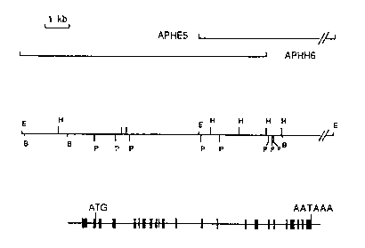
Figure 5 shows a structural organixation of the rat acyl-
peptide hydrolase gene. Figure 5A shows overlapping
recombinant phages containing the acyl-peptide hydrolase gene.
The overlapping genomic clones, APHE5 and APHH6, together
containin~ the entire acyl-peptide hydrolase gene, are
indicated by solid horizontal lines. Figure 5B shows the
restriction map of the acyl-peptide hydrolase gene and its
flanking regions. The EcoRI (F), BamHI (B), HindIII (H), and
PstI (P) sites are indicated by vertical bars. The S' (left)
to 3' (right) transcriptional orientation of this gene is
shown. Figure SG shows the exon-intron organization cf rat
acyl-peptide hydrolase gene. ~he location of the 23 exons
within the rat acyl-peptide hydrolase gene are indicated by
filled boxes. The locations of the translational initiation
codon, ATG, and the polyadenylation sign~l, MTAAA, are
marked by vertical lines.
- . ~ .:
. . -
. . . ~ . : - .
: : .
W0 90/08457 2 ~ 4 ~ ~ 9 5 Pcr/US9O/00137
-10-
Figure 6 shows a comparison of the amino acid sequenees
of acyl-peptide hydrolase and the DNF 1552 protein.
In the Figures, the amino acids have been designated by
single letters of the alphabet such that: A = Alanine, B =
Aspartic Acid or Asparagine, C = Cysteine, D - Aspartic Acid,
E - Glutamic Acid, F = Phenylalanine, G = Glycine, H =
Histidine, I = Isoleucine, K = Lysine, L = Leucine, M =
Methionine, N = Asparagine, P = Proline, Q = Glutamine, R =
Arginine, S - Serine, T - Threonine, V ~ Valine, W - Trypto-
phan, Y = Tyrosine, Z = Glutamine or Glutamic Acid.
DETAILED DISCUSSION OF THE INVENTION
Definitions
To aid in the understanding of the specification and
claims, including the scope to be given such terms, the
following definitions are provided.
IranscriPtion. ThP process of producing mRNA fro~ a
structural gene.
Translation. The process of producing a polypeptide from
mRNA.
Expression. The process undergone by a structural gene
to produce a polypeptide. It ;s a combination of transcrip-
tion and translation.
Plasmid. A circular double-stranded DNA m~lecule that is
not a part of the main chromosome of an organism containing
genes that convey res;stance to specific antib10tics. When
the plasmid is placed within a unicellular organism, the
characteristics of that organism may be changed or transformed
as a result of the DNA of the plasmid. For example, a plasmid
carrying the gene for tetracycline resistance (TetR) trans-
forms a cell previously sensitive to tetracycline into one
which is resistant to it. A cell transformed by a plasmid is
called a "transformant."
, ., . .. - . .
'.~ ~' ' ;, '`. ',
-
:: . ,
.. . . .
~. , ~ ,;; ; :
w 0 90/OB457 2 ~ PCT/US90/00137
Cloninq ~ehicle. A plasmid, phage DNA or other DNA
sequences which are able to replicat2 in a host cell. The
cloning vehicle is characterized by one or a small number of
endonuclease recogni~ion sites at which such DNA sequences may
be cut in a determinable fashion without loss of an essential
biological function of the DNA, which may contain a marker
suitable for use in the identification of transformed cells.
Markers, for example, are tetracycline res;stance or ampicil-
lin resistance. A cloning vehicle is often called a vector.
Recombinant DNA Molecules or Hvbrid DNA. A molecule
consisting of segments of DNA from different genomes which
have been joined end-to-end outside of living cells and have
the capacity to infect some host cell and be maintained
therein.
Operator. A DNA sequence capable of interacting with the
specific repressor, thereby controlling the transcription of
adjacent gene(s). ,
Promoter. A DNA sequence in which RNA polymerase binds
and initiates transcription of an adjacent gene(s).
Acyl-PePtide HYdrolas Q (APH). This term is meant to
include an acyl-peptide hydrolase(s) from any species, which
has the activity of releasing the N~-terminal acylated amino
acid from any protein or peptide in an in vivo or in vitro
system. The term acyl^peptide hydrolase is also used in this
invention to include any analogue, homologue, mutant or
derivative of a naturally occurring acyl-peptide hydrolase,
which cleaves the N~-acetylated amino acid from the N~-termi-
nal portion of a peptide or a protein. The term is also meant
to include fragments having less than the naturally-occurring
number of amino acids, such as partial fragments of natural
acyl-peptide hydrolases which retain the activity of cleaving
the acylated amino acid fr~m the N-terminal end of a protein
or peptide. The term is also used to include any product
which comprises the sequence of a naturally occurring acyl-
.. ~ : .
. . .
: ..... . . . . . ..
~: : ' ' ' ;' .
. .
,.
WO 90/0~157 PCl'tUS90~00137
2 ~ L~
-12-
peptide hydrolase or analogue thereof, together with one or
more flanking amino acids, which show acyl-peptide hydrolase
activity. The term acyl-peptide hydrolase also includes
synonyms such as acyl-amino acid releasing factor, acyl-amino
acid releasing enzyme, acyl-amino peptide hydrolase and
acetylaminoacyl-p-nitroanilidase.
Substantiall~ Pure Eorm. As used herein, the term
"substantially pure" or "substantially purified" is meant to
describe the protein which is substantially free of any
compound normally associated with the factor in its natural
state. The term is further meant to describe the factor which
is homogeneous by one or more purity or homogeneity character-
istics used by those of ordinary skill in the art. Fon
example, a substantially pure factor will show constant and
reproducible characteristics within standard experimental
deviations for parameters such as the following: molecular
weight, chromatographic techniques and such other parameters.
The term, however, is not meant to exclude artificial or
synthetic mixtures of the factor with other compounds. The
term is also not meant to exclude the presence of minor
impurities which do not interfere with the biological activity
of the factor, and which may be present, for example, due to
incomplete purification.
The molecular weight of rat liYer APH, as estimated by
gel filtration, is 290,000-320,000. There appear to be four
identical subunits, with one active serine per subunit. The
N~-terminus of the APH is acylated. APH appears to be a
serine protease, with a charge relay system involving serine,
histidine and carboxyl groups. The active serine is shown at
positions 620-627 of Figure 1 (diagonal-line filled box). The
amino acid sequence of this site is MGGSHGGF. The environment
of the actiYe site differs from other proteases of the trypsin
Family, due to the presence of histidine, and the lack of
aspartic acid. Althougn APH displays broad specificity for
,
;:.
w O 90/08457 2 ~ a ~ ~ PcTtusso/ool37
-13-
substrates, it cleaves Ac-Ala-, Ac-Ser-, and Ac-Met- contain-
ing peptides (the most csmmon N-terminal acetylated residues)
more effectively than other acyla~ed dipeptides. APH has very
low or no actiYity toward Ac-Trp-, Ac-Asp-, Ac-Glu, Ac-Arg-,
Ac-Phe, and Ac-Pro- containing peptides.
Acyl-Peptide Hydrolase ~APH) should be distinguished from
N~-acetyltransferase, which catalyzes the reaction in which a
protein accepts the acetyl group from an acetyl-CoA (Tsunasa~a
et al., Methods in Embrvoloav 106:165-170 (1984)). Acyl-
Peptide Hydrolase should also be distinguished from Amino-
acylase (Szajani, Acta Biochim. et BioDhys. Acad. Sci. Hunq.
15:223 228 (1980)) ~also known as ~-N-Acylamino acid hydrolase
- (Gade et al., Biochim. Biophvs.--Acta-662:86-93 (1981))]. --
Although APH has been isolated and purified from several
sources, there has been no sequencing to date of APH. The
present invention discloses that sequence (Figure 1).
The DNA sequence coding for APH may be derived from a
variety of sources. For example, mRNA encoded for AP~ may be
isolated from the tissues of any species that produces APH, by
using the Northern blot method (Alwine et al., Method EnzYmol.
6~:220-242 (1979))1 and labeled oligon~cleotide probes. The
mRNA may then be converted to cDNA by techniques known to
those skilled in the art. The probes may be synthesized based
on the kno~n amino acid sequence of APH peptides.
2$ Alternately, degenerative DNA probes may be used to
screen a DNA library of a species that produces APH, thereby
isolating a clone that contains the DNA sequence encoding APH.
The DNA library is created by the fragmentation, using one or
more restriction endonucleases of the genomic DNA, followed by
incorporation into vectors, and use thereof to transform host
cells, which are then plated and screened.
The DNA probe may be 1 abel ed wi th a detectable group.
Such detectable group can be any material having a detectable
physical or chemical property. Such materials have been well-
:, ,. . : , . ; - .
.., . - , .. . .. .
. ..., .., . . :
, . .. . .
WO ~û/08'157 ~ s 5 ~ 5 PCI/US~0/00137
-14-
developed in the field of immunoassays and in general most any
label useful in such methods can be applied to the present
invention. Particularly useful are enzymatically active
groups, such as enzymes (see Clin. Ohem. 22:1243 (1976)),
enzyme substrates (see British Pat. Spec. 1,54~,741~,
coenzymes (see U.S. Pat. Nos. 4,Z30,797 and 4,238,565) and
enzyme inhibitors (see U.S. Pat. No. 4,134,792); fluorescers
(see Clin. Che~. 25:353 (1979)); chromophores; luminescers
such as chemiluminescers and bioluminescers (see Clin. Chem.
25:512 (1979)); specifically bindable ligands; proximal
interacting pairs; and radioisotopes such as 3H, 35S, 32p,
125I and 14C. Such labels and labeling pairs are detected on
the basis of their own physical properties (e.g., fluorescers,
chromophores and radioisotopes) or their reactive or binding
properties (e.g., enzymes, substrates, coenzymes and inhibi-
tors). For example, a cofactor-labeled probe can be detected
by adding the enzyme for which the label is a cofactor and a
substrate for the enzyme. For example, one can use an enzyme
which acts upon a substrate to generate a product with a
measurable physical property. Examples of the latter include,
but are not limited to ~-galactosidase, alkaline phosphatase
and peroxidase.
A DNA sequence encoding APH may be recombined with vector
DNA in accordance with conventional techniques, including
blunt-ended or stagger-ended termini for ligation, restr;ction
enzyme d;gest;on to prov;de appropr;ate termini, f;lling in of
cohesive ends as appropriate, alkaline phosphatase treatment
to avoid undesirable joining, and ligation with appropriate
ligases.
To express APH, transcriptional and translational signals
reco~nized by an appropriate host element are necessary.
Eukaryotic hosts may be mammalian cells capable of culture in
vitro, particularly leukocytes, more particularly myeloma
cells or other transformed or oncogenic lymphocytes, e.g.,
., ~ ,
. ....
'.. . :
.
~.
~: ;
.~ ~
WO 90/~8457 2 ~ 5 PCI`/U~;90/00137
-15-
EBY-transformed cells. Alternatively, non-mammalian cells may
be employed, such as bacteria, fungi, e.g., yeast, filamentous
fungi, or the like.
Possible hosts for APH production are mammalian cells,
grown in vitro in tissue culture or in vivo in animals. Mam-
malian cells may provide post-translational modifications to
APH molecules including correct folding or glycosylation of
the correct sites. Mammalian cells which may be useful as
hosts include cells of fibroblast origin such as YER0 or CH0-
10 Kl, or cells of lymphoid origin, such as the hybridoma SP2/0-
AG14 or the myeloma P3x63Sgh, and their derivatives. Usually
the APH construct will be part of a ~ector having a replica-
tion system recDgnized by the host cell.
In a preferred embodiment, a prokaryotic cell is
15 transformed by a plasmid carrying the APH encoded gene.
Bacterial hosts of particular interest include E. coli K12
strain 294 (ATCC 31446), E. coli X1776 (ATCC 31537), E. coli
W3110 (F-, lambda~, prototrophic (ATCC 27325)), and other
enterobacteriacaes such as Salmonella t~Phimurium or Serratia
20 marcescens, and various Pseudomonas species. Under such
conditions, the APH will not be glycosylated. The prokaryotic
host must be compatible with the replicon and control
sequences in the expression plasm;d. A prokaryotic host w;th
a plasmid containing the cDNA encoded for APH has been
25 deposited on August 21, 1987 at the American Type Culture
Collection, Rockville, MD, USA, and given accession number
ATCC 67504.
In general, such vectors containing replicon and control
sequences which are derived from species compatible with a
30 host cell, are used in connection with the host. The vector
ordinarily carries a replicon site, as well as specific genes
which are capable of providing phenotypic selection in
transfDrmed cells. The expression of the APH encoded DNA can
also be placed under control of other regulatory sequences
,, . . . ;:
. .; . . ., - . .
. . ` .
WO 90/084S7 2 ~ PCI/US9û/00137
which may be homologous to the organism in its untransformed
state. For example, lactose-dependent E. coli chromosomal DNA
comprises a lactose or lac operon which mediates lactose
utilization by elaborating the enzyme ~-galactosidase. The
lac control elements may be obtained from bacteriophage lambda
plac5, which is infective for E. coli. The lac promoter-
operator system can be induced by IPTG.
Other promoter/operator systems or portions thereof can
be employed as well. For example, colicin E1, galactose,
alkaline phosphatase, tryptophan, xylose, tax, and the like
can be used.
For a mammalian host, seYeral possible vector systems are
available for expression. One class of vectors utilize DNA
elements which provide autonomously replicating extra-
chromosomal plasmids, derived from animal viruses such as
bovine papilloma virus, polyoma virus, adenovirus, or SV40
virus. A second class of vectors relies upon the integration
~f the desired gene sequences into the host chromosome. Cells
which have stably integrated the introduced DNA into their
chromosomes may be selected by also introducing one or
markers which allow selection of host cells which contain the
expression vector. The marker may provide for prototropy to
an auxotrophic host, biocide resistance, e.g., antibiotics, or
heavy metals, such as copper or the like. The selectable
marker gene can either be directly linked to the DNA sequences
to be expressed, or introduced into the same cell by co-
transformation. Additional elements may also be needed for
opt;mal synthesis of mRNA. These elements may include splice
signals, as well as transcription promoters, enhancers, and
term;nation signals. The cDNA expression vectors incorporat-
ing such elements include those described by Okayama, H., Mo L
Cel. Biol. 3:280 (1983), and others.
A wide variety of transcriptional and translational
regulatory sequences may be employed, Jepend;ng on the nature
. -
.. ,. ;:
WO g~/08457 2 ~ 3 ~ ~i PCI/US90/OOt37
-17-
of the host. The transcriptional and translational signals
may be derived from viral sourcesj such as adenovirus, bovine
papilloma virus, simian virus, or the like, where the
regulatory signals are associated with a particular gene which
has a high level of express;on. Alternatively, promoters from
mammalian expression products, such as actin, collagen,
myosin, etc., may be employed. Transcriptional initiation
signals may also be selected which allow for repression or
activation, so that expression of the genes may be modulated.
Of interest are regulatory signals which are temperature-
sensitive so that varying the temperaturP, expression can be
repressed or initiated, or are subject to chemical regulatisn,
e.g., metabolite.
Once the vector or DNA sequence containing the constructs
has been prepared for expression, the DNA constructs may be
introduced to an appropriate host. Various techniques may be
employed, such as protoplast fusioll, calcium phosphate
precipitation, electroporation or other conventionil tech-
niques. After the fusion, the cells alre grown in media and
screened for appropriate activities. Expression of the
gene(s) results in production of the APH.
The host cells for APH production may be immortalized
cells, pr;marily myeloma or lymphoma cells. These cells may
be grown in an appropriate nutrient medium in culture flasks
or injected into a synergistic host, e.g., mouse or rat, or
immunodeficient host or host site, e.g., nude mouse or hamster
pouch.
The APH of the invention may be isolated and purified in
accordance with conventional conditions, such as extraction,
precipitation, chromatography, affinity chromatography,
electrophoresis, or the like.
, : -:. :
. .: . .: , .
. .:.:. . . .
Wo 90/0~457 2 0 a~ 5 PCr/US90/00137
- l8-
Uses
APH, once produced and purified, can be used, for
example, in a pharmaceutical manufacturing environment tD
hydrolyze an N~-acylated peptide, or to amino-acylate the N~-
terminus of a peptide. The former is carried out in an
aqueous solution, and makes refractory proteins susceptible to
Edman sequencing. The latter may be performed in a near
anhydrous environment, and is useful in reducing degradation
of proteins to be used therapeutically. See the discussion
following A. Kllbinov, "Unconventional Catalytic Properties of
Conventional Enzymes," in B~sic Bioloqy of New DeveloDments in
Biotechnol w v, pp. 497-518 (A. Hollaender, ed. 1973), on the
use of enzymes in biphasic systems for organic synth~sis.
The near anhydrous environment will alter the substrate
specificity of APH, such that the amino-acylation of peptides
takes place. Substrate specificity of an enzyme in organic
solvents may be radically different from, and sometimes
opposite to, those in water (see 7aks et al., J. Am. Chem.
Soc. 108:2767-2768 (1986)). It has been shown that peptides
can be synthesized by trypsin and ~-chymotrypsin in solvents
miscible or immiscible with water (see Pugniere et al.,
Prote;ns _ _Structure~ Function, and Genetics 1:134-138
(198fi)). Porcine pan~reatic, yeast, and mold lipases have
been shown to vigorously act as catalysts in a number of
nearly anhydrous solvents. The activity of the lipases in the
or~anic media depends on the pH of the aqueous solution from
which the lipase is recovered. The maximum lipase activity in
the organic solvent coincides with the pH optimum of the
enzymatic activity in water (see Za~s et al., Proc. Nat'l
Acad. Sci. USA 82:3192-3196 (1985)~. It has also been shown
that a serine carboxypeptidase, such as carboxypeptidase Y
derived from yeast, can synthesize a peptide from the reaction
of an amino acid ester or amide or other substrate with an
- , ; ,
: ,: . . .,
-~ ;
, ~
, . .... , . , ~
. ;
WO ~0/08457 PCI-/US90/00137
2 ~
-19-
amino acid or other amine component (U.S. Patent No.
4,339,534)-
Enzymes such as APH can vigorously function as catalysts
in urganic solvents, provided that some basic rules are
followed. These rules include: (1) a proper choice of
solvent (with hydrophobic ones being the best if they do not
strip the essential layer of water from the enzyme molecule~;
(2) the use of an enzyme recovered from an aqueous solution of
the pH optimal for enzymatic activity; and (3) elimination of
diffusional limitations by vigorous agitation and fine
dispersion of the enzyme powder in the organic solvent (see
Zaks et al., 1986).
The reactants in the APH-catalyzed condensation reaction
are acceptor polypeptides, e.g., proteins with a free N~-
terminal group, and a substrate such as a benzyl alcohol
derivative of an acylated amino acid. Concentration of
substrate needs to be sufficient to drive the amino acylation
reaction. The solvent chosen is a hydrophobic one that does
not strip the essential layer of water molecules surrounding
the enzyme. The APH, antecedent to its placement in the
solvent, is recovered from an aqueous solution of the pH
optimal for enzymatic activity. Dispersion of the fine APH
powder in the solvent, and vigorous agitation is used to
pvercome diffusional limitations tZaks et al., J._Am. Chem.
Soc. 108:2767-2768 (1986j). Additionally, the organic
environment will facilitate extraction of the APH due to
enzyme insolubility in organic media (Zaks et al., Proc. Nat'l
Acad. Sci. USA 82:3192-3196 (1985))~
APH may be suspended in its fine hydrated powder form, or
may be immobilized on a carrier. The stability of enzymes
toward inactivating agents, such as the monohydric alcohols is
often enhanced by immobilization. It has been shown that
trypsin and ~-chymotrypsin, when immobilized on an insoluble
alumina-phosphocolamine complex, demonstrate remarkable
r,,
:'' '~ :' ', '; ,' " , , ' ~ ' '
', ' ' ., ~ ., ~, ' ` . ' ' . . . '. ' ' . , '
::' ' .. . ~ . .' . ; .
wo 90/On457 PCI/US90/0û137
204~5
-20-
resistance toward organic solvents, ;ncluding water-miscible
monohydric alcohols (Pugniere et al., 1986). APH may be
immobilized by methods known to those skilled in the art, on
beads and other carriers, which then may be used in batches or
columns.
Having now generally described this invention, th same
will be better understood by reference to specific examples,
which are included herein for purposes of illustration only,
and are not intended to be limiting unless otherwise speci-
fied.
EXAMPLES
Examsle 1--Extraction and Purification - of Acyl-Peptide
Hydrolase (APH)
Materials -- DEAE Sepharose CL-6B, FPLC columns (Mono Q HR5/5,
and Mono ~ HR5/5), Sephacryl S-300 superfine, Octyl-Sepharose,
and Polybuffer 74 were from Pharmacia. Spherogel CM-HIC
column (0.46 x 10 cm) was from Beckman. Hydroxylapatite
(Biogel HT) was from Bio-Rad. Glycerol was from BRL.
Reactigel 6X was from Pierce. Am;no acids (Ac-L-Ala) were
from Sigma. All other chemicals,were relagent grade or better.
En~vme Purification -- APH was purified from 300 9 of rat
li~er (male, CD strain) as described by Tsunasawa et al., J.
Biochem. (Tokyol 77:89-102 (1975), except for the substitution
o~ DEAE-Sepharose CL-6B and Sephacryl S-300 for DEAE cellulose
- 2~ and Sepharose 6B, respectively. The column sizes and
gradients were also changed. For hydroxylapatite chromato-
graphy, the starting gradient was S mM phosphate buffer
instead of 20 mM phosphate, and 10X glyeerol was used in the
gradient. Four mg of purified enzyme were obtained. During
DEAE-Sepharose CL-6B chromakography, an increase in total
aetiYity was observed. In order to confirm the homogeneity of
.
. . .
. :
WO 90/08457 2 ~ 5 PCr/US~0/00137
-21
the protein from the Sephacryl 5-300, additional chroma-
tography was carried out: (i) ion-exchange chromatography
with Pharmacia FPLC system on Mono Q and Mono S with various
buffers at pH's between S and 8; (ii) hydrophobic interaction
chromatography on Octyl-Sepharose and Spherogel CM-HIC; tiii)
chromatofocusing on Mono P with Polybuffer 74; and (iv)
affinity chromatography using Ac-L-Ala-- Sepharose, prepared
from Reacti-Gel 6X (Pierce) and acetyl-L-alanine. In no case
was further separation or increased aotivity observed. The
purification is summarized in Table 1.
Table-~
Purification of Acyl-peptide Hydrolase from Rat Liver
_
Specific
Activity Protein Aotivity Yield Purifi-
Step (unit)(mg) (unit/mg) (~) cation
1 Homogenate 194 44200 0.0043~ 100 1.00
2 12000 x g 194 39400 l0.00492 100 1.12
Supernatant
3 Ammonium Sulfate 150 25400 0.00591 77 1.35
(20-50%)
4 Heat Treatment 139 2520 0.0552 65 11.5
5 DEAE-Sepharose 208 29.3 7.10 108 1630
6 Hydroxylapat;te 148 5 90 25.1 76 5780
7 Sephacryl 5-300 118 4.04 29.2 61 6090
.
Examnle 2--Amino Acid Sequencing of Tryptic and Cyanogen
Bromide Fragments of APH
Materials -- APH was purified as in Example 1. Purity was
confirmed by SDS polyacrylamide gel eleotrophoresis by the
method of Laemmli, Nature ~2~:680-685 (1970).
UV measurements were obtained using a Hewlett-Packard
8450A UV Spectrophotometer. The amount of protein was
determined by the method of ~radford, M.M. (Anal. Biochem.
72:248-254 (1976)) using bovine serum albumin as a standard
. ~ . . . . .............................. .
. : .; . . . ;. .
. ; . ; ~ .. ~ . :
WO ~0/08457 2 f~ 9 ~ PCl/US90/00137
-22-
and expressed in nmol of rat liver acyl-peptide hydrolase
subunit, assuming that 1 nmol of enzyme refers to 1 nmol of
the Mr = 80,000 subunit of the enzyme (Kobayashi, K. and
Smith, J.A., J. Biol. Chem. 262:11435-11445 (1987)).Radio-
active samples were counted on a Beckman LS 3~01 scintillation
counter.
Cyanogen bromide, guanidine-HCl, 2-mercaptoethanol,
trifluoroacetic acid (TFA), were obtained from Pierce.
Acetonitrile (HPLC grade UV cut-off 188 nm) was from J.T.
Baker. Trypsin treated with N-tosyl-PheCh2Cl was purchased
from Worthington. Bradford protein assay reagent and
electrophoresis reagents were obtained from Bio-Rad, except
~-~ for molecular weight markers and Tris, which were purchased
from Sigma. Zwittergent 3-14 was from Calbiochem and [14-C]
iodoacetic acid (9.8 mC;/ mmol) was from New England Nuclear.
All other reagents were the purest grade that was commercially
available.
Amino Acid Analvsis -- The acyl-peptide hydrolase was dialyzed
extensively against 0.1 M acetic acid, lyophilized, and
hydrolyzed at 110- C for 24 hr and 48 hr in 6 M HCl containing
0.1% phenol. The amino acid composition was determined using
a Beckman Amino Acid Analyzer (see Moore, S., In: ChemistrY
and B _l w v of PeDt~des, (Meinhofer, J., Ed.), pp.629-652, Ann
Arbor Science, Ann Arbor, MI (1972)) (Tab7e 2).
~ . .................................. .. .
,
, . ~
~ . ,
W O 9~/08457 2 0 ~ r pcT/usso
-23-
~able 2
Amino Acid Composition of Rat Liver Acyl-peptide Hydrolase
The theoretical composition was determined from the primary
sequence deduced from the nucleotide sequence in Fig. 3. The
Sobserved composition was estimated by amino acid analysis of
the purified rat liver acyl-peptide hydrolase (N=3). The
observed composition was calculat~d assuming a subunit Mr ~
80,900.
Amino Acid Theoretical Observed
Asx 57 55
Thr 29 29
Ser 67 64
Glx 80 84
Gly 54 52-
~ ~ 15 Ala - 45 45
Val 61 61
Met 19 15
Ile 24 23
Leu 75 77
'20 Tyr 24 26
Phe 29 36
~is 19 19
Lys 30 31
Arg 34 34
Pro 50 65
Cys 19 NDa
Trp 16 ND
TOTAL 732
..
a Abbreviations: Asx a Asn ~ Asp; Glx = Gln + Glu; ND, not
determined
~ ~ ..
Acid -- Purified rat APH (3 nmol~ was dissolved in 0.5 M
Tris-HCl (pH 8.5) containing 7 M guanidine HCl/2 mM EDTA, and
reduced with 8-10 mM 2-mercaptoethanol under argon at a room
temperature for 12 hr or at 37-C ~or 3 hr. To the mixture
(0.19 ml), [14C~ iodoacetic acid (2.6 ~mol in 30 ~l 0.~ M
Tris-HCl (pH 8.5)/~ M guanidine HCl/2 mM EDTA) was added and
the reaction was carried out for 1 hr at 37C in the dark.
2-Merc~ptoethanol was then added to a final concentration of
0.2 M. The protein was desalted either by precipitating with
, . :
... ~ . . ~ .
: : . ,, ~ ;
- , - . .- .
- . . . .
., ." " ,`. .' ; '
WO 90/08457 P~/US90/00137
2 ~ a 9 S
-24-
four volumes of acetone/methanol (3:1 v/v) or by dialysis
against 0.1 M acetic acid and lyophilized in a Savant
~oncentrator/evaporator.
CvanoqLen Bromide Cleavaqe -- The carboxymethylated protein was
dissolved in 70% formic acid ~0.1-0.2 ml), to which 10-15 ~1
CNBr solution (100 mg/ml in 70YO formic acid) was added. The
mixture was incubated at room temperature for 24 hr and vacuum
dried after the dilution with water. The ONBr-cleaved peptide
fragments were purified by reversed-phase HPLC (RPLC) or by
lyophilization in a Savant concentrator/evaporator or further
fragmented by tryptic digestion.
Diqestion with Trvpsin -- The crude mixture of CNBr peptides
(3 nmol) were dissolved in 0.2 ml of 0.2 M ammonium bicar-
bonate containing 0.2% Zwittergent 3-14 and digested with
trypsin (50 pmol) treated with N-tosyl-PheCH2Cl for 20 hr at
37-C. The d;gest was vacuum dried and dissolved in 6 M
guanidine HCl in 0.1% TFA for RPLC pur;fication.
Purification of PePti-de Fraqments by Reversed-Phase HPLC--
The peptides were purified by RPLC on a Beckman HPLC system
344, using a C4 column (Vydac, 0.46 x 25 cm~ 10 micron
particle with 300 A pore size) for CNBr fragments or a Phenyl
column (Vydac, 0.46 x 25 cm, 5 micron particle with 300 A pore
size) for tryptic fragments. The crude peptide mixture was
applied to the column equilibrated with 0.1% TFA and eluted
with 0-80% linear gradient of acetonitrile in 0.1% TFA (for
CB-R and CB peptides) or with 0-60X acetonitrile in 0.1X TFA
(for CB/R peptides) in 160 min at a flow rate of 1 ml/min. A
mixture of tryptic peptides derived from a crude m;xture of
CNBr peptides was applied as described above, and eluted with
a 0-60Yo linear gradient of acetonitrile in 0.1% ~FA in 180
minutes at a flow rate of 1 ml/min. The elutions were
: ., . ., : .
- . . ~ . .. .~ . ..
WO ~0/08457 2 ~ P~/US90/OOt37
-25-
monitored both by 214 nm and 280 nm absorbance. Each peak was
collected manually, and, if necessary, further purified by
isocratic RPLC using the same column after being dried and re-
dissolved in 0.2 ml of 6 M guanidine HCl-0.1% TFA. The
optimum concentration of acetonitrile for separating the
peptides each fraction was estimated from the elution pattern
of the first HPLC (see equation of Wong et al., Proc. Nat'l
Acad. Sci. USA 82:7711-7715 (1985)).
Peptide Sequencinq -- Peptide sequence analyses were carried
out using an Applied Biosystems 470A Protein Sequencer and an
Applied Biosystems 120A Pth Analyzer (see Hewick et al., J.
Biol. Chem. ~ 7900-7997 (1981)) (Table 3).
. ., , '
, ~" , ',
.:. ,., .... , :, ~ , :
, ; .
W~ 90/08457 2 0 ~ i PCI/US90/00~37
? ~,
7 0 C >~ I o ~o ~ o ` o ~ o~ ~o to ~o
L E V
---- O c o ~ C V~
_ o ~ o
7 D. C u _ ~ ~ _ ~ u ~ u
" L 6'--
O ~ ' c ~ 7 ~ ~o
' C -- U ~7 ~ ~7
-- 6 0 ~ _ ~ _ --o ~, ~ O- '7 `O o ~ C~ o
L ~ O o ~
O U O L _ ,7 ~Ju a~ o ~ ~o ~ -- O ~
-- u c ~ "~~ --~ c --
-- Cl C u 6
-- O ~ `J-- U'~ t~ O~
~ o
L ~ ~ a ~7 u~ 7 ~ . co o 1~ -
- ~ a c~
O ~ ~ V ~ ` a ~ ~ ~ ~--
7 7 -- `O
7~ L~ ~ O -->~ ~O ~ ~ o ~ _
c~ ,_ ~ a- E L7 ~ >O~ n~ ~ o ~ _ ~o
'c 0. ~-- _ ~ _ V _ < _ V ~7 ~1 1.7 'S
<U ~ >~ UU
u> ~ ~-- o u --~ ~0 c C~ ~ o a _ ~ ~o
U C ~7 U ~ U ~ <t _ _ 1~ . L~ _ ~7 ~
,o ~ ~ 7~ C 'O `_ _. a r~ _ o c r~ ~ ~-- ~ ~o c r~
~7crO O O._ .C -- L -- :~ L~ _¦ C .~ L7 L~
c. a ~ o o ~ oC ~O _ t_ ~.a ~ C l`J C ~O ----
O .C 1o O _ ~ 7 ` ~¦`~ ~7-- ~7 ~ ~
LL~ -- O C O~L7 O ~ 0 ~ ~ ~ O L 1~1 ~ O C r~l
n.an.-o U O ~ ___co 0~0 --~ ---- c ~ --~
~ U ._ _7a V _.C L') -- O L7 1~1 _ V L7 .t
U _>--- Y C' C U ~ r~ U .0 7 _ ~ ~a ~V~ ~ _ a~ ~ ,,7
C~ O ' ~ ~ ' o~
~ _ O ~ _E ~7 ~_ O~ o ~_ ~ _ o ~ t~7 ~ ~ . `O C V~
oc ~ c. 0
7 ~ E ~ ~ O VC 0 7 ~, ~ 0'7 ~ U~ O/ Ql-~ a ~o o o~ ~o~
E ~-- c v
O ~ ~ 0 7~ ~ ~ ~ -- ~ ~~ C ~O ~ c7 ~7 ~ . 7 ~ c O O 0~ C~
7~ ~ _;
~ o ~ o Vl ~ LO C N ~ ~ O-- ~ N L ~ O r~ o O ~ O v~
o L7--~ E o _ N ~7 _ ~7 _ ~ _ _ ` v ~ C ~ U N ~r ~o
L
o _ i ~ v c7 O~ L. ~S ~ ~7 r~ --~ ~ ~ S o~ ~o ~ O o r~
o-- _rJ. <~ s < O _ L~--
C~ _-- Cr C co ~r 7 ~~ O ~ O C O~ N ~ u~ C -- o u~
O r ~ O L O NLU7 o7 C __ ~ O .C u~ O ~_ c
~ ~ > C _ L. o o ~ L7 ~o ~ o ~7 C~ .~ ~ 7 ' ~ ~ ~
~.7 cC-- o r ~ r < ~ o
-- L~ ~. U U ~ ~ >. N O C CO ~ O O ~ u~ ~ N ~ ~c _ ~ _
L7 a -- _ ~ o ~ o L7 E
~7-- a v ~ _
` ~ v ~ c~P~ ~L'~ o _ ~
2-- u - o accr O~ .~7a~ a o o _ oc
_ .~ o-- E ~ _ I u ~ c.: _ o ac ~ ~o ~o u
c ~ u c o ~ o c.7 m o~ m m m ca ~ ~7 m m m _
- u- L~ L- ~ _u ~1 ~ L~~ ~ ~ LJ ~ .
.. : -' ' ~ - . . . , :.
.. : ..
' ,: . . .
': :
, ~ .':. ~ ' ~
WO 90/0~4~7 2 ~ PCl"~US90/00137
2 7
¦ .C ~ X K X X X X ~
Z ~ co ~O O~ 'O. CO ~ C
O cc~ _ J
c~ ~ ~ Cl n
~O ~ _
~ cnU~ O
C~l 0 O~ .
<o~ ~ :
~ O~ ~L~ o
c~
O _ O-- O
c~ c~ J
O _ _ -- _
c~ - ' O
, . _. _._ O -- ~
>> ~ I~ ~ ~ ~: _
C _ C O~ O -- ~ ~
O.~ -- o
C O C~J O ~_ cn~ r ~o C
n ~ C~ ~0 ' c~ O
~ ~ C~o Co ~ U~0 1` C~ ~ ~ C C~l C
o ~ _ _ ~_ _ ~ C ---- oC ~
O Cl~ ~ --_O ~S ~ ~ ~ ~ Ci cn
ro _ No _o C~l 0 ,~ o 1~) U ~r c~J o ~ al _
m 3 ~ v ~ C co ~ r ~ -- ~ o c c~
~cc --~ -- o
C o ~Lo ~0 ~0 _~C~ ~0 ~ ~n _
I to c u o ~ ~ o ~ u n O ~ o _ _ c .
C O C~C_ ~ ~C~ = o~ C :"
E CL~o ~ o o ~ --1` ~. o . o cn ~o O
~ cn ~ ~ ~C tOcO ~ ~_ ~0 0
C~ ~, _ O , ~n _ O c O
--~~o = d o ~O dt~ o-- ~ o .-- cn o
o ~1 o ~o t'lo ~ o t ~ ~r tLr~ ~ r~ . o o .-- o o
L _ ~ ~ ~n
~ ~O O Cn ~0 ~ o~ ~ O~ ~ 0 _ _ _ _ O
C ~ C~ O O ~ ~
~O~-- t~ O ~ o ~t . C _
,., , _ _ _~ ~o _ S~ _ ~ ~C ~ ~ E --
L to ~ c~l CL_ t-- C O; O ~O O O C r~ .. o
o ~ o t~ ~ n ~ ~L'~ n _ _ o c ~ ~0 ~ ~ ~n C ~n O
_ _ ._ O ~ ~O ~ C~ t~J ~ O~ O ~ . L~
_ O o ~ ~n _ _ _ u~ ~ ~ 0 ~ ~0 tv tJ
O ~o ._ ~ U
' ' ., ~'', ,, . ~ ''' . ''` , ,' ' ' ' ':
WO ~0/0~457 ~ 5 ~ ~ Pcr/us~O/00137
-28-
Example 3--Preparation of Probes
Construction of Probes -- Two overlapping, degenerate
oligonucleotide probes, YS20.1 and YS17.2 (Figure 2A), derived
from the amino acid sequence of peptide OB18-R11-13c (Table 3)
were synthesized and used to screen a rat liver ~gtll cDNA
library. The oligonucleotide probes and primers were
synthesized with an Applied B;osystems 380A DNA synthesizer
using ~-cyanoethyl phosporamidites (Sinha, N.D. et al., Nucl. ;`
Acid Res 12:4539-4557 (1984)) and purified by polyacrylamide
gel electrophoresis according to the Applied Biosystems Manual
or by ethanol precipitation from a solution of oligonucleotide
containing 10 mM MgC12. YS17.2 and YS20.1 represent pools of
128-fold degenerative oligonucleotides. The YS17.2 and YS20.1
pools were 17 and 20 nucleotides in length, respectively. The
two probes overlap by 12 nucleotides, such that sequential use
of the probes to screen a DNA library would effectively screen
for a 25 nucleotide piece of APH encoded DNA.
ExamPle 4 -Creation and Screening of l;he cDNA Library and
Sequencing APH Encoded cDNA.
PreDaration of RNA -- Strain CD rat liver was qùick-frozen in
liquid N2 and thawed in yuanidine isothiocyanate, and the RNA
was purified by centrifugation through CsCl ~Ohirgwin et al.,
Biochem 18:5294-5299 (1979)). The yield was 600 ~9 of RNA.
Poly(A)+ RNA was selected by passage of the total RNA through
an oligo(dT) celluiose column (Aviv, H. et al., Proc. Natl
Acad. Sci. (U.S.A.~ 69:1408-1412 (1972)~. Forty five ~9 of
poly(A)+ RNA were obtained and shown not to be degraded by RNA
blot analysis of 1 ~9 of RNA by hybridizing with an actin cDNA
probe (Spiegelman, B.M. _t al., l._lluLL__5h~m 258:1~083-
10089 ((1983~).
:
' ' ' ' ~
W o ~O/08457 2 ~ PCT/~S9O/00137
-29-
PreDaration of cDNA Librarv -- Complementary DNA was synthe-
sized from 10 ~9 poly(A)+ RNA by the method of Gubler, U. et
al. (Gene 25:263-269 (1983)) 7 and cloned into the ~gtll
(Young, R.A. et al., Proc. Natl. Acad. Sci. tU.S.A.) 80:1194-
1198 (1983)), as described by Klickstein, L.B. (In: Current
Protocols in Molecular Bioloqy (Ausubel, F.M. et al., Eds.) pp
508.1. - 5.8.4., Wiley-Interscience and 6reene Publishing
Associates, New York, NY (1987)). The yield of recombinants
was 4 million from 100 ng of cDNA and 10 ~g of ~gtll vector
DNA. The library was amplified in E. coli strain Y1088
(~lacU169, ~ , suDF, HsdR~, HsdM+, metB, trDR, tonA21,
proC::Tn5 (pMC9)) and stored at 4-C.
Isolation of cDNA Ciones -- The library was plated at 25,000
plaques per 150 mm plate (for screening lo6 or fewer plaques)
or at 106 plaques per 225 ~m x 225 mm plate (for screening
more than 106 plaques), and duplicate fllters were lifted from
each ((Maniatis, T. et al., Molecular Cloninq: A LaboratorY
Manual, Cold Spring Harbor Laboratory, Cold Spr;ng Harbor, New
York ~1982)).
For screening with oligonucleotides, the oligonucleotides
were 5' end-labeled to a specific activity of 2-8 x lo8 cpm/~g
with ~-32P]-ATP and T4 polynucleotide kinase (Zoller, M. et
al , DNA 3:479-488 (1985)). The filters were hybridized with
oligonucleotide in 6xSSC, 0.1% SDS,0.1%SDS, 0.05% sodium
~5 pyrophosphate, 1x Denhardt's solution and 100 ~g~ml salmon
sperm ~NA at 65-C overnight- The Td max and Td min were
calculated for each mixture of oligonucleotides with the
formula: Td ~ 4(G+C)~2(A+T), as previously described for short
sequences (Suggs, S.V. et al., In: DeveloDmental Biol w Y Usin~
Purified Genes (Brown, D., Ed.), pp. 683-693, Academic Press,
New York, NY (1981)). The sequences were washed at progres-
sively higher temperatures in 6xSSC, 0.0~% sodium pyraphos-
phate, and 0.1% SDS until non-specific binding was reduced.
" . ,
,.,: , . . ..
,
,
' ~
WO 90/08457 2 ~ PCI'/US90/00137
-30-
For screening w;th cDNA probes, (XmnI-KpnI fragment from
APH5.2 or BanII-PstI fragment from APH36.1; Figure 2B) the
filters were hybridized overnight with nick-translated probes
in 50% formamide, 5xSSC, 5x Denhardt's solution, 10 mM sodium
phosphate, 0.1% SDS, 1 mM EDTA and 50 ~g/ml sonicated,
denatured salmon sperm DNA at 42'C. Filters ~ere washed in
0.2xSSC, 0.1% SDS, 1 mM sodium phosphate, pH 7.0, and 1 mM
EDTA at 55-C. The washed filters were exposed to Kodak XAR
film with an intensifying screen at -70-C. Phage yielding
duplicate signals were plaque-purified by additional rounds of
screening.
DNA Seauence Analvsis -- Restriction fragments from
-~ ~~~ APH5;2 and APH36.1 ~ere subcloned into M13mpl8 or M13mpl9 and
sequenced by the dideoxynucloetide chain termination method
(Sanger, F. et al., J. Molec. Biol. 94:441-448 (1975)). The
sequence of some clones was obtained by first constructing
deletion mutants using exonuclease III (Henikoff, S., Gene
28:351-356 (1984)). The cDNA insert of APH2.7 was subcloned
into Bluescript plasmid (Stratagene) and sequenced by the
dideoxy chain termination method, modified for double-stranded
sequencing by Guo et al. (Nucl. Acjds Res 12:3$7-394 (1983)).
The DNA sequence data were analyzed wil;h the University of
Wisconsin Genetics Computer User Group pro~rams (Devereux, J.
et al., Nucl. Acid Res. 12:387 395 (1384)).
Cloninq and Seauencina of cDNA Encodinq Rat Liver Acvl-Pe~tide
HYdrQla~e -- Twenty-Seven out of 450,000 recombinant clones
were found to hybridize with probe YS20.1. Twelve of these
clones were rescreened with the probe, YS17.2 to yield a
single clone, APH5.29 containing a 1.3 kb insert (Figure 2B~.
The DNA sequence of APH5.2 encoded the entir~ peptide sequence
of the tryptic peptide CB18-R11-13-c confirming that APH5.2
was an authentic clone. Since APH5.2 contained a poly(A)+
-
.. : . .
WO90/t)8'i57 2 ~ PCI/US90/00137
-31 -
sequence at its 5' end (Figure 2B, cross-hatched box),
probably artifactually created during the cons~ruction of the
cDNA library, an XmnI-KpnI fragment of APH5.2 was used to
rescreen one million clones from the same cDNA library, and
the APH36.1 clone containing a 2.5 kb insert was obtained
(Figure ~B). The protein sequence deduced from the DNA
sequence of APH36.1 contained all of the protein sequences in
Table 3, except for the amino terminal three residues of
peptide CB17-R13. However, its 5' end contained a 120 bp
fragment encoding rat serum albumin (Figure 2B, box with
diagonals).
In order to obtain the missing 5' sequence data, a 220 bp
- -- BanII-PstI fragment (Figure 2B) was used to rescreen the same
cDNA library. Five positive clones with different length
Poly(A) tails were obtained from 5 million recombinants. Four
cDNA clones, including APH2.7, started with the same nucleo-
tide sequence and contained an in-frame ATG codon at nucleo-
tides 6-8, while the 5' end of the fifth cDNA clone lacked 18
base pairs. A polyadenylation signal, MTAAA, was found at
nucleotides 2344-2349. Figure 3 illustrlates the complete cDNA
sequence for APH, as derived from APH5.2, APH36.1 and APH2.7.
PrimarY structure of_acv_ PePtide hYdrolase deduced from
cDNA --The complete DNA sequence was determined by combining
the sequences of APH36.1 and APH2.7 (Fig. 3). The DNA
sequence encodes a protein containing 732 amino acid residues,
assuming that the ATG at nucleotides 6-B is the translation
;nitiation codon. The deduced protein sequence contains all
the peptide sequences in Table 3 (Fig. 1). The protein has a
calculated molecular weight of 81,347, and the amino ac;d
composition based on the deduced protein sequence agrees
closely with the observed composition (Table 2). As deduced
from the DNA sequence, three lysyl residues were identified at
amino acid residues 118, 291, and 443, which correspond to the
:
~ . . . .
. ' ' . , ' ~.. '
~''': :
'~
WO 90/OB457 ~ 9 ~ PCT/US90/00137
-32 -
positions where Pth-Trp together with a late-eluting Pth-
derivative were observed (Table 3 and Fig. 1). Three N-
glycosylation consensus sequences (i.e., Asn-Xxx-Thr/Ser r
(Parodi, A.J. et al., Biochim. BioDhYs. Acta 559:1-37 (1979))
are identified at residues 134-136, 233-235, and 243-245.
HYdrophobicitY Analysis -- The hydrophobicity profile was
determined using the algorithm of Kyte, J. et al. (J. Molec.
~iQL 157:105-132 tl982)) with a window size of 8.
The deduced protein sequence of rat acyl-peptide
hydrolase was compared to the National Biomedical Research
Foundation and Swiss protein data~ases using the Wordsearch
and Bestfit programs from the University of Wisconsin Genetics
~ ~ Computer User Group programs (Devereux, J. et al., Nucl. Acid
Res. 12:387-395 (1984)), and the FASTP program based on the
algorithm of ~ipman, D.J. et al. (Science 227:1435-1441
(1985) ) . In order to identify possible active site regions in
rat acyl-peptide hydrolase, its sequence was compared ~ith
the peptide sequences, containing the active-site seryl,
histidyl or aspartyl residues, derived from known serine
proteases.
The hydrophobicity plot reveals that the protein contains
a hydrophilic region located between residues 80 and 220, but
it remains unclear whether this region has a specific role in
interactions with other proteins or in catalysis. The
computer-based search of the Nationa1 Biomedical Research
Foundation and the Swiss Protein databases revealed no
strongly homologous proteins. In addition, the comparison
between rat acyl-peptide hydrolase and short active site
serine-, histidine-, and aspartic acid-containing peptides,
derived from known serine proteases, failed to reveal any
significant similarities. Althou~h acyl-peptide hydrolase was
previously shown to be serine protease by inhibition experi-
ments, using diisopropyl fluorophosphate, acetylalanine
, ., . .~, : - , : :
.. . ..
w o 90/0~457 2 ~ PcT/~s9otool37
chloromethyl ketone, and other enzyme inhibitors (Kobayashi,
K. and Smith, J.A., J. Biol. Chem. 262:11435-11445 tl987);
~sunasawa et al., J. Biochem. (TokYo~ 77:89-102 (1975)), no
strong similarity between rat acyl-peptide hydrolase and
active-site peptides from other serine proteases were found,
suggesting that this enzyme may be a unique serine protease.
ExamPle 4--Cloning of the Entire Rat Acyl-Peptide Hydrolase
Gene
Materials -- Restriction enzymes, T4 ligase, T4 polynucleotide
kinase, E. coli DNA polymerase I and its Klenow fragment, AMV
: - reverse transcriptase, exonuslease III, DNase I, RNase-H,--T4
DNA polymerase, EcoRI methylase, calf intestinal alkaline
phosphatase, and nuclease Sl were from Boehringer Mannheim and
New England Biolabs. RNase A was from Sigma. The Bluescript
1~ plasmid, ~gtlO arms and packaging extract were from Strata-
gene. [~-32P]ATP, [~-32P]dCTP, and GeneScreen Plus membrane
were purchased from New England Nucle~r. [~-35S~[dATP~S was
from Amersham Corp. Oligo(dT) cellulose was from Collaborative
Research. Synthetic oligonucleotides were synthesized with an
Applied Biosystems 380A DNA Synthesizer using the silica-based
solid ph2se (Matteucci, M.D. et al., ?- Am. Chem. Soc.
103:3185-3191 (1981)) and ~-cyanoethyl phosphoramidite method
~Sinha, N.D. et al., Nucleic Acids Res. 12:4539-4544 (1984)).
PreDaration of Rat ~er DNA_and RNA -- The source of rat
genomic DNA and liver cytoplasmic RNA is adult Sprague-Dawley
rat liver. Liver DNA was prepared as described by Blin and
Stafford (Blin, N. et al., Nucleic Acids Res. 3:2303-2308
(1976)). Rat liver total RNA was isolated by guanidine
thiocyanate method, as described by Chrigwin et al. (Chirgwin,
~.M. et al., Biochemistrv 18:5294 5299 (1979~). Polyadeny-
lated RNA was purified by oligo(dT)-cellulose chromatography
,, .................. . . .. ,. . :~
- - . - ~ : . .. . ::
WO 90/08457 2 9 ~ PCI/US90/00137
-34-
(Aviv> M. et al., Proc. Natl. Acad. Sci. USA 69:1408-1412
(1972)).
RNA Blot AnalYsis -- RNA was purified as described above,
denatured at 65 ~C and transferred to 7etabind membrane
(Thomas, P.S., Proc. Natl. Acad. Sci. (U.S.A.~ 77:5201-5202
(1980)). Blots were hybridized ~ith the nick-translated XmnI-
KpnI cDNA tragment from APHS.2 in 50% formamide, 5xSSC, 5X
Denhardt's solution, 10 mM sodium phosphate, 0.1% S~S, 1 mM
EDTA and sonicated, denatured salmon sperm DNA (50 ~g/ml).
Filters were washed in 0.2xSSC, 0.1% SDS, 10 mM sodium
phosphate pH 7.0 and 1 mM EDTA at 55 ~C.
The RNA blot analysis of total RNA, using the XmnI-KpnI
.. ..
cDNA fragment derived from APH5.2 as prooe (Fig. 2B), revealed
that a single mRNA of 2.7 kb in roughly equivalent amounts
encodes acyl-peptide hydrolase in various rat tissues (i.e.,
spleen, muscle, lung, liver, kidney, and brain).
Isolation of Genomic Clones -- Two rat genomic libraries were
used to screened for APH gene. One l~brary was from Clone-
tech, which was constructed by partial EcoRI digestion of
Sprague-Dawley liver DNA and cloning into ~ phage Charon 4A.
A 2.4 kb EcoRI restriction fragment encoding rat APH derived
from APH36.1 was labeled by nick translation ~Sargent, T.D. et
al., Proc. Natl. Acad. Sci. USA 76:3256-3260 (1979)) with [~-
32P]dCTP to a specific activity of 108 cpm/~g and used as a
probe for screening this genomic library. The other library
which is constructed by partial HaeIII digestion and cloning
into ~ phage Charon 4A was a generous gift from Professor
James Bonner of California Institute of Technology ~Church,
6.M. et al~, Proc. Natl. Acad. Sci. USA 81:1991-1995 (1984)).
A 200 bp BanII-PstI fragment of APH36.1 labeled by random-
priming (Feinberg, A. et al., Anal. Biochem. 132:6 13 (1983))
with ~-32P]dCTP to a specific activity of 109 cpm/~g, was
,,
w o go/08457 2 ~ PcT/usso/ool37
-35-
used to screen this library. Approximately 1 x 10⁶ phages
from each library were screened by plaque hybridi7ation
~Church, G.M. et al., Proc. Natl. Acad. Sci. USA 81:1991-1995
(1984)). Positive plaques were purified, and phage DNA was
isolated (Maniatis, T. et al., ~olecular Cloninq: A Labora-
torv Manual, Cold Spring Harbor Laboratory, Cold Spring
Harbor, New York (1982)).
Analvsis of ~NA bv Restriction MapDinq and DNA Hvbridization-
-
The restriction map of the cloned gene was constructed by
digestion of phagP DNA with Yarious restriction endonucleases
~Maniatis, T. et al., Molecular Cloninq: A Laboratorv Manual,
. . .
Cold Spring Harbor Laboratory, Cold Spring Harbor, New Yor~
(1982)). For DNA blot analysis, DNA restriction fragments
were separated in an agarose gel, blotted onto GeneScreen Plus
membrane, and hybridized to 32P-labeled probe according to
manufacturer's recommendations. Probes are three 32P-labeled
rat APH cDNA fragments of APH36.1: a 5' 200 bp BanII-PstI
fragment, a 420 bp Kpn-EcoRI fragment, and a 2.4 kb EcoRI
fragment. For genomic DNA blot hybridization, the 2.4 kb
EcoRI fragment of APH2.7 was used as the probe.
DNA Seguencinq -- Restriction fragments of the rat genomic
clones were subcloned into Bluescript plasmid. Both orienta-
tions of the complete sequence, as well as upstream and
downstream regions, of the rat APH gene were determined by
Sanger's dideoxy chain termination method (Sanger, F. et al.,
Proc. Natl._Acad. Sc;. USA 74:5463-5467 (1977)), modified for
double-stranded sequencing (Guo, L.-H. et al., Nucleic Acids
Res. 11:5521 5539 (1983)), employing sequencing strategies of
the DNase I deletion method (Lin, H.-C. et al., Anal. B_ochem.
147:114 119 (1985)), exonucletse III deletion method (Heni-
koff, S., Gene 28:351-359 (1984)~, and synthetic oligonucleo-
: ,.: :: ~ ,
. ~ ,, ~. .
WO 90/OM57 P~/US90/00137
2~4~95
-36-
tide primers. Nucleotide sequence data were oompiled and
analyzed by the Genetics Computer Group Sequence Analysis
Software Package, version 5.0 (Devereux, J. et al., Nucleic
Acids Res. 12:387 395 (1984)).
Pr~paration of a cDNA LibrarY Containinq 5'-Untranslated
Renion of APH mRNA -- A 17 bp synthetic oligonucleotide, 5'
GTGACCTCCGGACCCAG 3', complementary to nucleotides 95-112 of
the APH2.7 was used as a specific primer to construct a cDNA
library in ~gtlO. The synthetic oligonucleotide (1 ~9~ was
annealed to 10 ~9 of poly(A)~RNA, and the first and second
strand synthesis of the cDNA was performed by the method of
Gubler and Hoffman (Gubler, U. et al., Gene 25:263-269
- - ~ (1983)). The ends of cDNAs were blunted with T4 DNA polymer-
ase, and internal EcoRI sites were methylated. The blunt-
ended cDNAs were ligated to EcoRI linkers, and following
EcoRI digestion the cDNAs were size-fractionated on a CL-4B
column (Wong, W.W. et al., Proc. Natl. Acad. Sci. USA 82:7711-
7715 (1985)). Then the cDNAs were ligated to ~gt10 arms, and
the recombinant phage DNA was packaged according to the
manufacturer's tStratagene) recommendations. Recombinant
phages were screened with 2 synthetic oligonucleotides~ 5'
M GTCCCGG M GTGAGG 3' and S' CTGACGCTCCATAGTCG 3', whose
sequences w~re derived from genomic (nucleotides 586-592, Fig.
4) and cDNA sequence (nucleotides 1-17 of APH2.7) respec-
tively. The phage DNA with the largest insert was purified
(Maniatis, T. et al., Molecular Cloninq: A laboratorY Manual,
Cold Spring Harbor Laboratory, Cold Spring Harbor, New York
(1982)), and the ~nsert was subcloned into Bluescript plasmid.
primer_Ext~nsion -- An oligonucleotide, 5' TAGGAGTGAGAAAATCA
3', complementary to nucleotide sequence in the first exon
~nucleotides 44-60, Fig. 4) was labeled at 5' end with [~-
32P~ATP and T4 polynucleotide kinase and then hybridized to 10
,, ' '
,,
.- ,
WO 90t08457 2 ~ PCl/US90/00137
-37
~9 of rat liver poly(A)tRNA in a solution of 0.1 M KCl, 5 mM
EDTA,a nd 5 mM sodium phosphate, pH 6.8. For hybridization,
the temperature of the solution was raised to 90C for 5 min
and returned gradually to 42-C. The RNA-DNA hybrids were then
subjected to reverse transcriptase reaction. After RNase A
digestion, phenol chloroform extraction, and ethanol precipi-
tation, the primer extension product was then electrophoresed
on an 8Yo polyacrylamide sequencing gel.
Isolation and_DNA Seauence of_the_Rat Acvl-DeDtide Hydrolase
Gene -- Initially, a rat genomic library, constructed in
Charon 4A fro~ a partial EcoRI digest of Sprague-Dawley rat
liver DNA was screened using the 32P-labeled rat liver APH
cDNA insert of APH36.1, as the probe. One million plaques
were screened, and twenty-eight hybridized with the probe.
All twenty-eight plaques were isolated and characterized by
restriction enzyme mapping and were found to be identical.
DNA blots of the restriction endonuclease digests derived from
each recombinant DNA were probed with the eDNA inserts of
APH36.1, the 200 bp BanII-Pstl fragment (5' end of APH36.1),
and the 420 bp Kpnl-EcoRI fragment (3tend of APH36.1), and
each contained a 9.7 kb EcaRI fragment, corresponding to the
3' end of the cDNA. One of these clones, APHE5 ~Fig. 5A), was
restriction enzyme-mapped, subcloned into Bluescript, and se-
quenced. A second rat genomic library, constructed by partial
HaeIII digestion of rat liver DNA and cloned into Charon 4A
(generously provided by Professor J. Bonner, California
Institute of Technology), was screened with the 200 bp BanII-
PstI fragment of APH36.1. Eight of one million plaques
hybr;dized with the probe, and their phage DNAs were isolated.
- 30 After restriction enzyme-mapping and DNA hybridization, one
clone, APHH6 ~Fig. 5A), overlapping with clone APHE5 and
extend.ng furthPr in the 5' direction was analyzed. The
.:, ,.:,................ .
vvo ~0/0~457 ~ 9 ~ PCT/~S90/00137
-38-
combined restriction map of APHE5 and APHH6 is shown in Fig.
5B.
DNA blot analysis of rat genomic DNA revealed two bands
corresponding to BamHI restriction ~ragments of about 4 and
9.4 kb. The size of the larger fragment agrees with the size
calculated from the restriction map, and the smaller fragment
extends beyond the 3' end o~ the map (Fig. 5B). Two bands
corresponding to EcoRI restriction fragments of about 7.5 and
9.7 kb were observed. The sizes of both fragments agree with
the sizes determined from the restrict~nn map (Fig. 5B). This
indicates that APH gene is present in a single form in the rat
genome.
For sequence analysis, the individual EcoRI, PstI or
BamHI fragments were subcloned into Bluescript, and individual
lS subclones were either subjected to limited unidirectional
digestion with exonuclease III followed by Sl nuclease
digestion or subjected to random, limited nicking with DNase I
followed by restriction enzyme digestion to generate a nested
set of deletions. Double-stranded plasmid DNA was prepared
from each deletion and sequenced by the dideoxy chain
termination method. For certain regions, the sequence was
determined by using specific synthetic oligonucleotides as the
sequencing primers. These two genomic clones were sequenced
in both orientations. The complete nucleotide sequence is
shown in Fig. 4. For simplicity in disrussing the genomic
sequence, a nu~bering system is used in which position +l
denotes the transcriptional initiation site of the APH gene.
As shown in Fig. 5C and Table 4, the precise locations of each
of the 5' and 3' exon-intron boundaries were defined by
aligning the genomic sequence with the cDNA sequence.
AnalYsis_ of the 5'-Untranslated Reaion of Rat Acyl-~ptide
~drolase mRNA -- Because the cDNA sequence lacked about 300
base pairs of the 5'-untranslated region, estimated by
,,. . , : ,. ~, , ~ .
- . ,;:. -, . : , .
.
w o ~o/08457 2 ~ 5 P~/US90/00137
-39-
comparing the size of APH mRNA and the size of cDNA, a cDNA
library containing the 5'-untranslated region of APH mRNA was
constructed as described above.
This library was screened with two 32P-labeled oligo-
nucleotide probes with sequences complementary to the first
seventeen bases of the cDNA clone APH2.7 or to nucleotides
586-602 of the genomic DNA (FigO 4). A 466 bp cDNA insert was
isolated, subcloned into Bluescript, and sequenced by the
dideoxy chain termination method. This 5'-extension sequence
contained the nucleotide sequence corresponding to nucleotides
37 to 262, 492 to 636, and 711 to 805 of the genomic DNA (Fig.
4). Therefore, the translational initiation codon, ATG, is
_ located at nucleotides 625-Ç27 (Fig. 4) since it is preceded
in frame by a termination codon, TAG, at nucleotides 568-570
and since there is no other ATG codon in between.
:, ,'., ::,
. . , . - .
W o 90/08457 2 ~ PcT/u~so/ool37
-40-
Table 4
Intron-Exon Junctions in the AcYl-PeDtide Hvdrolase Gene
exon size 5' splice 3~'~~ice ~ intron
no. of site site size
exon (bp)
1 262 GCTCACAgtcggct-----cccccagCTGGTTG 229
2 145 GCGTCAGgtgaggg~ tgcgcagGTGCTGC 74
3 133 CACACTGgtgtgta-----cttgcagAGTGGAC 452
4 127 GGGGGGAgtaagtg-----ttctcagGCTGCTG 746
94 CTTGGAGgtgagtc-----tcctcagGTCTGGG 92
6 76 GAGGATGgtgaggc-----catgtagACTGCTT 89
7 164 CATCM Ggtgcttg-----ttctcagGGGGACC ~ 141 -- -
8 138 ~GGTCAGgtcagca----~tttacagGCTTTTT 94
9 92 ATCGCAGgtgagga-----tttccagATCAGCT 76
41 M GTGTGgtaagtg-----ggcctag MCTACT 91
11 122 CTGCCTGgtgagtt------cttcagTACGACT 462
12 61 CTGGGAGgtaagag-----tttgcagAGAGCTT 1003
13 98 TCGGCAGgtaaaag-----gtttaagGAC~TGT 601
14 52 ACAGCTGgtgagca-----cctctagCGGGGTC 1104
89 M GCCTGgtgagta-----ttggcagAAAGTTG 358
16 139 CM TATGgtgagct-----cctgc:agCTGACCT 411
17 84 CCCCATGgtaggta-----tctgc:agGGGGACC 148
18 81 CTTCTGGgtaatgc-----ctttc:agTGM CTA 454
19 89 TGTCCAGgttgcag-----acttt:agTTTGCAG 93
191 CTGATTGgtgagtg-----tttalagGTGTATG 84
21 103 CCCTCAGgtactca-----tacccagGTAAAGA ~ 87
22 107 CTGTCCGgtgagtg-----cacal:agGCTCCTG 85
23 262
_
Determination of the Translational Initiation Site of the Rat
AcYl-~eDtide Hvdrolase Gene -- The S' end of the APH mRNA was
mapped by primer extension analysis. A 32P-labeled oligo-
nucleotide, 5' TAGGAGT~AGAAAATCA 3', complementary to
nucleotides 44-60 (Fig. 4), was hybrid ked with rat liver
poly(A~ RNA. This primer was extended with reverse tran-
scriptase in the presence of deoxynucleotides, and the length
of the extended product, determined as described above, was 60
nucleotides. Furthermore, if yeast poly(A)t RNA was substi~
.;. : .,. :: .
~ . .. . . . . . .
. ~ . . ~ ;.,
.. . . .
WO go/0~4~7 2 ~ PCI/US90/0~137
-41 -
tuted for rat liver poly(A)~ RNA, no extended product was
observed. Transcr;ption was found to begin with a T residue
on the DNA template, which corresponds to an A residue at
position 1 in Fig. 4. This A residue is situated 395 bp 5'
of the ATG translational initiation codon.
In summary, these examples show that the rat APH gene is
present in a single form. The complete sequence of the rat
APH gene was determined, including 2.58 kb of 5' flanking DNA,
2.75 exonic DNA, 6.94 kb of intronic DNA, and 1 kb of 3'
flanking DNA (Fig. 4). The exonic DNA data unequivocally
identified the translational initiation site, corresponding
the codon encoding the methionine at residue 1 of rat acyl-
peptide hydrolase, since there was no ATG codon positioned
between the in frame stop coding (Fig. 4, nucleotides 568-570)
and the translational initiation codon in exon 2 (Fig. 4).
These results also indicate that APH is not synthesized
as a precursor protein, since the pro~ein sequence of APH
following the NH2-terminal Met could be identified by
automated Edman degradation following CNBr cleavage.
As shown in Fig. 5C, rat APH gene, spanning 9.69 kb is
divided into 23 exons. ~he individual exons vary in s;ze
between 41 and 262 bp (Table 4). Th~ first intron interrupts
the 5'-untranslated sequence; all of the other introns were
within the protein coding region of the! gene. Table 4 lists
the sequences at the 5' and 3' junctions of each intron, and
these sequences are consistent with the consensus sequences
for intron-exon junctions of other eukaryotic genes (Sharp,
P.A., Cell 23:643-646 (1981~; Breathnach, R. et al., Annu.
Rev. Biochem. 50:349-3~3 (1981); Mount, S.M., Nucleic Acids
Res. 10:459-472 (1982)). All introns begin with the sequence
6T at the 5' boundary and end with the sequence AG to the 3'
boundary, and in all cases the intron sequences flanking the
5' and 3' boundaries are purine and pyrimidine-rich, respec-
ti~ely. The exon-intron organization of APH gene, presumably
-: :
.,.
. ~
-
.,. .,, :, . .
w 0 90/08457 2 ~ 5 ~ ~ PCTlUS90/OOt37
-42-
encoding a protease with active-site serine residue (Kobaya-
shi, K. and Smith~ J.A., ~ g~ brm_ 262:11435-11445
(1987))~ is much more comple% than either trypsin or chymo-
trypsin, which contain five and seven exons, respectively
(Rogers, J., Nature 315:458-459 (1985)). The residues of the
charge relay systems of ~hese enzymes are known to be encoded
by separate exons, but the distribution of the corresponding
residues in APH awaits additional studies. Based on an
extensive search of the National Biomedical Research Founda-
tion and Swiss Protein databases, as well as a comparison of
exon-intron organization of other serine proteases (e.g.,
trypsin, chymotrypsin, elastase, urokinase, kallikrein,
_ adipsin), acyl-peptide hydrolase is not clearly similar to any
of these serine proteases.
Analysis of the 5' flanking DNA of the APH gene revealed
a number of conserved sequences. These is a sequence 5'
TCATAAA 3', which could be a var;ant sequence for a ~TATA"
box, located at nucleotides -24 to -30. This is a customary
location for the TATA box, which is typically found 26-34
nucleotides upstream from the transcriptional initiation site
(Cordon, J. ~_3~, Science 209:1406-1414 (1980)). Another
sequence, 5'-TCAAT-3' (nucleotidcs -95 l~o -99), is found 95
nucleotides upstream from the transcriptlonal initiation site
and is similar to the "CC MT~ box sequence, which is usually
found 70-80 bases from the transcriptional initiation site.
Tn addition, a 6 bp sequence, GGGCGG, is repeated three times.
One repeat is located at the positions -81 to -76 and is
presumed to be within the promoter region o~ the APH gene.
The other two repeats, present in the reverse orientation as
CCGCCC, are located 5 nucleotides and 31 nucleotides down-
stream from the transcriptional initiation site. It is
reported that all the Spl-binding regions contain one or more
exact copies of this 6GGCG& sequence, which may be present in
; : ~ : - ,: ,
' . , ' . :, : , . , : . .:
;, , , ., ~
WO 90/08457 2 9 ~ P~/US90/00137
-43-
either orientation with respect to transcription (Dynan, W.S.
et al., Nature 316:774-448 (1985)).
A 200 bp sequence appears tandemly starting at 917 bp
upstream from the transcriptional initiation site. Compared
S with other sequences in the gene bank, the 3' two-thirds of
this sequence is similar to the mouse type 2 Alu repeat (80%
in similarity) (Kominami, R. et al., ~. Mol. ~iol. 165:209-?28
(1983)). A simllar sequence, but only as one copy, is present
in the junction of SV40 and Fisher rat DNA (Hasson, J.-F. et
al., J. Mol. Biol. 177:53-68 (1984)) and several other genes
(Min, H.Y. et al., Nucleic Acids Res. 14:8879 8892 (1986);
Osumi, T. et al., J. Biol. Chem. 262:8138-8143 (1987); Corden,
L.J. et al., Proc. Natl. Acad. Sci. USA 82:7934-7938 (1985);
Phillips, M. et al., J. Biol. Chem. 261:10821-10827 (1986)).
This repeat may have a regulatory role.
This gene may be specifically regulated by either cis-
and/or trans-acting regulatory factors. Such regulation may
be assoc;ated with prote;n synthesis or degradation.
Acyl-peptide hydrolases have been isolated from various
mammalian tissues, and their molecular properties and reaction
mechanism have been partially characterized. One aspect of
the present invention concerns the primary structure of rat
liver acyl-peptide hydrolase which has been deduced from the
nucleotide sequence of two cDNA clones isolated from a rat
liver ~gtll library (Fig. 3). This cDNA encodes a protein of
732 amino acid residues, and protein sequence analyses
derived from 19 CNBr and tryptic peptides confirmed the
identity of 292 residues. This enzyme has been shown to
consist of 4 subunits of identical s ke based on estimations
of Mr for the native protein and its subunits by gel filtra-
tion and SDS-PAGE, respectively (Kobayashi, K. and Smith,
J.A., J. Biol. Chem. 262:11435-11445 (1987)). Since all the
peptidP sequences obtained were found in the deduced protein
sequence, it is likely that the four subunits are identical in
, . ~
.-
: ,
.. ~
WO 90/08457 ~ 6s 5 ~ ~ Pcr/US9O/00137
-44-
their primary structure. A comparison of the deduced protein
sequence (Fig. 1) and the amino acid sequences deriYed from
automated Edman degradation (Table 3) reveals that the protein
contains three equivalently modified lysyl residues (residues
118, 291, 443), although ~he chemical nature of this modi~ica-
tion has not yet been determined.
There are three indirect lines of evidence that suggest
that the methionine residue at position 1 is indeed the NH2-
terminus of the protein. First, the calculated molecular
weight agrees closely with the subunit Mr estimated by SDS-
PAGE (81,347 versus 80,000 (Kobayashi, K. and Smith, J.A., J.
Biol. Chem. 262 11435-11445 (1987)), respectively). Second,
- the theoretical -amino acid composition is similar~~t-o thé~~
observed amino acid composition of purified acyl-peptide
hydrolase (Table 2). Third, the in~tiation codon, ATG,
corresponding to this methionine is in l:he right context for
an initiation codon, as described by Koxak, M. (Cell 44:283-
292 (1986)).
The NH2-terminal sequence of the deduced primary
structure of the en~yme is Met-Glu-Arg-Gln . Howe~er,
previous protein sequence analysis indicated that the NH2-
terminus of the protein is blocked (Kobayashi, K. and Smith,
J.A., J. Biol. Chem. 262:11435-11445 (1987)). If the
methionine were remove~ during translation by a methionine-
spec;fic am;nopeptidase, a residue located more C-terminally
would be expected to be blocked, in which case the sequence
of peptide CB1?-R13 (Glu-Arg-Gln---) could not be obtained.
Therefore, the methionine residue remains on the polypeptide
chain and undergoes an NH2-terminal modi~ication. Although
the chemical nature of this blocking group has not yet been
established, the well-documented occurrence of glutamyl
residues, as well as aspartyl and asparaginyl residues
adjacent to acetylated methion;nes suggests that the protein
probably is N~-acetylated. If this is the case, the Ac-Met of
WO 90/0$457 2 ~ PCl'/US90/00137
-45-
acyl-peptide hydrolase is apparently not cleaved in vitro or
in vivo by itself or other NH2-terminal processing enzymes
during its processing or intracellular sorting.
It has been demonstrated that acyl-peptide hydr~lase does
not effectively remove an acetylated amino ac;d from native or
denatured proteins in vitro (Kobayashi, K. and Smith, J.A., J.
Biol. Chem. 262:11435-11445 (1987); Gade et al. Biochim. et
BioDhYs. Acta 662:86-93 (1981)), although such residues are
effectively cleaved from N~-acetylated peptides (< 20
res;dues). Therefore, it seems likely that the in vivo
substrates for this enzyme may be short N~-acetylated peptides
resulting from protein degradation. However, a role for this
enzyme in the removal of N~-acetylated amino acids from other
polypeptide chains during co-translational processing cannot
be ruled out (Rubenstein, P.A. et al., J. Biol. Chem.
258:11354-113~0 (1983)).
RNA blot analysis indicates that a s;ngle 2.7 kb RNA
encodes acyl-peptide hydrolase in all the rat tissues
examined. Further, the amount of mRNA detected in these
tissues appears to be roughly equivalent, suggesting that
there are no tissue-specific regulation of acyl-peptide
hydrolase mRNA levels.
Exam~le 5-- Detection and Diagnosis of Small Cell Carcinoma
Four major types of lung neoplasms -- small cell
carcinoma (also referred to as "oat cell" carcinoma), squamous
carcinoma (also referred to as epidermoid carcinoma),
adenocarcinoma and large cell carcinvma -- account for 95% of
all primary lung neoplasms. Small cell carcinoma is of
substantial medical importance. It accounts for about 25% of
all lung neoplasms. Whereas other forms of lung cancer have
27-37% ~-year survival times, less than 1% of patients
suffering from small cell carcinoma survive 5 years from the
. . ~, .
.
- ~ , , ...
w o ~0/08457 PCT/~S90/00137
2 ~
-46-
time of diagnosis (See, for example, Harrison's Principles of
Internal Medicine, 11th Ed., Braunwald, E. et _al., eds.
(1987), pp.l115-1123, which reference is incorporated herein
by reference).
Presently, small cell carcinoma is generally detected
through routine chest radiograph; as many as 5-15~ of such
cancers are asymptomatic at the time of detection. The
disease is said to be in a limited stage when it is confined
to one hemithorax and regional lymph nodes; the disease is
said to be in an extensive stage when greater involvement is
observed.
Early detection of small cell carcinoma is associated
with a substantial increase in prognosis.- Five year cure
rates for li~ited stage disease are potentially 15-25%,
however, the potential 5 year cure rate for extensive stage
disease is only 1-5%.
Small cell carcinoma is treated with intensive chemo-
therapy and radiotherapy. The initial goal of treatment is to
obtain a complete regression of the tumor within 6-12 weeks of
therapy. Although the tumor often returns, the extent of
regression correlates to both median alld long-term survival.
Because of its metastatic potential, small cell carcinoma is
not generally treatable with surgery. However, if detected at
an early stage, surgical resection is possible, and is
associated with significantly improved cure rates.
Karyotypic studies have revealed a consistent deletion in
chromosome 3p (pl4-p23) among small cell carcinomas ~Whang-
Peng, J. et al., Science 215:181-182 tl981)). This observa-
tion has been supported by polymorphic RFLP marker studies
(Naylor, S.L. et al., Nature 329:451-454 (1987); Kok, K. et
al., Nature 330:578-581 (1987); Brauch, H. et al., N. Enql. J.
Med. 317:1109-1113 (19&7); Yakota, J. et al., Proc. Natl.
Acad. Sci. (U.S.A.) 84:9252-9256 (1987)). The frequency of
allele loss indicates that virtually all small cell carcinomas
. . . .
. , ,. ,
~..: ,
,, ~ .
WO 90/08457 2 ~ Pcr/us9OlOOt37
-47 -
contain a deletion for a portion of chromosome 3 (Naylor, S.L.
et_ al~, Genomics 4:355-361 (1989), which reference is
incorporated herein by reference).
The short arm of chromosome 3 has been implicated in
other lung cancers, in renal cell carcinomas, and in von
Hippel-Lindau syndrome (Kok, K. et al., Nature 330:578-581
(1987); Brauch, H. et al., N. Enal. J. Med. 317:1109-1113
(1987); Zbar, B. et al., Nature 327:721-724 (1987); Kovacs, G.
et al., Proc. Natl. Acad. Sci (U.S.A.~ 85:1571-1575 (1988);
Seizinger, B.R. et al., Nature 332:268-269 (1988), which
references are incorporated herein by reference. The loss of
activity of aminoacylase 1 in small cell carcinoma tumors and
the -fam;lial association of some of these-~dise~ases further
supports the correlation between these diseases and a deletion
in the small arm of chromosome 3 (Naylor, S.L. et al.,
Genomics 4:355-361 (1989)).
Recently, a DNA sequence (designated "DNF 15S2") was
cloned, and mapped to chromosome 3 (Gerber, M.J. et al., Amer.
~. Hum. Genet. 43:442-451 (1988), which reference is incor-
porated herein by reference; Naylor, 'i.L. et al., Genomics
4:355-361 (1989)). The cloned DNA was found to be capable of
identifying RFLP differences between normal DNA and DNA of
small cell carcinomas. In particular, polymorphisms were
identified using the TaqI restriction enzyme.
The amino acid sequence of the acyl-peptide hydrolase of
the present invention is substantially similar to the amino
acid sequence encoded by the DNF 15S2 probe sequence. Of the
621 residues of the DNF 15S2 protein, 67.6~o were identical to
those found in the acyl-peptide hydrolase of the present
invention (Figure 6~. Thus, nucleotide sequ2nces which encode
the acyl-peptide hydrolase of the present invention, or
fragments of this enzyme, may be used as probes to detect and
idsntify small cell carcinoma, and other cancers associated
with a deletion in chromosome 3.
-. , .
, ~
w o 9~/08457 2 ~ PCT/~S9~/00137
-48-
When used as a probe, such sequences are incubated under
conditions which permit them to hybridize to DNA or RNA of a
patient be;ng tested to determine the presence of small cell
carcinoma. Suitable hybridization methods are well-known in
the art (see, for example, Hames, B.D. and Higgins, S.J.
Nucleic Acid Hvbridization~ a Dractical aDDroach, IRL Press,
Washington, D.C. ~1985), which reference is incorporated
herein by reference.
After hybridization has been achieved, well-known methods
for detecting polymorphism (preferably restriction fragment
length polymorphism ("RFLP") analysis) may be employed to
determine whether a nucleic acid-containing sample contains a
polymorphism or sequence which is correlated to the ~resence
of small cell or other carcinoma. Many methods for performing
polymorphism detection analysis are known, and may be readily
adapted to employ the acyl-peptide hydrolase encoding
sequences of the present invention in the detection of small
cell and other cancers (see, for example, Wainscoat, J.S. et
al. 9 Hum. Genet. 75:384-387 (1987); Rabin, D. et al., Hum.
Genet; 75:120-122 (1987); Azuma, C. et al., ~mer. J. Obstet.
~ynQ~ol 160:734-736 (1989); Pakkala, S. et al., Leuk. Res.
12:757-762 (1988); Todd, S. et al., Genomics 4:53-59 (1989);
Chowdhury, M.K.U. et al., Theor. ADD1. l;enet. 76:25-32 (1988);
Yam, P. et al , Amer~ J. Hum. 6enet. 41:~67-881 (19873;
Freeman, S.M. et al., Hum. Immunol. 20:1-12 (1987); Yoffe, G.
et al., Ex~er. Hematol. 15:725 728 (1987); Jones, F.S. III et
al., Gene 39:77-84 (1986); Bernheim, A. et al., Proc. Natl.
Acad. Sci. (~.S.A.L 80:7571-7575 (1983); which references are
incorporated herein by reference).
Polymorphism detection assays have been used to detect
and identify cancers (Wada, M. et al., JPn. J. Canc. Res.
78:780-784 (1987~; Naylor, S.L. et al., Genomics 4:355-361
(1989); Gerber, M.J. et al., Amer. J. Hum. Genet~ 43:442-451
(1583); Kakehi, Y. et al., Int. J. Cancer 43:391-394 (1989);
: , :
'; ;' ' :.. '; : . ' ,
..
;: .
.. .
WO 90/08457 2 ~ 9 ~ PCI /US90/00137
-49-
Gum, J.R. et al., J. Biol. Chem. 264:6480-6487 (1989), which
references are incorporated herein by reference). Such assays
may be used as a general model for the assays of the present
invention.
Although the foregoing refers to particular preferred
embodiments, it will be understood that the present invention
is not so limited. It will occur to those ordinarily skilled
in the art that various modifications may be made to the
disclosed embodiments and that such modifications are intended
to be within the scope of the present invention.
.
,. . ....................... .. . . . . .
~: : '~, '(: ': -.: :
. .- ,, ~ . : :
WO 90/0~4S7 2 i~ 9 5 PCl/US90/OOt37
Into~ttt~ion~l Appllc5tlon No: PCT/
~ .
AlICROORGANlS MS
O~ ~t In ~o~l~on t~ltlt t~t ttt e~lu~ ~d tl~ n ttt~- ttn~ 1 ~1~ ~scn~lon I
IDat~tnnCA~o~ orDt~tt~t~tlT-
r~Ytt~t~ tt~t~ t~,t~-
t~t d t~l~l ht tttlAton ~
AMERIC~N TYPE C~LTURE COLLECIION
AUn~t~ d ~t~lhtr~ Int~tlltlon ~I el~l~t DOt~ e~t t~t e~r~l -
12301 Pæklawn Drive
Rockville, Mbry1artd 208S2
United States of Z~l.~riCa _
D t~ t~tO-II ' Aet~n l~tl~m~r
21 Allaust 1987 _ _ _ 6?504 _
Itt ~InClnO~tAI ItttDlCA710t a ~ (l~ttr U~n(t n tt t ~tDlltt~lq) Tlo~ tnloml~tllt~ll lt COnt~n~ld t~n ~ tl Wr l- n~te~ on- t O
. _ . _ . . .
. . _ __
pBR322 with partial rat li-~er acyl peptide hydrolase cDNA
in Escherichia coli DKl, pAPH 36.1
C DfUCnAr~D tltTAT~-- t'O~t IllttlCtl It~tDlCATlO~tt~ U tADtt ~11 D~ ~mtle (lon~ ~r~ not lo~ tllt ~lon~lx Stltl--)
._ .
_ __ _ __ _ _
O t frARATf ulllltl--ftllttC tOf l~tDlCATlOt~t~ t~'tn~11001-DDlle-ltl-~
Tl ~ n~le~llon~ b~lt~ tl-lo~ uù~nnid lo tll In~ r ~S~CI'Y 1~ o n- ~1 o ru~ ol mt Intllotlon~ ~ o
Ace~ on t~ ,m~ ol D-~o~0 a
_ _ _ _ _ .
~ ~ Tn,~ 1 ~t~ ,~ w,ln In~ In~- n~ on~ DDI C-I On ~n n rb~t tlo ~ ent~e~t ~t ~n~ r~eo ~ n~ olr~c.)
l~to~ ~-a o~r~c~" ~Q~ ~L
¦~ Tn~ o~l- ol OC-.D~ ~IOm In~ DDI.e-nU e.~ InUn ~In~ on~l ~ulon
~:~
_~ C
~ lm~ tl O~C~) _~
- -- _ I
o~n~ PCT AO ~t l~n~- T '-'1
. .
: . ~
': ' ' '~ : .'
. :: ,, .' - ::