Note: Descriptions are shown in the official language in which they were submitted.
2~~~J~~'~~.
~,~Q~THETIC MENT~CUa
Field of the Tnvention
The present invention is in the field of
implantable medical devices, and more particularly,
is directed to de~rices useful as prosthetic menisci,
and in vivo scaffolds for regeneration of meniscal
tissue and to methods for their fabrication.
Background of the Disclosure
The medial and.lateral menisci are a pair of
cartilaginous structures in the knee joint which
together act as a crucial stabilizer, a mechanism for
force distribution, and a lubricant in the area of
contact between-the tibia and femur. Without the
menisci, stress concentration occurs in the knee in
conjunction v~rith abnormal joint mechanics, and
premature development of arthritic changes occurs.
In the prior art, treatment of injured or
diseased menisci has generally been both by surgical
repair and by excision. With excision, regeneration
of meniscal tissue may occur. Additionally; it is
known that meniscal fibrochondrocytes have the
ability to migrate into a defect filled with a fibxin
clot and form tissue apparently similar to normal
meniscal fibxocartilage. When an adequate anatri~
scaffold is present within a meniscal defect, such
_2_
meniscal fibrocartilage may be formed. Meniscal
tissue is also capable of self-repair when exposed to
bleeding tissues, and additionally, it is also known
in the prior art that meniscal cells in tissue
culture are capable of cell division and matrix
synthesis. Replacement of an injured meniscus in an
otherwise healthy joint may prevent arthritic changes
and may stabilize the joint. In diseased joints,
replacement of the meniscus may reduce the
progression of the disease process, and may provide
pain relief. Allografting or meniscal
transplantation, is one method of replacement which
has been executed both in dogs and in humans.
However, this approach has been only partially
successful over the long term due to the host's
immunologic response to the graft, to failures in the
cryopreservation process, and to failures of the
attachment sites.
In alternative prior art replacement
approaches, menisci have been replaced with
prostheses composed of permanent artificial
materials. Such prosthesis have been constructed of
purely artificial materials in order to minimize the
possibility of an immunological response. In
addition, the use of such materials is believed to be
advantageous because it permits construction of a
structure which can withstand the high and repeated
loads which are encountered in the knee joint, and
because it can alter the joint mechanics in
beneficial ways that biological materials would not
tolerate.
_3_ ~~a~!~'~~
Fox example, a Teflon net has been used to
replace the resected meniscus of a dog upon which
fibrous ingrowth or regeneration was observed,
although accompanied by significant chondral
abrasion. A prosthetic meniscus has also been
constructed from resilient materials such as silicone
rubber or Teflon with reinforcing materials of
stainless steel or nylon strands (U.S. Patent No.
4,502,161) A maniacal component has also been made
from resilient plastic materials (U.S. Patent No.
4,085,466). In addition, reconstruction of maniacal
lesions has been attempted with carbon-fiber-
polyurethane-poly (L-lactide), but its success with
these materials is minimal (Leeslag et al.,
Biological and Biomechanical Performance of
Biomaterials (Christel et al., ads.) Elsevier Science
Publishers B.V., Amsterdam. 1986, pp: 347-352).
However, the replacement of maniacal tissue
with structures consisting of permanent artificial
materials generally has been unsuccessful,
principally because the opposing articular cartilage
of human and animal joints is fragile. The articular
cartilage in the knee will not withstand abrasive
interfaces, nor compliance variances from normal,
which eventually results from the implantation of
prior art artificial menisci. Additionally, joint
forces are multiples of body weight which, in the
case of the knee and hip, are typically encountered
over a million cycles per year. Thus far, prior art
permanent artificial menisci have not been composed
of materials having natural maniacal properties, nor
2~~~'~'~~.
_4-
have they been able to be positioned securely enough
to withstand such routine forces.
Therefore, what is needed is an improved
prosthetic meniscus composed of biocompatible
materials which are soft and lubricating.
Repair of other tissues such as skin and
nerve has been attempted using both synthetic and
natural materials. For example, Yannas et al.,
,fashioned endodermal implants, and artificial
epidermis aut of natural collagen and
glycosaminoglycans (U.S. Patent No. ~,Ofi0,081).
Nyiles et al. (Traps. Am. Soc. Artif. Intern. Organs
(1983) 2_x:307-312) reported the use of synthetic
resorbable polyesters fox peripheral nerve
regeneration applications, and the use of collagen
conduits as a scaffold for nerve regeneration.
However, even with the foregoing
technologies which have been applied to the
reconstruction of anatomical structures other than
knee joints; a structure suitable as a prosthetic
meniscus and constructed from totally resorbable
natural materials, or analogs thereof, has not been
developed in the prior art.
Accordingly, it is an object of this
invention to provide an improved meniscal prosthesis
which allows for normal joint motion.
Another object is to provide a meniscal
replacement or prosthesis which is biomechanically
-5-
able to withstand normal joint forces and is able to
function at those loads to protectwthe cartilage and
stabilize the joint.
Yet another object is to provide a
resorbable meniscal prosthesis which acts as a
temporary in VIVA scaffold for meniscal
fibrocartilage infiltration and regeneration.
Still another object is to provide a
meniscal prosthesis which is composed of
biocompatible materials having an organization
equivalent to that of the normal meniscus.
A further object is to pxovide a meniscal
prosthesis which is adapted for implantation by
standard operative techniques.
Another object is to provide a method of
regenerating meniscal tissue j=n vivo.
Still a further object is to provide a
method by which such prosthetic menisci can be
fabricated.
~O~fl '~~~.
-6-
Summary of the Inven ion
The present invention provides a
biocompatible and bioresorbable structure for
implantation into the knee joint which assumes the
form and role of a meniscus. This prosthetic
meniscus promotes and provides a scaffold for the
regeneration of tissue having the physical
characteristics of a natural meniscus.
to
The prosthetic meniscus of the present
invention is generally a dry, porous matrix of
biocompatible bioresorbable fibers, including natural
polymers or analogs or mixtures thereof. The matrix
is adapted to have in viva an outer surface contour
substantially the same as that of a natural
meniscus. Further, the matrix has pore size in the
approximate range of greater than 50 microns to less
than about 500 microns. With this configuration, the
matrix establishes an at least partially
bioresorbable scaffold adapted for ingrowth of
meniscal fibrochondrocytes. The matrix may have the
shape of a circumferentially extending wedge spanning
a predetermined angle greater than 0 degrees, and
less than or equal to 360 degrees, and having a
thickness in its central region which is less than
its thickness in its peripheral regions. In some
forms of the invention, the matrix may assume the
shape of a simple wedge, a crescent-shaped wedge with
a wide central region between two narrow distal tip
regions, or a circumferentially extending wedge
spanning an angle of 360 degrees and having a
depressed (concave) central region, for example.
r~
2~e~~~rl~.
_~_
The matrix is composed of biocompatible and
bioresorbable fibers, a portion of which may be
crosslinked. The fibers include a natural material
or an analog of a natural material such as a
biosynthetic analog. Tn a preferred embodiment o~
the invention, the fibers of the matrix are polymers
of, for example, natural molecules such as those
obtained from animal or human tissue. Natural fibers
useful for the same purpose include collagen,
elastin, reticul:in, analogs thereof, and mixtures
thereof.
In some forms of the invention, the fibers
may be randomly orientated throughout the matrix, or
may be ordered at specified regions. Alternatively,
the fibers may assume substantially circumferentially
extending or substantially radially extending
orientations throughout the prosthetic meniscus.
The matrix may also include
glycosaminoglycan molecules (GAGS) interspersed with
the fibers. GAGS are any mucopolysaccharide
molecules which provide lubrication and crosslinks
far the prosthetic meniscus of the invention. In the
preferred aspects of the invention, GAGS such as
cl:ondroitin 4-sulfate, chondroitin 6-sulfate. keratan
sulfate, dermatan sulfate, heparin sulfate,
hyaluronic acid, and mixtures thereof are a component
of the matrix. These GAGs may be uniformly dispersed
throughout the prosthetic meniscus as individual
molecules, or may be present in varying amounts in
different regions of the structure.
~"\
~~J~-~~.~.
_g_
Tn various forms of the invention, GAGS may
directi.y participate in covalent crosslinking
formation with the fibers, or may interact with the
fibe~:s mechanically in the form of entanglement or
thr%ugh interlocking mechanisms, forming stable
fiber-GAG complexes.
The matrix include about 75-100° natural
and/or synthetic fibers and about 0-25% GAGS by dry
weight, the proportions of which may be constant
throughout the structure or may be variable:
Tn a preferred embodiment of the invention,
the matrix has a density of about 0.07 to 0.50
g matrix/cm3 where "g matrix/cm3" is a unit conxioting
the number of grams in a cubic centimeter of the
matrix. Tn addition, it has an interfibrillary and
interfibrillary space of about 2 to 25 cm3/g matrix.
In another form of the invention, the
prosthetic meniscus may further comprise a mesh
composed of a bioresorbable, biocompatible material
which is attached to portions of the outer surface of
the matrix. Tie mesh aids in the successful
implantation of the prosthetic meniscus into the knee
joint by providing a temporary anchoring mechanism.
The invention also includes a method of
regenerating meniscal tissue ~n vivo. This method
includes fabricating a prosthetic meniscus and
implanting it into the knee aoint by surgical
procedures.
_g_
Further, the invention includes a method for
fabricating a prosthetic meniscus of the type
described above. Generally, the method includes
placing a plurality of fibers and/or fibers and GAGS
into a mold having a shape useful for knee joint
function, subjecting the fibers (and GAGS) in the
mold to two cycles of freezing and thawing,
contacting said fibers or said fibers and GAGS with a
chemical crosslinking reagent such that the fibers
then assume the shape of the mold, and lyophilizing
the resulting structure to obtain a dry, porous,
volume matrix.
The fibers may be laid down in a
circumferential orientation by rotating the mold as
they are placed therein. Alternatively the fibers in
the mold may be compressed with a rotating piston.
Radial orientation of the fibers is produced by
manually painting the fibers in a linear, radially
a0 directed fashion.
Specific densities and pore sizes may be
obtained in various regions of the matrix by
compressing the fibers or fibers and GAGs in the mold
prior to the second freeze-thaw cycle, subsequent to
the chemical crosslinking step. This may be
accomplished by applying pressure to a specific
region of the matrix with a piston of a predetermined
shape.
In a preferred aspect of the invention, the
crosslinking step is performed using chemical agents
which form intramolecular and intermolecular
~~~~~7~
-lU-
crosslinks. Useful chemical agents include, for
example, glutaraldehyde, formaldehyde, biocompatible
bifunctional aldehydes, carbodiimides, hexarnethylene
diisocyanate, bis-ionidates, glyoxal, polyglycerol
polyglycidyl ether, glyoxal, and mixtures thereof.
Particularly useful crasslinking agents are 1-ethyl,
3-(3-dimethylaminopropyl), polyglycerol polyglycidyl
ether, and glutaraldehyde.
In other aspects of the invention, an
,additional crosslinking step is performed by
lyophilizing the chemically crosslinked matrix and
then subjecting it to dehydrothermal crosslinking
procedures.
The invention will next be described in
connection with certain illustrated embodiments.
However, it should be clear that various
modifications, additions, and deletions can be made
without departing from the spirit or scope of the
invention.
2~~~1~'~~.
-11-
Brief Description of the Drawings
The foregoing and other objects of this
invention, the vaxious features thereof, as well as
the inven'~ion, itself, may be more fully understood
from the following description, when read together
with the accompanying drawings.,
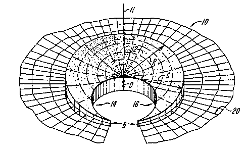
FIG. 1 shows a simplified diagramatic
representation of a human knee joint, with menisci in
native positioning;
FIG, lA is a diagrammatic representation of
a cut-away view of the knee joint showing the medial
and lateral menisci as they are positioned in viv
over the medial and lateral condyles.
FIG: 2 shows a perspective view of an
exemplary prosthetic meniscus in accordance with the
present invention;
FIG. 3 shows a perspective radial section of
the prosthetic meniscus of FIG. 2;
FIG. 4 shows a perspective view of an
alternative embodiment of the present invention;
FIG. 5 shows a sectional view along line 5-5
of the prosthetic meniscus of FIG. 4.
FIG. 6 shows a mold designed for the
fabrication of a prosthetic meniscus having a
cylindrical pad shape.
-12-
FIG. ~ shows a mold designed for the
fabrication of a prosthetic meniscus having a
crescent-shaped wedge form.
FIG. 8 shows a mold designed for the
fabrication of a cylindrical prosthetic meniscus.
FIG. 9 is a photographic representation of
,~ vivo rneniscal regrowth after a0~s resection and
implantation of the prosthetic meniscus.
FIG. 10 is a photographic representation of
an escplanted canine meniscus.containing a section of
scaffold, arid demonstrating ,~ vitro regrowth of
maniacal tissues into the scaffold.
FIG. 11 is a photographic representation of
two canine knee joints three months after surgical
resection.
FIG. 12 is a series of graphs showing the
hydrodynamic profiles of the proteoglycan aggregates
in the regenerated meniscus (FIG. 12B) compared to
the resected rim alone (FIG. 12D), and compared with
control samples including the remaining fibxocartilage
post-resectian is the medium (FIG. 12A) and the
remaining fibrocart3lage post-resection in the
associative extract (FIG. 12G).
-13-
Deserin ion of the Invent~nn
It has been discovered that a prosthetic
meniscus fabricated from biocompatible and
bioresorbable fibers can be surgically implanted into
the knee joint so as to provide normal joint motion
and strength. This prosthetic meniscus also acts as
a scaffold for regenerating meniscal tissue whose
ingrowth is encouraged by the physical
characteristics of the implanted device.
FIG. 1 shows a diagramatic representation of
the normal positioning of medial meniscus 7 and
lateral meniscus 8 in the human knee joint 3 between
the femur 2 and tibia 4. These menisci, when
compressed between the femur 2 and tibia 4, become
tough except at their points of attachment. FTG. lA
shows the in vivo structure of medial meniscus 7 and
lateral meniscus 8 in the knee joint 3. The menisci
conform to the shapes of the surfaces between which
they are positioned; thereby resulting in two
distinct in vivo forms. For example, the medial
meniscus 7 has a relatively open crescent shape,
while the lateral meniscus 8 has a relatively closed
crescent shape,
An exemplary prosthetic meniscus 10 is shaven
in FIG. 2. The prosthetic meniscus 10 is a generally
wedge-shaped, porous dry matrix or scaffold which
extends circumferentially or laterally at least in
part about a central axis 11. In the preferred form,
the prosthetic meniscus l0 has the shape of a
crescent-shaped wedge, extending circumferentially
_1~_ ~~~~ ~'~1
about the sale 11, and comprising a relatively wide
central region 12 between two narrow distal regions
14 and 16. In the preferred form, the wedge has
maximum height A at its peripheral edge of
approximately 0.4 inches, a height D at its central
point of approximately 0.2 inches. and a maximum
radial dimension C of approximately 1.0 inches. The
crescent shaped wedge subtends an angle B about axis
11 substantially in the range of about 135 to about
155 degrees, and preferably of about 150 degrees.
In the embodiment illustrated in Fig. 2, the
prosthetic meniscus 10 includes a mesh member 20
extending from its peripheral edge. The mesh member
is composed of a biocompatible, bioresorbable
15 material, and provides a readily used means for
anchoring the array 10 in place. The mesh member 20
may function in this capacity until sufficient tissue
ingrowth occurs to then provide that function. By
way of an example, the mesh member 20 may be a #1
20 mesh screen composed of absorbable suture materials
such as polyglyconate, Dexon, or polydioxane (PDS)
woven into a mesh. Non-absorbable suture materials
such as Goretex may also be used.
FIGS. 4 and 5 show an additional embodiment
of the present invention which is similar in
composition to the prosthetic meniscus depicted in
FIG. 2. More particularly, FIG. 4 depicts a right
circular cylinder-shaped meniscus 22, extending fully
about axis 11, i.e. where angle S equals 0 degrees.
(i.e. the meniscus subtends 360 degrees.) FIG. 5
shows a sectional view along line 5-5 of the meniscus
-15-
shown in SIG. 4. The device illustrated in Pigs. 4
and 5 show the shape of the meniscus 22 when
implanted; that is, the height D at areas 11 is less
than the peripheral height A of the device. Prior to
implantation, the device 22 may in some cases not
have this relationship but upon implantation, the
normal loads applied by the body force this
conformation.
In alternative forms of the invention, still
other shapes may be used. Por example, it is not
r
required that the wedge be symmetrical. These
embodiments may have densities of collagen fibers and
dispersions of GAG molecules and crosslinks,
permitting accommodation of differing stress levels,
rates of ingrowth, and resiliency. Differing
densities may be obtained in vivo where a device
having uniform density is implanted, and body loading
causes non-uniform compression of the device.
The prosthetic meniscus may be fabricated of
any biacompatible. bioresorbable fibers which include
a natural material ar an analog thereof; pseferably
polymeric in structure, which can provide mechanical
strength and protection and lubrication while
encouraging tissue ingrowth ~e.g., collagen,
reticulin, elastin, cellulose, or biosynthetic
analogs thereof). These fibers may be ordered in
substantially cirCumferentially-extending or
substantially radially-extending orientations. with
the density of fibers being substantially uniform
throughout the matrix. Alternatively, the matrix
fibers may be unordered. In either the ordered or
2~~~~~'~~.
-lE-
unordered configuration, the density of the fibers
may be non-uniform. In the non-uniform
configuration,relatively high densities of fibers may
be established at anticipated points of high stress
by local application.
In an alternative aspect of the invention,
the intrafibrillary and interfibrillary space is
relatively high, a condition which promotes ingrowth
of regenerated meniscal tissue. For example, the
density of the meniscus may be in the range of about
10-25 g matrix/cm~. Alternatively, the
intrafibrillary and interfibrillary space is
relatively low, a condition which provides
cushioning, lubrication, and mechanical support fox
the knee joint and which retards tissue and cell
ingrowth, thereby diminishing the rate of scaffold
resorption (e. g., density is in the range of about
2-ZO g matrix/cm3).
The temporary stability of the shape of the
structure when in vivo, and the rate of meniscal
resorption; are both attributed to the effective
crosslinking formation between at least one portion
of the fibers. The crosslinking reagents used may be
any biocompatible bifunctional reagents which
interacts with amino groups, carboxyl, or hydroxyl
groups on a single fiber (intramolecular crosslinks),
or the fibers or on the fibers and the GAGS,
~0 resulting in covalent bond formation between adjacent
molecules (intermolecular crosslinks): Useful cross-
linking reagents include aldehydes, hegamethylene
~~J~i~~~
-17-
diisocyanate, bis-imidates, polyglycerol polyglycidyl
ether, and carbodiimides.
The crosslinked device maintains sufficient
degree of hydrophilicity and elasticity which
simulates the properties of the natural meniscus,
i.e., ability to sustain mechanical stress and to
protect and lubricate articular surfaces. In
addition, the structure provides an ideal environment
for cell infiltration and extracellular matria
synthesis and deposition resulting in regeneration of
natural meniscal tissue.
GAGS may be dispersed throughout the
fibers. Alternatively, they may act as
intermolecular crosslinks between fibers. These GAG
crosslinks are composed typically of at least one of
the group of molecules consisting of chondroitin
4-sulfate, chondroitin 6-sulfate, keratin sulfate,
dermatan sulfate, heparin sulfate, and hyaluronic
acid. The dispersion of GAG crosslinks is preferably
uniform, but may be more concentrated at anticipated
points of high stress, typically at the distal
regions 14 and 16, and less concentrated in the
central region 12 (FIG. 1). In such configurations,
the GAG concentration may be in the rangy of about
0-25% in the distal regions Z4 and 16, a:;;d 3r~ the
range of about 0-10% in the central region 12.
However, when uniform, the dispersion of GAG
throughout the prosthetic meniscus may be, for
example, in the range ~f about 1-15%.
~~~~e~1
--18-
Intermolecular crosslinkages can also be
established through a dehydrothermal process (heat
and vacuum) which results in peptide bond formation
between an epsilon amino group of lysine or
hydroxylysine and a carboxyl group of aspartic or
glutamic acid.
The crosslinked device has a relatively high
thermal stability between about 55-85° C, preferably
between about 65-75° C, for sufficient ~ vivo
stability. This may be achieved through manipulation
of the crosslinking conditions, including reagent
concentration, temperature, pH, and time.
In a one embodiment the prosthetic meniscus
is constructed mainly of Type I collagen fibers
without GAG crosslinks. Type I collagen fibers may
be obtained from the Achilles tendons of animals.
~Iowever, the fibers may also be obtained from animal
skin or from the. skin or tendon of humans. The
tissues are treated with a series of mechanical and
chemical means to either totally remove the
non-collagenous materials or reduce them to a minimal
level. In the preferred processing steps, the tendon
or skin is mechanically disintegrated into fine
pieces useful for further processing. The
disintegration may be achieved by grinding the tissue
at liquid nitrogen temperature, or by cutting the
tissue into small pieces with a sharp knife. In
certain applications, the tendons are mechanically
disintegrated along the fiber direction in order to
maintain the length of the fibers for mechanical
strength.
-19-
Salt extraction of tendon at neutral pH
removes a small portion of the collagen molecules
that are newly synthesized and have not yet been
incorporated into the stable fibrils. Salt also
removes some glycoproteins and proteoglycans that axe
associated with collagen through electrostatic
interactions. Other salts such as KCl and the like
can be used as a substitute for NaCl.
lipids that are associated with the cell
membranes or collagenous matrices may be removed by
first extracting with detergents such as Triton
X-100, followed by extracting with ether-ethanol
mixtures. The concentration of Triton X-100 is
usually about 2-4%, but is preferably about 3%. The
preferred mixture of ether-ethanol is usually at
about a 1:1 ratio (vlv). The period of extraction is
usually from 8 hours to 96 hours, as is preferably
from about 24 to 48 hours.
Further extraction may be accomplished by
matrix swelling conducted at two extreme pHs. Both
acidic and basic swelling weakens the non-covalent
intermolecular interactions, thus facilitating the
release of non-covalently attached glycoproteins,
glycosaminoglycans (GAGs), and other non-collagenous
molecules through the open pores or the collagenous
matrices.
The swelling of matrix at alkaline pH is
done by treating the collagen ~t high pH with
Ca(OH)2, NaOH, or the like, for a period of about
8-96 hours. alkali extraction in the presence of
~~~~t~'~~1
-20-
triple-helical stabilizing salts such at (CH3)NC1,
NT-I3S04, or the like reduces the potential risk of
denaturation of the collagen. Alkali treatment
dissociates the non-cross-linked glycoproteins and
GAGS from the collagen matrices. The alkali, also
removed the residual lipids through saponification.
The acid swelling may be conducted at a low
pH in the presence of acetic acid, HCl, or the like.
Like the alkali treatment, the acid swelling removes
non-cross-linked glycoproteins and GAGS.
The non-triple helical portions of the
molecule (telopeptides) are involved in
intermolecular crosslinking formation. They are weak
antigens and are susceptible to attack by proteases,
such as pepsin, trypsin, and the like. Prolonged
digestion with such proteases dissociates the fibrils
(fibers) into individual molecules. However, if the
digestion process is properly controlled such that
maximal telopeptides are removed without complete
dissociation, the immunogenic properties of the
fibrils can be reduced to a minimal Level without
compromising the mechanical stxength. For example,
to isolate molecular collagen, the digestion of skin
or tendon with pepsin is usually conducted at an
enzyme:collagen ratio of about 1:10 for about 24-96
hours at below room temperature. In comparison,
fibrils may be obtained by limited pepsin digestion
achieved at a ratio of about 1:100 (enzyme:collac~en)
for about 24-96 hours at 4° C.
-21- '~~J~~~~~
Collagen fibers obtained according to this
methodology are then used to fabricate the prosthetic
meniscus of the present invention. However, it must
be appreciated that collagen obtained from other
sources, such as biosynthetically-produced collagen
or analogs thereof - may also be used in the
construction of the prosthetic meniscus.
In one embodiment, the prosthetic meniscus
further includes an adhesion molecule or adhesive
portion or analog thereof which is incorporated
within the network of fibers, and which aids in
maniacal tissue regeneration. Useful adhesion
molecules include peptides such as fibronectin see
e.g., U.S. Patent Plc. 4,589,881, 4,661;111 and
4,578,079), a portion of which can be conjugated to,
for example, chondroitin sulfate.
The method of fabrication includes molding
the collagen fibers into a predetermined shape using,
for example, the mold forms described below in
conjunction with FIGS. 6-8. The fibers may be placed
randomly in the mold, or may be oriented in specific
directions to achieve a meniscus having specific
structure characteristics. Other components such as
GAGS which may participate in the crosslinking
reaction, can be mixed in with the fibers in a random
or non-random fashion before the structure is
subjected to various crosslinking and dehydrating
procedures including various chemical and/or
dehydrothermal methods. Adhesion molecules or
adhesive fragments or analogs thereof may be added to
the structure before the final drying step by soaking
the structure in a solution containing that molecule,
2~~~~~~~.
-az-
or by specifically coupling it to an existing fiber
or crosslink. For example, the adhesion portion of
fibronectin may be crosslinked to chondroitin sulfate
at a concentration of 3 peptide molecules per
molecule chondroitin sulfate by soaking the
prosthetic meniscus in a 50 mg/ml solution thereof.
By following the processes described in the
above examples set forth herein below, a prosthetic
ZO meniscus of the form shown in FIGS. 2 or 3 may be
constructed having the characteristics listed below
in TABLE 1.
TABLE 1
Physical Characteristics
height A = 0.20 - 0.40 inches
angle B = 25 - 45 degrees
radius C = 0.5 - 2.0 inches
height D = 0.05 - 0.10 inches
Density - 0.07 - 0.'5 g/cm3
Tntra- and Interfibrillary space = 2-25 cm3/g
matrix
Constituents
fiber (collagen) content = 75-100%
GAG content - 0-25%
The following non-limiting examples describe
rnei:hods of fabrication and in vivo testing of the
prosthetic meniscus of the invention.
-23-
EXAMPLE 1
Mold Fabrication':
A mold 100 useful for fabricating the
prosthetic meniscus is made of implantable stainless
steel or biocompatible plastics such as teflon,
polypropylene, delrin, or combination of these
materials. The mold 100 is composed of three pieces
102, 104, and 106 as shown in FIGS. 6-8.
By way of example for the disk-shaped
meniscus illustrated in FIGS. 4 and 5, the mold l0O
of FIG. 6 is used: The first piece 102 is disk-Iike
and has a diameter substantially equal to that of the
desired meniscus. Piece 102 is perforated to allow
liquid to pass through under pressure. The inner
surface 103 of piece 102 has the desired shape of one
side of the meniscus-to-be-formed.
The second piece 104 is a hollow cylinder
which has the same inner dimension as the first piece
102. The third piece 106 is a cylindrical piston
which has an outer diameter slightly Iess than the
inner diameter of piece 104. The "top", or crown,
surface 108 of piston 106 has the desired shape of
one side of the meniscus-to-be-formed.
For the meniscus of FIG. 3, the mold of FIG.
7 is used where the shape of piece 102; and
cross-section of piece 104 have the shape of an
angular segment. Fbr a flat circular disk meniscus,
the mold l00 of FIG. 8 is used where pieces 102 arid
104 are the same as in FIG. 6 and piece 106 is
2~~~~'~1
-24-
similar to that piece in FIG. 108 but has a flat
crown surface 108.
During fabrication of the meniscus 10, the
piece 102 is first assembled within piece 104, as
shown in FIGS. 6-8. The constituent fibers (in a
fluid) are placed against the surface 103 of piece
102. Then the crown surface 108 of piston 106 is
driven toward surface 103 along a compression axis
106a until the fibers are compressed, the fluid is
driven out through piece 102, and the desired axial
dimension of the compressed fiber array is attained.
The mold is then frozen in preparation for chemical
crosslinking.
EXAMPhE 2
Pre~aaration of Purified Type T ~ollaaen
A) Tissue:
Bovine, porcine, or sheep Achilles tendon is
obtained from USDA-approved slaughter houses. The
preferred age of the animals is between 12-18
months. The tissue is kept cold during the
purification process except where specified to
minimize bacteria contamination and tissue
degradation.
B) Mechanical Disintegration:
The adhering tissues of carefully selected
tendons are first scrapped off mechanically. The
tendons are then minced or cut into fine pieces and
-25-
washed in excess quantities (10 volumes) of cold
water to remove residual blood proteins and water
soluble materials.
C) Salt Extraction:
The washed tendons are extracted in ten
volumes of 5% NaCl, 0.01 M Tris, pH 7.4, for 24 (+/-
4) hours to remove salt soluble materials. The salt
extracted tendons are repeatedly washed in about ZO
volumes of water to remove the salt.
D) hipid Extraction:
The material is extracted in 3% Triton X-100
for 24 (+!- 2) hours. The detergent is removed by
extensive washing with water. The material is then
extracted in 3-4 volumes of ether-ethanol (1:1
vol/vol) for 24 (+/- 2) hours to further minimize the
lipid content. The lipid extracted material is
extensively washed in water to remove the ether and
ethanol.
E) Matrix Swelling:
The material is then subjected to two
extreme pH extractions to remove non--collagenous
materials: Alkaline extracti~n is conducted with 3-4
volumes of 0.2 M NaOH at pH 12.5 - 13.5 at room
temperature (RT) in the presence of 1.0 M (CH)NCl for
24 (+/- 2) hours with mild agitation.
~~~~~~'~1
-26-
Following alkaline extraction, the pH is
neutralized with HC1 and the material is washed writh
water. The pH is then adjusted to 2.5 - 3.U by
adding concentrated acetic acid to a final
concentration of 0.5 M. The acid extraction is
continued for 24 (+/- 2) hours with agitation.
F) Limited Proteolytic Digestion:
The acid swollen tendon is then subjected to
a limited proteolytic digestion with pepsin (enzyme :
collagen = 1 : 100) for 24 (+/-) 2 hours. The pepsin
and telopeptides are removed through dialysis.
The swollen fibrillar material is then
coacervated by adjusting the pH to its isoionic point
with 1 M NaOH or HC1 or by adjusting the ionic
strength to 0.7 with NaCl. The agg~cegated collagen
fibers are harvested by filtration, and the filtered
material extensively washed with cold buffered
sblutiono The highly purified type I collagen may be
stored (-20 ~0 -40° C) until used.
EXAMPLE 3
Device I Fabri a~-; n.,
A) The collagen content of the highly
purified type I collagen fibrils from E°~AMPLE 2 is
determined either by gravimetric methods or by
determining the hydroxyproline content assuming a
I~.S% by weight of hydroxyproline in type I
collagen. The amount of purified material needed to
~~~~~'~1
-27-
fabricate a given density of a meniscus device is
then determined and weighed.
B) A solution of fibrillar collagen is
carefully fit into a mold of specified dimensions,
e.g, according to the exemp7.ary meniscus described
above in conjunction with FIG. 2-5 (see EXAMPLE I and
FIGS. 6-8 far the description of molds). Collagen
fibers are laid down in random manner or in an
oriented manner. In the oriented manner,
circumferential orientation of the fibers is produced
by rotation of the piston about its principal axis as
the material is compressed in the mold; radial
orientation is produced by manual painting of the
collagen fibers in a linear, radially directed
fashion.
C) The fibers are frozen at -20° C, turned
out of the mold, and thawed at RT.
D) The fibers are then resuspehded in
phosphate buffered saline, put back into the mold in
the desired orientatian(s), and compressed with the
piston.
E) The compressed fibers are then refrozen
at -20 C and then thawed at RT.
F) The resulting structure is crosslinked
by soaking in a 0.2°s glutaraldehyde solution, pFi 7.6,
for 24 (+/- 0.5) hours. Each glutaraldehyde-
cross-linked meniscal device is subsequently rinsed
_28_
repeatedly in 500 ml of phosphate buffered saline
(PBS) solution, pH 7.4, for 4, 8, 24 and 48 hours.
G) The rinsed matrix is then lyophilized.
EXAMPLE 4
Device iT Fabri~atann
A)-G) (same as in EXAMPLE 3)
H) The lyophilized matrix is sub3ected to
dehydrotherrnal crosslinking by vacuum and heat. The
vacuum is first applied to reduce the residual water
content to a minimal level (same structural water,
I5 about 3°a, may still be associated with collagen
triple-helix as part of the structure stabilizing
factor). The heat is increasing in steps to 110° C
(ø/- 5°), and continually applied at 110° C under
vacuum for 24 (-~/- 2) hours.
EXAMPLE 5
Device TIT Fabrication
A) (same as in EXAMPLE 3)
B) The collagen material is dispersed in
0.01 M HCl solution at pH 2-2.5. Predetermined
amounts of various GAGS are weighed and dissolved in
water. For example, far a given density of 0.25
g/cm, the collagen content will be 0.244 g, the
hyaluronic acid content will be 0.003 g; and the
chondroitin sulfate content will be 0.003 g for a
2.5% GAG content. The GAG solution is mixed in with
-~g_
the collagen solution and placed in the mold in the
desired orientation as described iri EXAMPLE 2.
C)-G) (same as in EXAMPLE 3)
EXAMPLE 6
Device IV Fabrics ; n
A)-C) (same as in EXAMPLE 3)
D) (same as in EXAMPLE 3 except that the
fibers laid down are not compressed.
E)-G) (same as in EXAMPLE S)
EXAMPLE 7
Device V Fabrication
A)-E) (same as in EXAMPLE 3)
~0
F) The molded collagen is crosslinked in 5%
polyglyc~rol polyglycidyl ether in 50% ethanol and
0.1 M Na~C03 at pH 10.0 ~or 29 (+/- 2) hours. The
crosslinked device is rinsed ~or 4, 8; 24 and ~S
hours, each. with 500 m1 of PBS, pH 7.~.
G) (same as in EXAMPLE 3)
-30-
EXAMPLE 8
vi ~ VT Fabrication
A)-E) (same as in EXAMPLE 3)
F) The molded collagen is crosslinked in
the presence of 1-ethyl-3-(3-dimethylaminopropyl)
carbodiiznide (10 mg/g matrix) in 0.9% NaCl, pH 9.7 at
room temperature for 24 (+/- 2) hours. The addition
of carbodiimide is made every 3-4 hours, and the pH
is adjusted to 4.7 after each addition of
carbodiimide.
(same as in EXAMPLE 3)
EXAMPLE
Device VIT Fabri an on
(A) - (D) same as steps (A) - (D) as
described in Example II.
(E) For attachment purposes, a mesh of
absorbable polyglyconate suture material, matched to
~5 the size o~ the mold, is laid in the dispersed
collagen such that it protrudes from the structure's
periphery to form a skirt which may extend over the
tibial plateau. This mesh provides both immediate
attachment sites and long term fibrous ingxowth.
-31_ ~0~~~'~~
Ex~M~x,E 10
Testincr
The prosthetic menisci were evaluated ,fig
vivo using animal models and in vi r to determine
ability to function or to serve as a regeneration
template for normal meniscal tissues.
1. In vivo studies
Seventeen prosthetic menisci (device
IIT typed were implanted into eleven immature
Yorkshire pigs. Seven joints underwent a two-thirds
subtotal resection of the medial meniscus with
replacement by a matched prosthetic meniscus; two
joints underwent a two-thirds subtotal resection
alone; two joints received a similar subtotal
meniscectamy with the resected portion immediately
replaced with suture fi$ation; and two joints
received total meniscectomy alone.
Evaluation of all joints was made at 3 or 6
weeks. All arthrotomies healed well, and all animals
progressed to full weight bearing. At final
evaluation all prosthetic material had been partially
or completely resorbed without evidence of joint
destruction or cartilage abrasion. ~Teovascular-
i~ation was observed as the basic healing mechanism
in both the prosthetic implanted menisci as well as
in the controls, and in all joints there was evidence
of early meniscal regeneration. The prosthetic
meniscus material conformed to the appropriate joint
shape. In addition, there was no clinical evidence
of implant rejection over a 6 week period.
Histologically, there was acute inflammation followed
by neovascularization and extensive fibroplasia with
early hyalinization of the newly formed collagen.
(See FIG. 9)
,~, ~zvo studies of the invention 3n mature
dogs have demonstrated induced meniscal regeneration
through the prosthetic material. Normally, the
mature canine stifle is known to not regenerate a
meniscus and is known to develop significant
arthritic changes. However, sia weeks after
meniscectamy and implantation of scaffolds in
accordance with the present invention, there occurred
significant regeneration of the meniscus through the
scaffolds. The scaffolds provided joint protection,
as determined by diminished cartilage erosions,
osteophyte formation and affinity for India ink.
FIG. 11 is a photographic representation of
two canine knee joints three months after surgical
resectian. The joints were protected by the
prosthetic implant with subsequent regrowth of a new
meniscus. The joint on the right in FIG. il
underwent an SO~a m~n3scal resection alone. The
dramatic articular cartilage protection is
highlighted by India ink.
New collagen and glycosaminoglycan formation
was evidenced histologically, by Alcian Blue and
Masson's Trichrome stains.
The hydrodynamic size and chromatographic profiles of
the proteoglycans synthesized within both the
meniscal implants and the controls were similar when
analyzed on a Sephacryl S-500 column as shown in FIG.
12. FIG. 12 is a series of graphs showing the
hydrodynamic profiles of the proteoglycan aggregates
in the regenerated meniscus (FIG. 12E) compared to
the resected rim alone (FIG. 12D), and compared with
control samples including the remaining fibrocartilage
post-resection in the medium (FIG. 12~) and the
remaining fibrocartilage post-resection in the
~associativs eztract (FIG. 12C).
2. in vitro Studies
Menisci were aseptically harvested fram
mature dogs, trimmed of all adherent tissue, and
placed into Gey°s balanced saline solution. Each
meniscus was bisected in the eoronal plane and 3 mm
full-thickness circular defects were made in each
maniacal half. The defects were filled with a ~ mm
diameter plug of one of two prototypes of a complex
collagen-based biosynthetic scaffold (prosthetic
meniscus). The menisci were placed in sig well
culture plates containing six ml of Dulbecco°s
modified Eagle's medium supplemented with 10% fetal
bovine serum, sodium ascorbate, and 0.1%
penicillin/streptomycin. Cultures were maintained at
~~~~:~'~1
-33A-
37°~ in a humidi~ied atmosphexe ~~ 1~~ c~Z/90~5 air,
~ed three times par week, and placed in Fresh culture
-34-
wells every week to prevent the formation of explant
cell cultures. At intervals of one, four, and six
weeks after initiation of culture, three menisci from
each group were removed, faxed, and evaluated with
serial sections and staining.
The results (shown in FTG. 10) demonstrated
increasing cellular migration and invasion over
time. There was no apparent toxicity from the
ZO material. The morphologic characteristics of the
migrating cells were more fusiform and elongated than
native fibrochondrocytes. The depth of cellular
penetration into the scaffold appeared to be limited
by the density of the prosthetic complex.
The present invention may be embodied in
other specific forms without departing from the
spirit or essential characteristics thereof. The
present embodiments are therefore to be considered in
all respects as illustrative and not restrictive, the
scope of the invention being indicated by the
appended claims rather than by the foregoing
description, and all changes which come within the
meaning and range of equivalency of the claims are
therefore intended to be embraced therein.
what is claimed is: