Note: Descriptions are shown in the official language in which they were submitted.
2~8~1
PERIODONTAL PROBE
This invention relates to a periodontal probe. In
particular, this invention relates to a periodontal probe
having a calibrated tip for diagnosing periodontal disease and
gingivitis.
Periodontal disease is the most widespread disease
in the world. It is basically an inflammatory disease of the
gums which spreads to and destroys the supporting bone of the
teeth. In time, teeth may abscess, become loose or painful
and either fall out or are removed by a dentist. Fortunately,
the dental profession has continually developed more effective
2 ~
methods to treat periodontal disease, but these treatments are
dependent upon the patient seeking treatment by an appropri~te
party, usually a dentist.
The disease is largely silent as characterized by an
absence of symptoms, much like high blood pressure. For
example, there may or may not be bleeding or pus around the
teeth. Advanced cases frequently cause systemic problems due
to the massive amount of infection present.
The sole cause of periodontal disease is dental
plaque which is a bacterial substance present in the mouth.
However, due to the fact that every person has a different
genetic background, some people are resistant to the disease
while others are extremely prone. Additionally, the problem
is enhanced by infrequent dental visits and non-diagnosis of
the disease.
The periodontal probe is the only significant
clinical tool used for checking a person's periodontal disease
status. Conventional probes have either lines or marks to
indicate the depth that the probe penetrates between the tooth
and the gum. A non-diseased condition is reflected by a probe
depth of from 1 to 3 millimeters between the tooth and gum.
A deeper insertion indicates a problem, and the depth of the
insertion corresponds to the amount of bone loss.
Additionally, at a probe depth of 5 millimeters, it is
virtually impossible to remove calculus or foreign debris from
the roots of diseased teeth. At this critical depth, a
patient will likely require surgical treatment which may be
costly and uncomfortable.
Conventional periodontal probes having uncolored
lines or marks indicating probe depth are difficult to see
when used by a dentist. Because of the inability of
conventional probes to clearly and accurately measure probe
depth, dentists occasionally insert a flexible gutta percha
.
- 21D~8~
point into periodontal pockets and take a radiograph to see
the anatomy of the pocket, since gutta percha can be
visualized on x-ray. This procedure may be used to decide if
surgical treatment is required. This procedure is obviously
very tedious and exposes the patient to additional x-rays.
A primary or first stage of periodontal disease is
gingivitis which is detected by eliciting any bleading while
probing. One method of diagnosis of gingivitis is the use of
pieces of balsa wood which are sold, for example, by Johnson
& Johnson under the trademark "STIM-U-DENT." One problem with
the use of pieces of balsa wood is that they are too large and
rough to be used accurately.
Further, many bac~ teeth have two or more roots.
Frequently, bone is lost during periodontal disease between
the roots. This area of the tooth where the roots divide is
referred to as the "furcation." When bone is lost between
roots, there exists a "furcation involvement." The depth of
furcation involvement is of paramount importance in
determining the prognosis and required treatment of the tooth.
Without a means of accurately assessing the depth of these
areas, it is difficult for dental practitioners to make a
proper diagnosis, particularly with respect to a surgery or no
surgery decision.
The dental instrument art lacks a probe that the
dental professional can use to easily diagnose periodontal
disease and gingivitis and simultaneously be used to easily
determine if surgical treatment is likely required. In
addition, a method for routinely diagnosing and monitoring
periodontal disease and gingivitis, and at the same time
assessing if surgical treatment is likely reguired does not
curre~tly exist.
.
2 ~
It is therefor~ fi obje_t of the present invention
to provide a periodontal probe useful for the early detection
of periodontal disease and gingivitis.
It is also an object of the present invention to
provide a periodontal probe useful to determine if surgical
treatment is likely required.
Another object is to detect furcation involvement by
means of a periodontal probe which has the ability to easily
assess the depth of furcation involvement in all directions,
thus enabling the medical practitioner to determine if
surgical treatment is likely required in a manner not
previously possible, specifically because conventional probes
cannot clearly indicate the exact probe depth where surgical
treatment is generally required.
Yet another object is the diagnosis of gingivitis by
demonstrating the bleeding point.
Another object of the invention is to provide an
easy to use, disposable periodontal probe for the use of
dentists and physicians to routinely check for periodontal
disease and to determine if surgical treatment is likely
required.
It is still another object of the invention to
provide a method for the detection of periodontal disease.
In accordance with one aspect of the present
invention these objects are achieved by a periodontal probe,
comprising:
(a) an elongated member having a distal end with a
tip thereon;
(b) the distal end being tapered to the tip and
having a plurality of condition indicating portions
comprising:
2 ~
(1) a first portion indicating a non-diseased
condition disposed between the tip and a second portion,
(2) the second portion indicating a moderately-
diseased condition disposed between the first portion and a
third portion;
(3) the third portion indicating a diseased
condition wherein surgery is required;
(4) a narrow line being positioned in the
second portion between the first and third portions, with the
narrow line defining a boundary between two areas, a first
area indicating a diseased condition likely not re~uiring
surgery located between the first portion and the narrow line
and a second area indicating a diseased condition likely
requiring surgery located between the narrow line and the
third portion.
In accordance with another aspect of the present
invention these objects are achieved by a method for
diagnosing periodontal disease comprising the steps of:
ta) inserting a periodontal probe, at the juncture
between a tooth and gum, comprising:
an elongated member having a distal end with a
tip thereon;
the distal end being tapered to the tip and
having a plurality of condition indication portions
comprising:
(1) a first portion indicating a non-diseased
condition disposed between the tip and a second portion,
(2) the second portion indicating a moderately-
diseased condition disposed between said first portion and a
third portion;
(3) the third portion indicating a diseased
condition wherein surgery is required;
- 5
:
2 ~
(4) a narrow line being positioned in the
second portion between the first and third portions, with the
narrow line defining a boundary between two areas, a first
area indicating a diseased condition likely not requiring
surgery located between the first portion and the narrow line
and a second area indicating a diseased condition likely
requiring surgery located between the narrow line and the
third portion.
(b) examining the distal end and ascertaining which
of the three portions or narrow line is visible at the
juncture;
(c) removing the periodontal probe from between the
tooth and gum; and
(d) repeating steps (a) through (c) at least once.
Other objects, features and advantages of the
present invention will become apparent from the following
detailed description and accompanying drawings wherein:
Figure 1 is a side plan view of a periodontal probe;
Figure 2 is a magnified view of a section of the
distal end of the periodontal probe shown in Figure 1;
Figure 3 is a perspective view of a periodontal
probe inserted at the juncture between a healthy tooth and
gum;
Figure 4 is a perspective view of a periodontal
: probe inserted at the juncture between a diseased tooth and
gum which does not likely require surgery;
Figure 5 i8 a pergpective view of a periodontal
probe inserted at the juncture between a diseased tooth and
gum which likely requires surgery; and
2~56~
Figure 6 is a perspective view of a periodontal
probe inserted at the juncture between a diseased tooth and
gum which requires surgery.
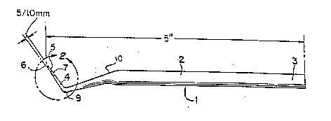
Referring to Figure 1, there is shown a periodontal
probe 1 comprising an elongated member 2. The elongated
member 2, which is of FDA compliant non-toxic plastic material
suitable for oral use, has a proximal end 3 and a distal end
4. Such material can be polypropylene homo-polymer,
polypropylene co-poly~er, high density polyethylene homo
1~ polymer or polyethylene co-polymer or polybutylene
terephthalate or nylon or ABS (acrylonitrile, butadiene,
styrene, ter polymer) or acrylic multipolymer, or polymer
blends or alloys; which may incorporate a 5% to 45% talcum,
mica or fiberglass filler. In the preferred embodiment the
material is either 40% talcum filled polypropylene homo
polymer or polybutylene terephthalate, with filled
polypropylene homo polymer being the preferred of the two. In
order to obtain the desired flexibility, material is used
which has a flex modulus (tangent) in the range of about 1.3-
9.2 x 10⁵ p.s.i. with a preferred modulus of about 5.1 x 10⁵
p.s.i.
The distal end 4, which is flexible, has a first
portion 5, indicating a non-diseased condition, which is
disposed between a tip 6 and a second portion 7. The second
portion 7 indicates a moderately-diseased condition which may
require surgery and is disposed between the first portion 5
and a third portion 8.
The third portion 8 indicates a diseased condition
wherein surgery is required and is disposed adjacent to the
second portion 7. A narrow line 9 is located at about the
;'
- 7
.
-` 2~6811
midpoint of the second portion 7 and indicates that a di eased
condition exists that likely requires surgery.
The elongated member 2 has a first bend 10 disposed
between the proximal end 3 and the distal end 4. A second
bend 9 is disposed between the tip 6 and the first bend 10.
It should be noted that the elongated member can have any
number of bends at any desired angle which can achieve the
desired function of enabling the dental probe to be held by
hand and enabling the easy insertion of the distal end at the
juncture between a tooth and gum. ~he purpose of the bends in
the elongated member is to offset the distal end at an angle
which facilitates easy insertion and examination of the dental
probe. A desired embodiment is achieved when the first bend
is directed downward forming an obtuse angle at the bottom
side of the elongated member and the second bend is directed
upward forming an obtuse angle at the top side of the
elongated member.
Referring to Figure 2 there is shown a magnified
view of a section of the flexible distal end of the dental
probe shown in Figure 1 in which the first portion 5 is about
3 millimeters in length, the second portion 7 is about 4
millimeters in length, and the third portion 8 is about 3
millimeters in length, all portions together extending a
length of about 10 millimeters from the tip 6. Additionally,
the narrow line 9 is disposed about 5 millimeters from the tip
6, at approximately the midpoint of the second portion 7.
When the distal end is inserted at the juncture between a
tooth and gum up to a length of about 3 millimeters, the first
portion remains visible indicating a non-diseased condition.
Insertion of the distal end beyond about 3 millimeters but
less than about 5 millimeters results in the second portion
being partly visible indicating a diseased condition not
likely requiring surgery. Insertion of the distal end beyond
-- 2 ~
about 5 millimeters but less than about 7 millimeters
indicates a diseased condition that likely requires surqery.
Insertlon of the distal end beyond about 7 millimeters
indicates a severely diseased condition requiring surgery.
Figure 2 shows the second portion 7 extending from about the
3 millimeter mark to about the 7 millimeter mark, shows the
narrow line 9 at the 5 millimeter mar~ and the third portion
8 extending from the 7 millimeter mark to the 10 millimeter
mark.
Additionally, the first, second and third portions
can be contrastingly color-coded to aid the user when
attempting to ascertain which of tbe three portions is visible
during the examination. The narrow line can be contrastingly
color-coded with the second portion to highlight if the narrow
line is visible during examination. In one example of color-
coding, the first portion can be colored green, the second
portion can be colored white, the third portion can be colored
red and the narrow line red. In effect, any three colors
which contrast each other can be chosen. The color is
obtained by the use of a non-toxic compliant ink.
The dental probe shown in Figure 1 is about 5 inches
in length, but can be any length that is convenient for hand-
held use. The dental probe shown in Figure 2 tapers from the
first bend 10 to the tip 6 which has a width of about 1/2 of
a millimeter. The tip at the distal end can have any width
that is suitable for inserting the distal end at the juncture
between a tooth and gum.
The present invention also includes a method for
diagnosing periodontal disease. This method includes the
steps of inserting a dental probe at the juncture between a
tooth and gum. The distal end is then examined to ascertain
which of the three portions is visible at the juncture and
ultimately whether a diseased or non-diseased condition
_ g _
',
., .
. - .
.
.
2~3 ~
exists. Furthermore, if the second portion is visible the
distal end can be further examined to determine if the narrow
line is visible. This method creates a simple yes-no
situation for determining disease status as well as whether
surgery is, may be, or is not required. The dental probe is
then removed from between the tooth and gum and gently
inserted at another juncture. ~his process can be repeated
until all the tooth/qum junctures are examined.
Referring to Figure 3, there is shown the dental
probe 1 inserted at a juncture between a tooth and gum. The
first portion 5 remains visible indicating a non-diseased
condition. The second portion 7, narrow line 9 and third
portion 8 are also visible as a result of the dental probe not
being able to penetrate at least about 3 millimeters.
Referring to Figure 4, there is shown the dental
probe 1 inserted at a juncture between a tooth and gum in
which the second portion 7, narrow line 9 and third portion 8
remain visible indicating a diseased condition not likely
requiring surgery.
Referring to Figure 5, there is shown the dental
probe 1 inserted at a juncture between a tooth and gum in
which only part of the second portion 7 and the entire third
portion 8 remain visible indicating a diseased condition
likely requiring surgery.
Referring to Figure 6, there is shown the dental
probe 1 inserted at a juncture between a tooth and gum in
which only the third portion 8 remains visible indicating a
diseased condition requiring surgery.
The method of detecting gingivitis is substantially
the same in that the probe is used to detect any bleeding.
The Examiner can ascertain whether the depth of or furcation
involvement exceed the critical 5 millimeter mark.
-- 10
`` 2 ~
The present method can also include the step of
coding the three portions with contrasting colors for ease of
identification. In addition, the narrow line can be color
coded to contrast with the second portion increasing its
visibility. The specific portion of the distal end visible
for each tooth and gum area examined can be charted with an
examination chart showing the teeth and gums in their position
relative to one another. In this method, the entire mouth can
be charted to determine where the troubled spots are.
Additionally, the chart can be used to indicate whether or not
bleeding occurred. Therefore, at a glance, the patient can
see from the chart where the pockets are or where bleeding
occurred. A person using this method can conceivably chart
individual patients after a period of time to see what changes
may have occurred following either professional care or self-
treatment, for example, by improved tooth brushing and dental
flossing. The probe as stated above, can be made from plastic
or metal and can be disposable, depending upon the desired
use.
While several embodiments of the invention have been
described, it will be understood that it is capable of still
further modifications, and this application is intended to
cover any variations, uses, or adaptations of the invention,
following in general the principles of the invention and
including such departures from the present disclosure as to
come within knowledge or customary practice in the- art to
which the invention pertains, and as may be applied to the
essential features hereinbefore set forth and falling within
the scope of the invention or the limits of the appended
claims.
-- 11 --