Note: Descriptions are shown in the official language in which they were submitted.
.....,.,." ~ 'J u~~ I~JI
PATENT
METHOD AND APPARATUS FOR
COMPUTING TOMOGRAPHIC SCANS
BACKGROUND OF THE INVENTION
Field of the Invention
The present invention pertains generally to the use of
a detachable x-ray detector array in combination with an
existing radiation therapy x-ray simulator to produce
computed tomographic image reconstructions. More par-
ticularly, the present invention relates to an apparatus
for producing a computed tomographic scan from the width-
collimated fan beam produced by an existing x-ray simulator
and to a method for transforming the data produced by the
- detector array of the apparatus into a back projected image
of the target object.
Introduction
X-ray computed tomography (CT) is a technique for
obtaining cross-sectional reconstructions of three dimen-
sional objects using x-rays. In the simplest example of
CT
imaging, a narrow beam of penetrating x-rays is scanned
across an object or patient in synchrony with a radiation
detector on the opposite side of the patient. If the beam
is monoenergetic or nearly so, the transmission of x-rays
through the patient is given by the equation
I = Io exp(-~Cx) [l]
where the patient is assumed to be a homogeneous medium
with the attenuation coefficient ~,. If the x-ray beam is
intercepted by two regions with attenuation coefficients
~.1
and ~2 and thicknesses xl and x2, the x-ray transmission
is
characterized as
I = Ioexp[-(hlxl + f~2x2) ]
This formula is generalized to many (n) regions with
different linear attenuation coefficients with the argument
of the exponent
n
E /lixi = (~,l~lx1 + I-c2x2 + . . . ~.~,nXn) [ 3
1=1
1
suBST~TuzE s~:-~~
iPEanS
CA 02060181 1994-OS-15
~i"lUS 9 ~ / ~ '~ 3 ~_~
IPEA~I~S ' . ~ r r X991
PATENT
n
I = EIOexp [ - (I~ixi) ~ [ 4 l
i=1
Separate attenuation coefficients cannot be determined with
a single transmission measurement because there are too
many unknown values of ui in the equation. However, with
multiple transmission measurements at different orienta
tions of the x-ray source and detector, the separate
coefficients can be distinguished so that a cross-sectional
display of coefficients is obtained across the plane of
transmission measurements. By assigning gray levels to
different ranges of attenuation coefficients, a display is
obtained that represents various structures in the patient
with different x-ray attenuation characteristics. This
gray scale display of attenuation coefficients constitutes
a CT image.
The first CT systems were introduced in approximately
1971 by the EMI Corporation of England for use in medicine.
These early systems used an x-ray source mounted in a
gantry with detectors. The patient was inserted between
the x-ray source and the detectors and the joined x-ray
source and detectors were moved about the patient to obtain
projection rays through the patient. These values were fed
to a computer which then reconstructed a cross sectional
5 image of the plane through which the pencil beam of x-rays
passed. During this translational scan of perhaps 40 cm in
length, multiple (e.g., 160) measurements of the x-ray
transmission were obtained. Next, the angular orientation
of the scanning device was incremented one degree and a
second translational scan of 160 transmission measurements
was performed. This process of translational scanning at
one degree increments was repeated through an arc of 180
degrees so that 28,800 x-ray transmission measurements were
accumulated. Those measurements were then transmitted to
a computer equipped with a mathematical algorithm for
reconstructing an image of attenuation coefficients across
the anatomical plane defined by the scanning x-ray beam.
2
SUBSTITUTE S';
CA 02060181 1994-OS-15
.,.
P~1'lUS ~ x/04 374
IPEA/US 1 ~ ~"~~~, -.
Although this approach yielded satisfactory images of
stationary objects, considerable time (4-5 minutes) was
required for data accumulation and the images were subject
to motion blurring. Soon after the introduction of pencil
beam scanners, fan-shaped x-ray beams were introduced so
that multiple measurements of x-ray transmission could be
made simultaneously. Fan beam geometries, with increments
of a few degrees for the different angular orientations
(e, g., a 30-degree fan beam and 10-degree angular
increments), reduced the scan time to 20-60 seconds and
improved the image quality by reducing the effects of
motion. Computed tomographic scanners with x-ray fan beam
geometries and multiple radiation detectors constituted the
second generation of CT scanners.
In late 1975, the third generation of CT scanner was
introduced. These scanners eliminated the translational
motion of previous scanners, using rotational motion of the
x-ray tube and detector array or rotational motion of the
x-ray tube within a stationary circular array of 600 or
more detectors. With these scanners, data accumulation
times as fast as two seconds are achievable.
Both stationary and rotating anode x-ray tubes are
used in CT scanners. Many of the translation-rotation CT
scanners have an oil-cooled, stationary anode x-ray tube
with a focal spot on the order of 2 X 16 mm. The limited
output of these x-ray tubes necessitates a sampling time of
about 5 msec for each measurement of x-ray transmission.
This sampling time, together with the time required to move
and rotate the source and detector, limits the speed with
'~ which data can be accumulated with CT units using translat-
ional and rotational motion.
To reduce the sampling time of 2-3 msec, most fast-
scan CT units use rotating-anode x-ray tubes, often with a
pulsed x-ray beam, to achieve higher x-ray outputs. Even
with rotating-anode tubes, the heat-storage capacity of the
3
CA 02060181 1994-OS-15
SUBSTITUTE SHEET
IPEA/US
PCTIUS ~ ~ / 0 ~ 3 7 4
IPEA/US - ~ ~.. ~- ~ ~~~'_ ____
PATENT
anode may be exceeded if cooling periods are not observed
between sets of successive images.
After transmission through the patient, the x-ray beam
is collimated to confine the transmission to a slice with
a thickness of a few millimeters and to reduce scattered
radiation to less than one percent (1%) of the primary beam
intensity. The height of the collimator defines the
thickness of the CT slice. This height, when combined with
the area of a single picture element (pixel) in the dis-
play, defines the three-dimensional volume element (voxel)
in the patient corresponding to the two-dimensional pixel
of the display. A voxel encompassing a boundary between
two tissue structures (e.g., muscle and bone) yields an
attenuation coefficient for the pixel that is intermediate
between the values for the two structures. This "partial
volume artifact" may be reduced by narrowing the collimator
to yield thinner slices. However, this approach reduces
the intensity of the x-rays incident upon the detector and
the detector signals are subject to greater statistical
fluctuations, thus introducing more noise into the
displayed image.
To reduce the detector response time, all detectors
used in CT scanning are operated in current rather than
pulse mode. Also, rejection of scattered radiation is
assigned to the detector collimator rather than to pulse
height analyzers. Detectors for CT scanning are chosen on
the basis of detection efficiency (greater than 50%), short
response time and stability of operation, and are either
gas-filled ionization chambers or solid scintillation
~ detectors. Scintillation detectors include NaI (TI) and
CsI crystals and newer bismuth germanate (BiGeO) detectors
chosen for their high detection efficiency and low fluores-
cence decay time. On information and belief, most
ionization chambers in current use contain xenon pres-
surized up to 25 atm to improve the x-ray detection ef-
ficiency. With any detector, the stability of response
4
CA 02060181 1994-OS-15
SUBSTITUTE SNEEt
~FEA/US
lUS 9G/04374
pit
lPEA/US
X91
PATENT
from one transmission measurement to the next is essential
for the production of artifact-free reconstruction images.
With a pure rotational source and detector geometry, for
example, detector instability gives rise to ring-shaped
artifacts in the image. Minimum energy dependence of the
detectors over the energy range for the CT x-ray beam also
is important if corrections for beam hardening are to be
applicable to all patient sizes and configurations.
All of the early CT systems were designed and built
only to perform CT studies. The concept of using other
types of radiation sources that had not been specifically
designed for CT imaging was initiated in the mid 1970's.
Several of these efforts utilized existing x-ray
therapy simulators. An x-ray simulator is a device that
duplicates a radiation treatment unit in terms of its
geometric, mechanical and optical properties, but uses a
diagnostic x-ray tube as the source of radiation to simu-
late the properties of the treatment beam. A simulator
allows the beam direction and the treatment fields to be
determined while encompassing the target object with the
simulator's irradiation. Since the simulator's emissions
are generally less intense and less energetic than the
emissions of therapy devices, there is a reduction in the
target object's exposure to radiation.
The combination of a detector system and an x-ray
therapy simulator provides the necessary front end of a CT
system. Application of the requisite information process-
ing techniques and algorithmic reconstruction processes in
combination with the simulator cum detector system enable
production of CT images.
Reconstruction Algorithm
The numbers computed by the reconstruction algorithm
are not exact values of attention coefficients. Instead,
they are integers, termed CT numbers, which are related to
attenuation coefficients. On most newer CT units, the CT
numbers range from -1,000 for air to +1000 for bone, with
5
SUBSTITUTE SNEET
CA 02060181 1994-OS-15
IPEA/US
PCTnS ~/~~+37~___
__ _-,.
~~r'.,»91
PATENT
the CT number of water set at 0. CT numbers normalized in
this manner are termed Hounsfield units and provide a range
of several CT numbers for a one percent (1%) change in
attenuation coefficient.
To portray the CT numbers as a gray scale visual
display, a storage oscilloscope or television monitor may
be used. This viewing device contains a contrast enhan-
cement feature that superimposes the shades of gray
available in the display device (i.e., the dynamic range of
the display) over the range of CT numbers of diagnostic
interest. Control of image contrast with the contrast
enhancement feature is essential in x-ray computed
tomography because the electron density, and therefore the
x-ray attenuation, are remarkedly similar for most tissues
of diagnostic interest. These electron densities vary from
3.07 x 10²³ elec/cc for fat tissue to 5.59 x 10²³ elec/cc
for the densest tissue, bone. Lung tissue has a much lower
electron density, 0.83 x 10²³ elec/cc, because of the
alveolar and branchial spaces.
Known reconstruction algorithms are one of four types:
(1) simple back projection - In this method,
each x-ray transmission path through the body is
divided into equally spaced elements, and each element
is assumed to contribute equally to the total at
tenuation along the x-ray path. By summing the
attenuation for each element over all x-ray paths that
transect the element at different angular orien-
tations, a final summed attenuation coefficient is
determined for each element. When this coefficient is
' combined with the summed coefficients for all other
elements in the anatomical section scanned by the x-
ray beam, a composite image of attenuation coef-
ficients is obtained. Although the simple back
projection approach to reconstruction algorithms is
straight forward, such an algorithm produces blurred
images of sharp features in the target object.
6
CA 02060181 1994-OS-15
~UgS'~TL ~ :~ F' =t ~
I~fiIUS ~ v l ~ ~+ 3 7 ~+
. .. _ . .___
PATENT
(2) integral equations - This reconstruction al-
gorithm uses a one dimensional integral equation for
the reconstruction of a two-dimensional image. In the
convolution method of using an integral equation, a
deblurring function is combined (convolved) with the
x-ray transmission data to remove most of the blurring
before the data are back-projected. The most common
deblurring function is a frequency filter that removes
the high-frequency components of the x-ray transmis-
sion data. These components are responsible for most
of the blurring in the composite image. One of the
advantages of the convolution method of image
reconstruction is that the image can be reconstructed
while x-ray transmission data are being collected.
The convolution method is the most popular reconstruc-
tion algorithm used today in computed tomography.
(3) Fourier transform - In this approach, the x-
ray attenuation pattern at each angular orientation is
separated into frequency components of various
amplitudes, similar to the way a musical note can be
divided into relative contributions of different
frequencies. From these frequency components, the
entire image is assembled in "frequency space" and
then reconstructed by an inverse Fourier transform
reconstruction process into a spatially correct image.
For high-resolution images, the Fourier transform
reconstruction process requires a computer of con-
siderable capacity.
(4) series expansion - In this technique,
variations of which are known as ART (algebraic
reconstruction technique) and SIRT (simultaneous
iterative reconstruction technique), x-ray attenuation
data at one angular orientation are divided into
equally spaced elements along each of several rays.
These data are compared to similar data at a different
angular orientation, and differences in x-ray at-
7
SUBSTITUTE SHEET
CA 02060181 1994-OS-15
PCTI~S ~ ~ / ~ ~ 3 7 4
IPEA/US ~ ~. ~ ;~,~f- _
PATENT
tenuation at the two orientations are added equally to
the appropriate elements. This process is repeated
for all angular orientations, with a decreasing
fraction of the attenuation differences added each
time to insure convergence of the reconstruction data.
In this method, all x-ray attenuation data must be
available before the reconstruction process can begin.
While the various algorithms used for CT image reconstruc-
tion each have their own limitations, the quality of the
overall procedure is dominated by the quantity and quality
of the measured transmission data. The quantity of data is
restricted by the specific scanner design and by
limitations placed on time and computer resources. Phenom-
ena which tend to degrade the quality of the measured data
include:
a) Geometrical errors such as misalignment or motion
of the scanning system or patient motion,
b) Instability of the x-ray source,
c) Statistical fluctuation of the measured signal,
d) Polychromaticity (non-monochromaticity) of the x-
ray beam,
e) The finite dimensions of the scanning aperture,
f ) Residual signal due to the time response function
of the detector system (afterglow).
If, as a result of these factors, the projection values
derived from the measured data do not adequately represent
the line integrals of the linear attenuation coefficients
within the slice being scanned, even the most perfect
reconstruction algorithm will give rise to a distorted
image. Each of these factors and the manner in which they
are addressed in the case of the method of the present
invention is set out in the following paragraphs.
Geometrical Error
The most practical approach to error introduced by
scanner misalignment or motion lies in the construction and
8
CA 02060181 1994-OS-15 SUBSTITUTE SHE~1'
IPEA/US
P~CTIU S ~ ~ / G ~ ~3 7 4
~____
IPEAIUS ~ ~ ~' ~ ~ ~9~1 '
PATENT
maintenance of the scanner itself, including periodic
testing of the mechanical registration. Patient motion is
less controllable but can usually be minimized by proper
patient support and through the use of the fast (fan beam)
scanner. If patient motion is monitored, such as through
the use of a transducer arrangement or a laser beam reflec-
tion method, then data correction is feasible. The correc-
tion amounts to the shifting of data in the computer or can
be done by altering the algorithm parameters which define
the position of the data.
X-Ray Source Instability
The instability of the x-ray source is generally
corrected for by adjusting the measured data in accordance
with the signal measured by a reference detector. Another
approach is to monitor the electronic parameters of the x-
ray source, such as kVp and mA, and to make the corrections
to the measured data from this information. The reference
detector method, while not extremely sensitive to kilovolt-
age variations, is simpler and more easily utilized through
electronic hardware. Computer correction in accordance
with the present invention makes possible the use of lower
cost x-ray power supplies and kVp monitoring to correct for
changes in effective beam energy.
Statistical Fluctuation
Systemic error may arise in such forms as drift and
gain variations in the detector system and associated
electronics, or in the form of background or bias currents.
If these variations can be monitored and quantified,
corrections can be implemented through hardware circuitry
or computer correction. If such correction is not
feasible, this type of error becomes superimposed upon
random error from such sources as photon statistics and
electronic noise. Electronic noise is a function of the
detector and associated electronics while factors affecting
the number of photons which can be counted include:
radiation source output, detector efficiency, source-
9
CA 02060181 1994-OS-15
SUBSTITUTE S~:~~T
~pEA/US
p~"f'I1.IS °~1~4374
1.~
IPEA~US T ~ i7EC 19~~
PATENT
detector geometry, scan time per measurement, transmission
through the patient, and detector aperture size. Random
error cannot be dealt with by software methods, but
minimizing this type of error is a major consideration in
the design of any scanner system.
Polychromatic Effects
Since flux-rate requirements based on statistical
considerations for any reasonable scan time normally rule
out the use of other sources of radiation, an x-ray tube is
the only practical photon source for CT scanning. As a
result, a spectral distribution of photons is involved
rather than photons of a single energy. Since lower energy
photons have higher attenuation coefficients than higher
energy ones, the beam becomes progressively "harder" as it
traverses an increasing patient thickness. A "harder"
beam, having a lower effective attenuation coefficient than
a "softer" one, introduces a degree of inconsistency into
the measured data used for reconstruction. In the absence
of compensation for this effect, e.g., utilizing a fixed
length water bath or software corrections applied at the
preprocessing stage, this effect leads to a distorted image
characterized by a general increase in reconstruction
coefficients from the center to the periphery of the cross-
section.
The object of preprocessing the measured transmission
data before reconstruction is to "linearize" the logarithm
of the ratio of the incident to transmitted intensity. For
a monoenergetic beam traversing a homogeneous medium, this
logarithm is a linear function of increasing thickness,
while for a polychromatic beam this function is no longer
linear. If the characteristic attenuation of a particular
incident x-ray beam by an increasing mass thickness of
water is known and if it can safely be assumed that the
materials encountered within the body have attenuation
properties similar to those of water, the measured data can
be corrected to an idealized monoenergetic (linear) re-
SUB~"'',TE ~~EET
~VI
CA 02060181 1994-OS-15
~~ E,iJS
P~1"lUS ~v/~~+374
.. _ . .___ _
IPEA~US -' ~ ~; PC ~~~~
sponse for some suitable energy. Since the corrected data
is then logarithmically linear, that data can be utilized
by the reconstruction algorithm to produce a spatially
consistent image that is independent of patient size. This
correction may also be implemented by assuming an average
composition of tissue and bone instead of water for the
various degrees of attenuation.
Finite Dimensions of Scanning Aperture
The qualities of resolution and image sharpness are
l0 closely associated with aperture size. Theoretical devel-
opment of the reconstruction algorithms is based upon an
infinite amount of infinitely thin transmission data which,
in practice, is approximated by a finite number of trans-
mission measurements of an x-ray beam of finite dimensions.
The minimum aperture size is limited by photon statistics
for a given x-ray output, geometry and scan speed.
The slit height, perpendicular to the linear motion of
the scanner, acts to make the resultant reconstructed
coefficient a type of average coefficient over many thin
transverse planes. This average is not inconsistent with
the assumption of infinitely thin rays since the theoreti-
cal development of these algorithms is limited to two
dimensions. Distortions due to this vertical smearing are
somewhat minimized because of the homogeneity of the human
5 body over short vertical distances. In some respects, this
smearing is advantageous in that a larger volume is consid-
ered in each cross-sectional slice, hence fewer slices need
be reconstructed to include the entire volume of interest.
However, in the interpretation of the two-dimensional
reconstruction, the resultant coefficient need not pertain
to only one type of tissue, especially for small structures
and near boundaries. This necessity is particularly
important in relating these coefficients to effective
atomic number, density, chemical composition, or specific
tumor types.
11
~~$~TITUTF SNEET
CA 02060181 1994-OS-15
IPEAIUS
PCTIU S % ~ / ~ ~ 3. 7_ ~
____
IPEA/US . . ..
PATENT
The slit width, parallel to the linear motion of the
scanner, introduces a compromise between the algorithm
development and practical measurement. The approximation
that the average intensity transmitted along the width of
the aperture is the same as the relative intensity along a
central ray is partially responsible for the lack of
sharpness noted along many boundaries and can significantly
degrade resolution. If the slit width is less than the
linear increment between samples, computer enhancement of
the data is not feasible. However, if the slit width
exceeds this increment, then information is available from
which the intensity for an aperture of a width approaching
that of the linear increment can be calculated in accor-
dance with the present invention. This technique is
comparable to reconvolving the point spread function from
other imaging devices, except the correction is applied to
the measured data before an image is formed rather than as
a modulation transfer function enhancement done as a post
processing procedure on the final reconstructed image. The
deconvolution is along discrete steps, the measured data,
and not a continuum, therefore leading to the limit of data
resolution, the linear increment.
Time Response of Detector System
The time response is an important consideration in the
5 choice of detector systems. If signal decay due to an
impulse of radiation on a detector is slow, then it would
appear from the measurements alone that some radiation was
still incident on the detector some short time after
radiation exposure. Likewise, a particular measurement
during a scan may be partially due to radiation incident on
the detector from some time prior to that measurement. In
the case of the linear scanner considered here, this
temporal smearing due to the detector response may be
related to a type of spatial response of the detector by
the time spacing of the measurements or scan speed. In
this way, the time response of the detector is considered
12
CA 02060181 1994-OS-is SUBSTITUTE SHEET
IPEA/US
v ~~J~.~~ '-v
IPEA/US ? n r ~ ~ )991
PATENT
similar to or as part of the aperture transmission function
and can be corrected for in the same manner.
Applicability to Fan Beam Scanners
For fan beam scanners, many of these correction tech-
niques still apply. Furthermore, the temporal response of
the detector is related to measurements made with the same
detector as a function of angle (time). The aperture
itself is less likely to overlap than in the case of the
linear scanner, however, the effective aperture due to
cross-talk and patient scatter may also be handled by these
preprocessing techniques. Modifications applicable to
patient scatter may include making a correction to the
measurement of interest as a function of intensity attenu-
ated (including scatter) rather than intensity transmitted
to a nearby detector along with the distance of this
detector from the detector of interest. In any case, this
type of scatter correction is just an approximation. The
corrections pertaining to geometrical errors, instability
of the x-ray source and the polychromaticity of the x-ray
beam are applicable to either type of scanner.
Some of these techniques are known to be in commercial
use, especially in regard to polychromatic correction.
Hardware approaches to some of the problems are also
common, such as careful construction of the scanner and use
5 of patient supports and restraints to minimize geometrical
error, reference detector methods to compensate for x-ray
source variation, temperature compensating amplifiers to
reduce drift, increasing x-ray filtration to reduce poly-
chromatic effect, increasing x-ray output to allow for
smaller apertures, and the selection of detectors to reduce
afterglow. Even so, the present method for reducing these
factors which tend to degrade the quality of the data is
beneficial and in some cases absolutely necessary.
Alternative approaches to the construction of CT
systems involve combining radiation sources and detection
systems and the mounting of permanent detection systems on
13
CA 02060181 1994-OS-15 :L.:
,.
~~ ~~/~~+374
n . . ~ n . ,J J r
n, r r~~~y
IPEA/US PATEN
linear accelerations (linac). These systems were developed
to employ CT in patient positioning and as an aid in
therapy planning, and utilized collimation of the linac
output to produce a fan beam configuration in the CT
system. Results with these systems can be made to coincide
with other CT systems which use pencil beam geometry in the
primary beam.
CT reconstruction has also been performed using a C06o
teletherapy unit in conjunction with a GE Maxitron. In
this application, rather than rotating the radiation
source, the subject was rotated in front of the stationary
radiation source to obtain the projection data for the
cross-sectional reconstruction.
In spite of these many improvements and alternate
approaches, there remains a need for improved methods for
reducing the effect of these error factors on a back
projected image, as well as for actually back projecting
the image of the target object. There is also a need for
an apparatus for implementing these methods, and especially
an apparatus which is capable of being mounted in com-
bination with an existing x-ray simulator without changing
the function of that apparatus. Such an attachment makes
possible a highly desirable economy and flexibility of
application, especially when combined with the improved
5 methods of correcting for the effect of the above-listed
error factors and back projecting the image of the target
object. It is, therefore, an object of the present inven-
tion to provide such an apparatus and such a method.
Another object of the present invention is to provide
a method and apparatus which improves the resolution of a
CT image by providing an improved method for back project-
ing that image.
Another object of the present invention is to provide
an improved method for correcting for the errors in the
calculated back projected image caused by off center
14
sussT~TVZ~ ~~ ~~
CA 02060181 1994-OS-15
~~~A/i.lS
CA 02060181 1998-O1-07
rotation of the source of the x-ray beam and/or the detector array.
Other advantages of the present invention will be apparent to those skilled in
the art
from the following description of the presently preferred embodiments thereof.
SUMMARY OF THE INVENTION
These advantages are achieved by providing an apparatus for back projecting a
CT
scan comprising a linear detector array, the mounting of which may be adapted
for mounting
to the film holder of an x-ray simulator normal to the central axis of the fan
beam produced
by the simulator without altering the original function of the x-ray source of
the simulator,
which comprises a plurality of individual radiation detector elements. Each
individual
detector element produces an output signal having an amplitude proportional to
the energy
intensity of an x-ray incident thereon, the intensity of an incident x-ray
being proportional
to the density of the target through which the incident x-ray passes before
striking the
individual detector element. Also provided may be means for collecting the
output signals
of each individual detector element at a plurality of incremental angles as
said detector array
and the source of the fan beam are rotated around the target object and
translating said
signals into a back projected computer tomographic scan of the target object.
Translation may be accomplished by correcting for the spread of the x-rays in
the
fan beam and for the differences in the intensity of the x-rays comprising the
fan beam
depending on the position of the individual detector element in the detector
array relative to
the central axis of the fan beam. Translation also involves scaling the output
signal to
account both for the relative distance from the source of the fan beam to the
target object and
for the distance from the source of the fan beam to the detector array and
transforming the
output signal from each detector element into an output signal representing
the output
signal that would have been produced by each detector element had the incident
x-ray
originated from a parallel beam instead of a fan beam. Finally, translation
involves
converting the transformed output signal at each incremental angle of the
detector array into
a gray scale value for a picture element having a specific set of coordinate
relative to the
CA 02060181 1998-O1-07
coordinates of the detector array and outputting the gray scale value to an
appropriate display
means for displaying the tomographic scan.
Also provided may be a method of producing a computed tomographic scan of a
target object with an x-ray simulator comprising the steps of projecting an x-
ray beam
through a target object, detecting the x-ray beam incident on a detector
positioned on the
other side of the target from the source of the x-ray beam, defining polar and
Cartesian
coordinate systems to describe the geometry of the x-ray beam, target object,
and detector
and locating the position of the source of the x-ray beam, the center of
rotation axis, and the
detector on the polar and Cartesian coordinate systems for each incremental
angle as the
source of the x-ray beam and the detector rotate relative to the center axis
of the target
object. The output signal from each detector may be then transformed into an
output signal
representing the output signal which would have been produced by that detector
had the
incident x-ray originated from a parallel beam source rather than a fan beam x-
ray source
and the transformed output signal may be converted at each incremental angle
of the detector
into a gray scale value for a picture element having a specific position on
the Cartesian
coordinate system. The gray scale values are then output to an appropriate
display device.
In accordance with an aspect of the present invention there is provided a
computed
tomography apparatus comprising an x-ray radiation therapy simulator including
a beam
source for producing a width-collimated x-ray fan beam and a table supporting
an object in
the fan beam produced by the source so that the beam exposes substantially the
entirety of
the object; a detector selectively mountable to said simulator at a location
in the fan beam
on the opposite side of the table from the beam source and which, when removed
therefrom,
does not alter the original function of said simulator; said detector
including a plurality of
individual radiation detector elements mounted to said simulator on a line
having an axis
which is substantially perpendicular to the central axis of the fan beam, each
of the detector
elements producing an output signal having an amplitude proportional to the
intensity of the
radiation incident thereon; said simulator including means for rotating the
beam source
together with said detector mounted thereto along a substantially circular arc
about a center
for taking a plurality of exposures of the object at angular increments along
the circular arc;
and a computer operably connected to said detector programmed for receiving,
storing, and
16
CA 02060181 1998-O1-07
processing the output signals of each of the detector elements at each angular
increment,
calculating the ratio of the output signals from radiation incident upon each
of the detector
elements traversing the object to the output signal from the same detector
element for the same radiation in the absence of a target object, transforming
the ratio into
the frequency domain for filtering, and convolving the filtered signal to
produce a value in
the spatial domain representing the output signal of each detector element had
the detector
element detected a parallel beam of radiation from the source at each angular
increment
having a central axis which is contiguous and co-linear with the central axis
of the fan beam.
In accordance with another aspect of the present invention there is provided
an
apparatus for attaching to a radiation therapy x-ray simulator for computing a
CT image, the
simulator comprising a radiation source for producing a fan beam, a film
holder mounted
substantially perpendicularly to the central axis of the fan beam produced by
the radiation
source, a table for supporting a target object between the radiation source
and the film
holder, and means for rotating the radiation source and film holder around the
target object
supported on the table comprising a linear detector array adapted for mounting
to the film
holder of the x-ray simulator with the long axis of said array substantially
perpendicular to
the central axis of the fan beam produced by the radiation source of the
simulator without
altering the ability of the simulator to function as a simulator; said
detector array comprising
a plurality of detector elements, each individual detector element producing
an output signal
having an amplitude proportional to the energy intensity of the x-ray
radiation incident
thereon, the intensity of the incident x-ray radiation being proportional to
the density of the
target object through which the incident x-ray radiation passes before
striking the individual
detector element; and computer means for scaling the output signal from each
detector
element to account for the relative distance from the radiation source to the
target object and
for the distance from the radiation source to said detector array,
transforming the output
signal from each detector element into the frequency domain for filtering,
convolving the
filtered signal to produce a signal representing the signal that would have
been produced by
each detector element had the incident radiation been a parallel beam instead
of a fan beam,
and converting the transformed signal at each incremental angle into a gray
scale value for
a picture element having a specific set of coordinates relative to the
coordinates of said
detector array for output to an appropriate display means.
16a
CA 02060181 1998-O1-07
In accordance with another aspect of the present invention there is provided a
method
of back projecting the image of a CT scan comprising the steps of (a) exposing
a target
object to an x-ray fan beam; (b) producing a signal representing the intensity
of a beam of
the x-ray incident upon a detector after passing through the target object;
(c) scaling the
signal in accordance with the signal which would have been produced by the
detector
producing the signal had the x-ray not passed through the target object; (d)
transforming the
scaled signal into the frequency domain for filtering; (e) convolving the
filtered signal to
produce a value in the spatial domain representing the signal which would have
been
produced by the detector had the beam been a ray comprising a parallel beam
having a
central axis which is contiguous and co-linear with the central axis of the x-
ray fan beam;
and (f) repeating steps (a)-(e) at a plurality of incremental angles around
the target object to
produce a back projected image of the target object.
In accordance with another aspect of the present invention there is provided a
method
of back projecting the image of a CT scan comprising the steps of (a) exposing
a target
object to an x-ray fan beam; (b) producing a signal representing the intensity
of a beam of
the x-ray incident upon a detector after passing through the target object;
(c) scaling the
signal in accordance with the signal which would have been produced by the
detector
producing the signal had the x-ray incident thereon not passed through the
target object; (d)
convolving the scaled signal to produce a value representing the signal which
would have
been produced by the detector had the x-ray beam been a ray comprising a
parallel beam,
the central axis of which is contiguous and co-linear with the central axis of
the fan beam;
(e) repeating steps (a)-(d) at a plurality of incremental angles around the
target object; and
(f) producing a back projected image of the target object by assigning a gray
scale value to
the attenuation coefficient calculated from the back projected, convolved,
scaled signal at
each angle for each picture element defined by the intersection of the fan
beam and the target
object.
16b
P~'TlU S ~ ~ I a ~ 3 7 4
__
tPEA~US ~ ~ ~ E C 1991
PATENT
BRIEF DESCRIPTION OF THE DRAWINGS
Figure 1 is a schematic, sectional view of an x-ray
simulator having an apparatus constructed in accordance
with the present invention mounted thereto.
Figure 2 is a schematic figure representing the
interconnection of the component parts of the apparatus of
the present invention and the processing of the data by
those parts.
Figure 3 is a flow chart of the processing of the data
in accordance with the method of the present invention.
Figure 4 is a schematic representation of the geometry
of the coordinate system in which the x-ray source, target
and detector array of the present invention are located.
Figure 5 is a schematic representation of the geometry
of the back projected image computed in accordance with the
method of the present invention.
Figure 6 is a graphical representation of an exaggera-
tion of a measured center of rotation shift for an x-ray
simulator rotating 360 . The off-center shift Sj is plotted
as a radius from the center versus the angle in degrees.
Figure 7 is a graphical representation of a method for
measuring the center of rotation shift as a function of an
angle j.
DETAILED DESCRIPTION OF THE PREFERRED EMBODIMENTS
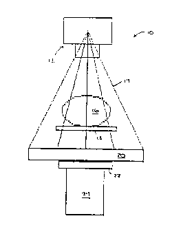
5 Referring to Fig. 1, there is shown a schematic repre-
sentation of an x-ray simulator, indicated generally at
reference numeral l0. X-ray simulator l0 is generally com-
prised of the x-ray tube 12 used for radiation therapy
simulation which produces a fan beam, indicated at refer-
ence numeral 14, incident upon a target object 16 posi-
tioned on table 18. X-ray beam 14 is also incident upon
the detector array (not shown) of the apparatus 20 of the
present invention. The apparatus 20 is comprised of the
linear detector array (see Fig. 2), the mounting of which
is adapted for fitting into, mounting next to, or covering
the film holder 22 of x-ray simulator 10 normal to the
17
CA 02060181 1994-OS-15 SUBSTITUTE SHEET
IPEA/US
~~'t"IU~ ~ ;,1~~374
. _ . __.
IPEA/US ~ ~ ~ E 0991
PATEN
central axis of the fan beam 14 produced by the source 12
without altering the original function of simulator 10.
The film holder 22, or cradle, is mounted on top of the
image intensifier tube 24. The x-ray beam is collimated by
using the shutters or collimators (not shown) provided by
the x-ray therapy simulator 10. The beam is preferably
shaped with a width of approximately one centimeter and a
spread determined by the length of the detector array of
the apparatus 20 of the present invention. If the project-
ed shadow of the target object 16 to be scanned is less
than the maximum width of the detector array, the length of
the beam is decreased so as to just encompass the diameter
of the target object 16.
Referring now to Fig. 2, there is shown a schematic
representation of an exemplary apparatus used in connection
with the practice of the method of the present invention.
The detector array 26 utilizes an image intensifying screen
(not shown) placed over a linear photodiode array of detec-
tors. In a presently preferred embodiment, the photodiode
array 26 is comprised of eight to sixteen light sensitive
modules (not shown), each composed of a silicone chip with
127 photodiodes (not shown). The photodiodes are used in
a capacitive storage mode (with reverse bias) so that the
light emitted by the intensifying screen discharges the
5 photodiode capacitors. The scintillator material used is
ytterbium-gadolinium oxide, a screen material commonly used
in diagnostic radiology. other scintillator materials may
likewise be used to advantage in accordance with the method
and apparatus of the present invention. This screen may
also be made of different thicknesses to improve photon
collection efficiency.
An external sync trigger circuit 28 is provided for
interfacing the x-ray simulator 10 with an external sync
circuit 30 to trigger the timing circuitry of the apparatus
20 of the present invention when the x-ray tube 12 starts
to emit the fan beam 14. External sync circuit 30 provides
18
$UBST1TUTE SHcET
CA 02060181 1994-OS-15 IPEA/US
P'~'lU ~ '~ ~d l ~ y ~ 3 7 4 _ . _
IPEA/uS 1 p D E C 1991
PATENT
the synchronization signal to the preprocessor 32 and
determines the integration period of the detector array 26.
The external sync circuit 30 consists of an input (not
shown) to provide software control of offset and gain
calibration reset signals to the preprocessor 32, a contin-
uous clock circuit (also not shown) to provide the synchro-
nization and integration period for the detector array 26
during gain calibrations, and a variable clock circuit
(also not shown) which utilizes an EPROM to provide the
synchronization and integration period for detector array
26 during data acquisition. Preprocessor 32 receives the
analog signal from the detector array 26 and, under control
of the external sync circuitry 30, digitizes the data and
outputs the data to the microcomputer interface circuit 34.
Computer interface circuit 34 converts the data
received from the preprocessor 32 to a format that is
compatible with the input-output (I/O) board 36 of the
particular microcomputer utilized for processing of the
data as described below. The microcomputer I/O board 36,
after receiving the data from microcomputer interface
circuit 34, inputs the data to the memory of the micropro-
cessor 38, which is preferably a 68030 or 80386-based
microcomputer. In a presently preferred embodiment, an
Apple Mac II Plus (tm) with eight megabyte memory, large
5 hard disk, 80-140 megabytes, floppy disk and CRT displaying
a 480 x 512 pixel image (or, alternatively, a special CRT
to display a 512 x 512 image) having a 25 and preferably 30
megahertz internal clock is used to advantage. In addi-
tion, the output of the microcomputer I/O board 36 provides
the software control signals to the external sync circuit
30 for offset and gain calibrations, the continuous clock
and variable clock circuits. The microcomputer 38 performs
the data manipulation routines described below to produce
the CT image, outputting the processed data to an appropri-
ate display means such as the high resolution monitor which
provides a gray scale or color display of the reconstructed
19
CA 02060181 1994-OS-15 S~~S~~~~~~ Si~C
IPEA/tJS , .
CA 02060181 1998-O1-07
PATENT
CT image, the film recorder (matrix camera) 42 :rhich
provides a hard copy output (film) of the imago on the high
resolution monitor 4o, and/or the large capacity storage
device 44 which stores the image data as wall as patient
information.
A general schematic outline of the data manipulation
process is sat out in Fig. 3. ey the term "data" it will
bs understood that reference is made to the processed
output signals of each individual detector or photodiode
l0 coaprising detector array 26, which is proportional to the
intensity of the x-ray incident thereon. Preprocessor 32
integrates the output signal of each photodioda and reads
out the resultant signal upon external command controlled
by aicrocomputer 38. In so doing, the data is corrected,
:soothed, and liltered as shown in the step represented by
box ~6 in Pig. 3. This filtering step is required because
the individual detector elsaants, or photodiodes, bacoas
detective and may malfunction either partially or complete-
ly. A correction algoritha is provided which identities
those faulty detector elements through comparison with the
neighboring detector elements, the narrow separation
between detector slas~nts utilized allow the generalization
that the responses for adjacent detector elements should ba
relatively close to each other, and replaces the bad data
2s with a corrected, interpolated value. A smoothing al-
gorithm is provided for extending this method by setting a
limit, or preset salectivn criterion, on the variation of
adjacent individual detector elements and interpolating
response values for those detector elements whose variation
trvm the neighboring detector elements exceeds that presa-
lactad criterion. Recognition of when an individual
detector element malfunctions is determined experimentally
by exposure to x-rays both in air and through a phantom
target (not shown) and the history of the detector array.
35 Characterising sack step in more detail, the data is
first smoothed in accordance with the formula
~0
P'C1'1US % ~/~~3~4_
~i
tpEA/US 10 D E C 1991
PATENT
P(2) - (P(1) + P(3))/2 [5J
where P(1), P(2), and P(3) represent the signal outputs
from each of three successive detector elements in detector
array 26. The smoothing is extended by selecting a maximum
variation between adjacent detectors based upon the detec-
tor response history. For example, a cutoff limit of 0.2
or 0.3 times the adjacent value may be selected so that if
the reading from a particular detector element varies from
its neighbors by more than 0.2 or 0.3 times the output of
the neighboring elements, that output signal is replaced by
the average of the output signal from the neighboring
elements on either side of the bad element. In the event
that several detectors in a row have malfunctioned, a
scaled averaging method is used to correct the data. The
difference between the last good output signal at one end
of the malfunctioning row of detector elements and the
first good output signal at the other end is divided by the
number of detector elements malfunctioning and the resul-
tant quotient is used as a constant which is sequentially
and iteratively added to the last good output signal to
provide a replacement for the next output. The correction
is added to this value for the next reading and so on, up
to the first good output signal. For example, given the
following output signals, 110, 125, 255, 255, 255, 255,
5 140, 141, the correction is applied as follows:
C = (140 - 125) /5 = 3
In this example, there are five intervals from the last
good output signal at one end of the malfunctioning detec-
tor elements and the first good output signal on the other
end of the malfunctioning detector elements. The corrected
output data is 110, 125, 128, 131, 134, 137, 140, 141.
Other corrections account for variations in x-ray
output. These variations can be caused by current and/or
voltage variations. Metering circuits on the x-ray simula-
for 10 are used to monitor these parameters, and the output
of those circuits is used as an input to preprocessor 32.
21
~~ ~ST!~UTE SHEET
CA 02060181 1994-OS-15
CA 02060181 1998-O1-07
PATENT
Current variations are corrected in linear fashion: i!, !or
instance, the current drops from 5 mA to 4 mA, the detector
elements aro corrected as (5/4) x P(i). Voltage correc-
tions are non-linear. The correction factor is formed as
a power function o! the voltage, i.a., VN, where N is a
number in the range 1.5 S N S 3. N is determined experi-
mentally and is idiosyncratic to the system being con-
trolled. Suppose the voltage drops from 120V to 110V and,
for that range, it has been determined that N = 2. The
to correction is made as follows:
P(i)~~ _ [120/110]Z x P(i)
Additional corrections are performed using interpola-
tion procedures to optiaize accuracy in the calculated
intensities used in back projection when correlating the
detector in the detector array 26 that intercepts a back
projected, or parallel beam, ray 50 which passes closest to
the point in the target object 16 being reconstructed. The
coordinate system in the back projection process is defined
from i = 0 at the first (-x) detector to Np - 1, where Np is
the number o! detectors. For example, using a 2048 detec-
tor array 20 with a detector spacing of 6; = 0.045 cm, the
total length DL of the line of detectors in detector array
2o is
DL ~ 2048 x 0.045 ca = 92.16 cm.
The factor 2048 is used because the detector line is based
on the spacing of the individual detectors and there are
2047 spaces between the 2048 individual detectors in
detector array 20. The center o! the detector line is
taken as zero. Tha x coordinate along this line then
varies from
-92.16/2 to 92.16/2.
The x coordinate of the its' detector is
xi = (-46.08 cm + (i + 0.5)) x (0.045 cm),
e:g., !or the 1135th detector,
x1135 = (-46~08 + (1135 + 0.5)) x (0.045) = 5.0175 cm.
22
CA 02060181 1998-O1-07
PATENT
The coordinate on the detector line sphere the ray
passes closest to the reconstruction target point is desig-
nated Lbp (back projected). once this coordinate is
determined, it is possible to calculate which individual
detector will rsceive the ray passing through Lbo. Let
Nbp ' ((Lpp + 46.08)/ 6, + 0.5J (6J
and Nbp ~ IHT ( Nbp J
where "IHT(J" indicates the integer of the number in the
brackets in equation (6J and the !actor 0.5 is an end
correction such that in the limit vhere Lbp is approximately
equal to -46.08, the formula will return the integer value
of one (.1) .
Round oft error in calculating this integer is sini
mized by linear interpolation. The fractional part of N~
is .
INT ( Nbp J - Nbp ~ tbp . ( ~ J
Sn the simplest round otl, i.e., i! tbP < 0.5, Hbp
INT(Nb~). It tbD 2 0.5, Nbp ~ INT(Nbp) + 1. For exempla, it
Nbp~ 756.58, the detector output taken would be pl(757).
ZO To be more exact, interpolation yields
P1 ~ (1 - t~) P1((NbpJ) + tbp Pl((NbpJ + 1).
In the numerical exaaple, P1 ~ (1 - .5Z8) Pl(756) + .528
P1(757).
The term "saoothing~ (steps 46 and/or 7Z o! Fig. 3)
=5 refers to the filtering or the data by both smoothing and
ripple filtering. Smoothing is accomplished using a set of
reconstructed values which may be represented as
M(1, 1) IK(l,Z) I4(I, 3) . . . .. . . . .M(1,N)
M(=~l) M(Z,2) M(=,~).........M(Z,N)
30 H(3,1) M(3,Z) M(3,3).........M(3,N)
M(N,1) M(N,2) M(N,3).........M(N,N)
I7
CA 02060181 1998-O1-07
PATENT
Here, N ~ 2k where k = an integer, i.e., N = 128, 256, 512,
etc. sing four point smoothing for nearest neighbor (j
denotes the row and i denotes the column):
M'(j.i) ~ [M(j-1.i)+M(j+i.i)+M(j,i-1)+M(j.i+1))/4 [9A)
The top, bottom, and adjacent left and right neighbors are
used with equal weight. For rive point smoothing for
nearest neighbor
[M(j,i)+M(j-i,i)+M(j+iiti)+M
(j.i-1)+M(j,i+1))/g [9B)
M(j,i) is added in along with the tour nearest neighbors.
For weighted neighbor smoothing with the central value
given the greatest weight, nine point smoothing is used:
M'(j.i) ~ [1~(j-i.i-1) + ZxM(j-i,i) + ixM(j-i,i+1)
+ 2xx(j, i-1) + 4xM(j. i) + 2xM(j, i+1) [9c)
is + 1xM( j+1, i-1) + 2~( j+1, i) + 1~( j+1, i+1) )/is.
Thfs weighting systea can ba varied with different weights
applied to dilterent locations depending upon the circua-
stances as known to those skilled in the art.
Ripple filters are used to eliminate the wave intro
ZO duced into the reconstruction by employing Fourier methods.
The first step in application of such a filter is to obtain
x-ray transmission values for a water (uniform target
density) phantom. With known reconstructed CT values,
these true values era compared to observed CT values. Tha
25 ripple filter factor for each picture element is given by
the true value/observed value. To achieve correction, the
value for each picture element is multiplied by the filter
factor.
There are many other different types of filters which
30 may be used to advantage depending upon the circumstances
as known to those skilled in the art who have benefit of
this disclosure. Such filters might include, for instance,
edge enhancement rilters, high frequency suppression, bone
enhancement filters, soft tissue enhancement filters, and
35 so on.
24
PCTJUS ~ i -~ ~+r~
~P~p,IUS 1 C D ~ C 199
PATENT
The smoothed, corrected data from step 46 is convolved
at step 47 and then back projected at step 48. Convolution
47 and back projection 48 are accomplished in accordance
with an algorithm which is derived as follows. Referring
to Fig. 4, there is shown the geometry for the fan beam 14,
the parallel beam 50 and the coordinate system used to
describe the algorithm of the present invention. In this
system, x-axis 52 is on a line 54 through the center of
rotation 56 and perpendicular to the y-axis 58 which passes
through the center of rotation 56 on a line from the x-ray
source 12 to the center of the detector array 26 which is
parallel to the x-axis 52. The distance from the x-ray
source 12 to the center of rotation 56 is D, reference
numeral 60. The separation between the center of rotation
56 and the plane of the detector array 26 is a distance V,
reference numeral 62. The x-ray source 12 and detector
array 26 are incrementally rotated about the center of
rotation 56 through angles T as shown at reference numeral
64 in Fig. 5. The increments may be equal in size or
unequal. When equal increments are used, T = 180/N or T
- 360/N, where N is the number of increments, and the
system may be rotated either through a semi-circle or the
entire arc of a circle.
The projection angle, dT~, indicated at reference
numeral 66, is given, in the equal increment case, by
Tj = 90° + jdT where 0 s j s N - 1,
[10]
and ST is the increment used to increase the projection
angle. The x-ray source coordinates for each j are given
by
x~ = D Cos (T~)
[11]
SU~,;-._v__ ; ~_t~T
CA 02060181 1994-OS-15
CA 02060181 1998-O1-07
PATENT
y> > D sin(T~) ,
[12J
Tha angle, To = 90°, defines the initial configuration of
the system before rotation, a.g., at j = 0. At this angle,
To, the coordinates of aach of the detectors in detector
array 20 are:
Xd(0, i) _ -D~2 + (i + 0.5) bi
[13J
ya(o, i) _ -v
[14J
where DL = detector array length in cm., 61 = DL/Np detector
spacing in ca., No =~numb~r of detsctors in the array, and
0 S i S ND - 1.
The 0th detector is considsred the left most (-y) in
detector array 20. The spacing is actually the distance
between the centers of each detector in detector array 20.
For Tj > 90~, the coordinates of each detector in
detector array 20 are given by:
xe(j, i) = xd(o, i) cos (j, 8T? - ya(o, i) sin (j, aT~
tiSJ
ya(j, i) ~ xd(0, i) sin lj, aT~ + ya(0, i) cos (j, dT~
[isJ
In Figure 4, the parallel rays 50 are constructed by back-
projection from the detector array 26. Ths intersections
0! the parallel 50 and tan bean 14 rays define the circle
of reconstruction 68.
In defining the circle of reconstruction 68, the next
step 70 (see Fig. 3) in manipulation of the data is ac-
complished by microprocessor.-38 using the following steps.
The diameter of the reconstruction circle 68, Dry, along the
line 54 is given by the scaling relationship,
26
CA 02060181 1998-O1-07
PATENT
Dre~D s DG~W
[17]
wham t~ - D + V and is the distance from the source 12 to
the center of detector array 26. The Jacobian of the
scaling transformation from the line of the detector array
~6 to the line 54 along the diameter of the reconstruction
circle 68 is
Mi ~ D~ ( M .
(18)
The distance between successive back projected parallel
beans or detector rays 50 at the level of the diaseter of
l0 the reconstruction circle 68 is.
6i ~ ' 6i MI
~i9)
The scaling factor M1 allows tranatorsation of the data
talon along the detector line to appropriate values along
the parallel line 54 through the center of rotation 56 and
lying along a diaaeter of the reconstruction circls 68.
To norsalize the projscted data fro' the incident
begs, tan bees projection data is taken for x-ray transsis-
sion through air and through target objects. Let
ptj.s) ~ log (1,(~.ll~rot~.i)~
[20)
where raij~i~ is the i ~ detector reading at angle j in air
and rp~ j~s~ is the ith detector reading at angle j for x-
rays tranasitted through the patient or other target. Tha
ranges of the integer indices are the same as defined
above.
Por each angle T~, the one dimensional discrete Fourier
transform of the projection data era calculated as:
Z7
PtrIIUS ~ v~U'~37~
. .__.
~PEA~US ~ ~ ~ ~' C 1991
PATENT
ND 1
b[FTZ(j,b)] - ~ p(j,i)cos(2nbi/ND)
i-o
[21J
ND-1
c[FT2(j,c)] - ~ p(j,i)sin(2nci/Nd)
i=o
[22J
where b and c are the indices defining incremental distance
in the horizontal and vertical dimensions, respectively, in
the portion of the coordinate system in the circle of
reconstruction 68 as more fully described in Fig. 5 and
infra in connection with the discussion of equations [49J
and [50], and 0 < bi, ci < ND - 1.
These transforms are multiplied by the ramp frequency
filter. In the notation of discrete Fourier transfor
mations, these are:
GI(j,b) - GI(j,Nb-b) = b[FTI(j,b)]
[23J
G2 (j, c) = G2(j,N~-c) - c[FT2(j,c)]
[24J
where 0 <_ b,c <_ Nb~~ [25]
and Nb~~ = ND/2 (ND even) [26]
Nb,~ _ (ND - 1)/2 (ND odd). [27J
The application of filters as outlined above is
facilitated by working in the frequency domain. It is
useful to modify the product transforms using spatial and
frequency domain notation. Here, the step-wise, discrete
functions G1(j,b) and G2(j,c) are rewritten as continuous
functions,
GI (j, f) - f FTI(j,f)
[28]
28
CA 02060181 1994-OS-15 5~;~'~i'il~lE SEE
IPEA/US
CA 02060181 1998-O1-07
PATENT
G2 ( j , t') = f F'T2 ( j , ~
(29J
whars, as batore, 0 S j 5 No - 1 and t, the frequency, is
determined by the saapling theorem which states that whars
a function h(x) is defined oust the range 2Rr~ (diameter of
the reconstruction circle 68) then the Fouriar transform of
h(x), FI(r), is fully described by points 6P ~ 1/2Rr~ apart.
Conversely, it the range of interut of H(F) is Zrx then
h(x) may ba sampled at intervals not greater than 6;
1/Zlx. Thus, in equations (ZSj and [29J, P ~ O,Pl,
x0 1~,...lx, and
l" ~ i / ( Z 8l~ Nyqui s t frequency, and
(30j
6F ~ 1/ZRse lVyqu~at frequency spac~Eng.
(31J
The inverse Pouriar transform of equations [Z3) and
[Z4] may be performed to obtain the modified projection
is ray, Pi(j,t) directly. However, this technique does not
yield results as good as those obtained by utilizing the
results of equation (36), infra.
The rasp frequency filter, ~t~ (absolute value of t),
appears as the Jacobian of the transformation from rsctan
ZO gular coordinates to polar coordinates. Equations [Z37 and
-[24j are the frequency domain counterparts of the con-
volution in the spatial domain of the two functions P(j,i)
and H(j,i). The convolution thaorea is used to obtain:
~,~i
PZ (j. i) ~ ~ P(j, il) X(j. j-il)
,~o
[32j
2s where P1(j,i) ~ moditisd projection rays,
P(j,i) ~ original projection ray values
Z9
CA 02060181 1998-O1-07
PATENT
H(j,i) ~ inverse Fourier transform of the ramp
filter,
0 s i s ND-1 and
o s j s ND-1.
The inverse Fourier transform of the ramp filter is given
in R,t notation as:
rr
S (R, f) ~ a r ~ t~ cos ( 2 RRf) o f
~'0
[33]
R = radius in real space.
The sine tern does not appear because the ramp filter is~an
even function in frequency space and the contributions from
the negative and positive parts of the frequency spectrum
account for the !actor, two, multiplying the summation.
It is useful to treat the inverse transform of the
ramp filter in the limit as a continuous function. This
treatment is accomplished by using the integral rather than
the discrete tranalorm, or converting the summation in
equation [33] to its integral analog;
rr
S(R. ~ ~ j (~~ exp [2uiRf] d t
'rr
[34]
where i is the imaginary square root of -1. The integral
ZO in equation [34] is a truncated version of the transform of
the absolute filter function with cutoffs at tFM. Conse-
quently, no frequencies greater than +Fh or less than -FH
will be found in real (configuration) space in the function
S(R,t) convolvad with the projection data; i.e., no tre-
quencies outside the tFM band will be found in the convolved
projection data.
Integration by parts is used to solve this integral to
obtain:
[35]
CA 02060181 1998-O1-07
PATENT
S(R, f) ~ 2F~,sin (2nRF~,) / (2nR) ~ 2 (cos (2nRF,~) -1) / (2nR)'
Equation [35) reduces to:
S(R, f) = 2FN sin (2~RF,~) / (:cR) - sin' [nRFw] / (~R)'
[36]
S(R,f) is convolvsd with the projection data, p(r,t) where
t is the angle o! the projection line with the + y axis 58
measured counter clockwise and R is the distance of the
projection ray from the origin (see Fig. S); thus R ~ KdR,
where R is a general index defining the distance o! the
point in question from the origin o! the coordinate system.
Lower case letters, "p" and "t", are used to indicate
variables in conf iguration :pace and capital letters. are
used to represent variables in frequency space. Accor-
dingly,
p1 (R, t) = p(R, t) * S(R, !J
[37J
where "*" denotes convolution.
The conceptual development leading to equation [ 37 ] is
used to derive the hybrid reconstruction algorithm used in
the method of the present invention. Returning to the
discrete representation, the Fourier inverse transform or
the ramp function, (f~ is written as:
~r
Sam (k) _ [1/di (F~)2] ~ (f~ [exp(2x~fk/F~) ~ exp(-2sffk/Fx)
r
[38j
Again, 6i is the spacing in the detector array 26 or the
distance between back-projected parallel rays 50, f denotes
the frequency, and Fx is interpreted here as the number of
frequencies in the frequency band.
Since ~f~ is an even function, equation [38J may be
rewritten as:
31
CA 02060181 1998-O1-07
PATENT
tr
Su.e(k) ~ (1/ 6~ (F'~)'j ~ t cos (2~~k/F'") .
.o
(39]
This siaplitication in equation [39j results from the tact
that ~t~ is an even function about the origin. Ths factor
two results from changing the limits from -Fx 5 F 5 FN to 0
S S F S FK and is abaorb~d in the cosine definition.
When k ~ 0, equation f39] beco~~s
S~IO) ~ I (F'~?2 -1 J / 46; (g~)2
f~0]
In tha limit, as FK approaches ~~, equation f40] siaplitias
to
S~"e ~ 1 ( 46~)
f41]
When k is different than aero, equation f39] b~coaes
(Pw i) ain' fs (P"+1) k/ (2P,a 1 - (P"+i) sin' f ~c (Pr 1) k/2F,
2l~;sia' (kF")
(42]
In the Bait, as FK approaches m, the expression in equation
f ~ Z ] bscomu
Sake ~ f-sin' (ek/Z) J /bi (sP,',) J .
(43J
when k is avan and nonzero, Sa~,~ ~ 0. When k i: odd and
nonzero,
s~,~ ~ -1 (8i (xFx)'J .
(dal
Combining the conditions, the expression for Sdiac to be
ZO used in the convolution is
31
P~1"/US ~'v1~~37~.
~p~~us 1 ~~ a ~ c X991 ___
PATENT
Saisc = 1/ (48i) - 1/ [ai (nFM) ] .
[45]
Equation [32] is rewritten as:
ND 1
PI (j, 1) _ [P( j, 1) / (4a1) ] - [1/ (n2a.z) ] ~ P(j, il) a
11'Q
[46]
where ( i-il) is odd only. Equation [ 46 ] reduces in the real
space convolution summation to:
ND-1
P1 ( j, i) - [P( j, i) / (4ai) ] - [1/ (n2ai) ] ~ P( j, il) / (1-il) 2.
il~o
[47]
where (i,il) is odd only, 0 < i < ND -1, and 0 <_ j <_ ND -1.
The di in the convolution summation has cancelled one ai in
the denominator of both the first and second terms of
equations [46] and [47], while the first term is now
entirely outside the summation. The odd only summation for
(i,il) is caused by the sine term being zero when (i,il) is
even and +1 when (i,il) is odd.
Back projection geometry is shown in Fig. 5. The x,y
increments are given by:
ax, ay = 2Rrc/Np
[48]
where Np = number of pixels in one dimension, i.e., 32, 64,
128, etc. and Rr~ = radius of reconstruction circle 68:
X = -Rrc + ( aRrc/2 ) + baRlc
[49]
y = Rzc - ( aRzc/2 ) + CaRzc
[50]
and b,c = 0, 1, 2, ...NP-1.
33
CA 02060181 1994-OS-15
",
CA 02060181 1998-O1-07
PATENT
Tha linear coefficient for each x,y or b,c is:
am
~(b,C)~~ P1 (j. is (x,y))BTZ,
0
[51]
where N ~ number of angle increments and 6T1 is n/N for
both 180 and 360 degrees of rotation. Here, is(x,y) is
determined at sack angle, and j is the number of the
parallel ray 50 at the projection angle j which lies
closest to the point x,y. The absorption coefficient is
calculated from the following relations. The angle ~ of
the ray of interest is defined by:
~ ~ tan'1 (Y. x) .
to .
[sad
Because ~ is used in conjunction with Tj, ~ must be defined
in the same fashion as Tj, or from 0 to 360 degrees. If x
< 0 and Y > 0, then 90° 5 ~ 5 180~. If x < 0 and 'y < 0,
than 180~ 5 ~ 5 270~. It x > 0 and y ~ 0, then 270° 5 ~ 5
360~. Let
L~=W1s Sit1 f [Tj-~1 / [D-rsCOS (T~-~) ] )
[53]
whsrs rs ~ (x~ + ysjl~? [54j
and is (x,y) ~ INT[(Le + DL/2)/i + 0.5].
As before, "INT[]" is the "integer part of". The factor
0.5 appears in equation [55] because when L8 ~ -Dy/2,
is(x,yj must ba zero.
In the back projection process, a coordinate system is
defined from i ~ 0 to Nfl -1 running from the lafthand (-y)
aide of the detector line, ND is the number of detectors,
and the datector array Z6 is marked off in the same fashion
as a meter stick. Zn the back projection process, the
point along the detector line is determined where the
parallel ray 50 passing closest to the point in the object
16 being reconstructed falls. This is Le. With Le defined,
34
CA 02060181 1998-O1-07
PATENT
the number of the detector corresponding to Lg is deter-
mined. For example, suppose Lg was calculated as 15.463
using equation (53) and assuming the detector array 26 is
comprised of 2048 detectors and is of the length 92.16 cm
as calculated above, the number of the detector lying on
that point is given by
is ~ INT((15.463 + 69.12/2)/0.045) + 0.5) ~ 1112.
This calculation indicates that the ray 50 from detector
1112 passed nearest the point (x, y) that is being
reconstructed.
The quantity Le is defined differently for the hybrid
reconstruction algorithm of the present invention than for
the parallel beam geometry. At each angle, T~, a line is
passed from the x-ray target position xTj,yTj through the
point x,y and trom~thers to an intersection with the x-ray
detector line. Thus, Ls is defined as the distance t troa
the center of the detector line. For each value o! x,y,
there are N back projections.
In practice, a rectangular grid with N, x N, dimen
sions is reconstructed and all values of ~b~e lying outside
the circle of reconstruction 68 are set equal to zer~~.
' Using the rectangular grid for reconstruction greatly
simplifies any smoothing routines when they are utilized.
Further, interpolation may be used when a ray 50 doss not
pass through a point in the back projection.
Referring now to Fig. 6, which is a graphical repre-
sentation of an exaggeration of a measured center of
rotation shift for an x-ray simulator rotating 360~, the
oft-center shift S~ (shown at 76) is plotted as a radius
from the center of rotation 56 versus the angle in degrees.
When the x-ray beam which should tall on the central
detector in the detector array 26 does not pass through the
center of the circle of reconstruction 68, two errors may
occur. The first error is a linear displacement of the
detector array 26 parallel to the central axis of the tan
beam 14. The second error is the magnification or demagni-
~TILIS ° ~ l ~ ~: 3 '7 4
_ . _ ___
IPEA/US _1_ ~ D E C 1991
PATENT
fication of the distance between detector arrays (8i) used
in the hybrid reconstruction algorithm described above.
The latter error occurs when the center of rotation 56 is
shifted in a direction perpendicular to the line on which
the linear detector array 26 is positioned (e.g., the
detector line). Further, both errors may occur at the same
time.
Referring to Fig. 7, a simplified method of measuring
the shift in the center of rotation is illustrated graphi-
cally. A round rod (represented schematically at reference
numeral 74 in Fig. 5) which is relatively opaque to x-rays
is placed at the nominal center of rotation shown by line
78 in Fig. 7, and data obtained using the method of the
present invention is shown as curve 84. An eccentricity in
the center of rotation causing a shift in the direction
parallel to the central axis of the projected fan beam 14
results in projection data for the rod 74 with a center
shown by the dotted line 80 with projection data 82. The
curves 82 and 84 shown in Fig. 7 represent the
reconstructed projection data taken over each angle T~.
From this data, the shift, shown at reference numeral 76 in
Figs . 6 and 7 , in the number of detector widths for each
angle is measured. This shift is used to correct equation
[44] as follows:
.5 i5(x,y) - INT[(L8 + DL/2]/i - sj + 0.5], [56]
where s j is equal to the shift in number of detector widths.
Magnification or demagnification of di is ignored. When
the center of rotation shift is present, the geometry must
be altered so that at the maximum shift in either direc-
tion, the shifted detector rays 50 at the edge of detector
array 26 will still pass through the target object 16.
Having described a preferred method in accordance with
the present invention, a method of conducting a CT scan
using that method will now be described. Initially,
eccentricities in the rotation of x-ray tube 12 must be
determined and the individual detectors comprising detector
36
SUBSTITUTE SHEET
CA 02060181 1994-OS-15 ~pEA/US
G~/~'~374
. _ . ,__.
-_
IPEA/US ~ ~ D E C 19 _
9i
PATENT
array 26 calibrated. A kit (not shown) comprised of -
several rods in a holder, aluminum filters, and a CT
phantom are provided for that purpose. Placing the center
of the phantom for determining motion eccentricities at the
nominal center of rotation 56, a scan is performed to
identify any shift in the center of rotation 56. That scan
also enables verification of the angle encoder output,
whether that angle encoder (not shown) is integrated with
the apparatus hardware (see Fig. 2) or output from those x-
ray simulators which include such an encoder.
The aluminum filters are then used to determine the
effective energies and x-ray output for the nominal operat-
ing voltage of the x-ray simulator. That data is used for
correcting individual detector output at different voltages
as at step 46. With a second phantom comprised of differ-
ent materials, scans are performed at different voltages.
The results are used to set up a Hounsfield scale for the
different energies.
Once the initial measurements have been accomplished
to load the correction algorithms into microcomputer 38 and
the detectors of detector array 26 calibrated, the
patient/target object 16 is positioned and the desired
current and voltage parameters selected. The computer
program is then initiated to acquire CT data, and on
.5 computer ready signal, x-ray output is initialized first
and the x-ray rotation started. For j=0 to 359, where j is
the angle in degrees, the computer is signaled to acquire
data at approximately one degree intervals (or any other
odd or even increments as selected by the operator). For
i=0 to ND-1, where ND is the number of detectors comprising
detector array 26 and is operator selectable, PP(j,i) is
acquired where PP(j,i) represents the output of the analog
to digital converter (ADC) in microcomputer I/O board 36
for the detector i at the angle j. Note that the number of
increments (angles) multiplied by the number of detectors
will be the number of individual data points and that, for
37
SUBSTITUTE SHEET
CA 02060181 1994-OS-15 ~pEA/~S
CA 02060181 1998-O1-07
PATENT
instance, where 0~ PP(j.i) S 4095, 4095 corresponds to a
lZ bit ADC, 8191 co=responds to a 13 bit ADC, 16,383 corre-
sponds to a 14 bit ADC, and so on.
Each of the quantities PP(j,i) is then multiplied by
a correction number obtained from the calibration procedure
(stsp 46) to adjust the data to the values that would have
been read it the individual detector was operating per
tactly. This correction also remedies the beam hardening
artifact as discussed above. A reference detector out is
also used to correct for variation in the output of the x-
ray unit as a function of the angle j.
PP(j,i) is the input to the hybrid reconstruction
algorith~/convolution routine (steps 4~ and 48), and alter
convolution, Fourier artifact filters are applied as
is described above. for c ~ 0 to by - 1 and b ~ 0 to Hp - 1,
the back projection is dons (step 48) and then repeated at
the next b, next c.
Reconstructed linear attenuation coefficients are
then converted to gray scale values using the Hounsfield
scale derived from scanning the phantoms described above
in connect ion with the setup procedures . A water phantom
is scanned and the reconstructed coefficients are
assigned a value of 1000, with air being 0. Other
values, such as for bone, are then automatically given a
value in this scale. In this fashion, the Hounsfield
scale is potentially wider than that or prior known CT
scales, enabling detect ion of verist ions in CT values not
currently available. By way of example, if the scan of
a water phantom gives a value of 0.18 per cm for an
average linear attenuation coefficient of water, 1000 is
assigned as the value for water and 1000/ . 18 ~ 555. 55 is
obtained as the scale factor for the unit being used. As
is known in the art, 0.36 x 5555.55, or 2000, is assigned
a gray scale of 0 to represent the densest scale for this
system, or twice the density of water, and 2000/255 =
7 . 843 is the diet ribut ion of the gray scale number vs . CT
numbers:
38
~TIUS ~'J1~43'l4~_~
~P~A~~S _ , ~~~ 199i _.
PATENT
0 CT scale = air = 255 gray scale
1000 cT scale = water = 127 gray scale
2000 CT scale = twice density of water = 0 gray scale
The CT values may be displayed along with gray scale
numbers if desired.
Although described in terms of the above presently
preferred embodiments, it is not intended that the scope of
the invention be limited thereto. Instead, it is intended
that changes in the specifics set out above which do not
depart from the spirit of the invention described herein be
included within the scope of the following claims.
39
CA 02060181 1994-OS-is SUBSTITUTE SHEE~
IPEA/US