Note: Descriptions are shown in the official language in which they were submitted.
2 ~
~,
~ITLE: TUNNELED EPIDURAL_CATHETER
~ACKGROUN~ OF THE INVENTION
This inven~ion relates to long-term epidural catheters
adapted for use in pain mana~ement, e.g. to relieve intractable
pain in cancer patients.
Heretofore, a number of dlfferent methods have been
utilized to relieve intractable pain. These include palliative
or "curative" therapy ~i.e. surgery, radiation therapy or
chemotherapy), systemically administered narcotics,
transcutaneous electrical stimulation, nerve blocks, rh~zotomy,
radiofrequency, induced lesions, epidural or dorsal column
electrical stimulation, and central nervous system neurosurgical
intervention, e.g. cordotomy, thalamotomy, acupuncture, and
hypnosis.
While systems for relieving cancer pain by the
administration of morphine using an indwelling system have been
disclosed in the literature for some time, e.g. in "cancer Pain
Relieved by Long-Term Epidural Morphine With Permanent
Indwelling Systems for Self-~dministration", by C. Poletti et
al, Journal of Neurosur~ery, Vol. 55, October, 1981, pp
581-584, lt has only been relatively recently that the
tr_atment of intractable pain by epidural infusion of a
narcotic has gained acceptance in a nwmber of medical centers.
More recently, the responsibility for the treatment and
control o~ pain has been moving from the surgeon and general
practitioner to the anesthesiologist. As anesthesiologists are
broadening their practice outside the operating room suite,
they are managing acute pain in the post operative areas and
chronic pain in the clinics.
In the past five years, approximately one thousand pain
clinics have been established, about 60% of which are headed by
anesthesiologists. A majority of these pain clinics treat
cancer pain and many are a~filiated with a cancer treatment
center.
patO5123 2
~7~
One or -he treatment modalities gaining in popularity
~or terminal cancer pain management i5 the tunneled epidural
catheter. This procedure provides better analgesia without
fxequent injections or cumbersome I.V. equipment, present fewer
complications, and are generally bet~er tolerated by the
patient. Epidural narcotic administration works well because
there are opiate receptors located all along the spinal cord.
Thus, the narcotic can act directly on the receptorS, producing
localized analgesia without more blockage. This in turn allows
for lowex dosages and minimizes cerebral and systemic efiects.
The tunneled epidural catheters currently on the market
are what may be termed a "two-piece" catheter consisting of a
first or distal piece for introduction into the epidural space,
e.g. as close to the dorsal midline as possible, and a second
or proximal piece which is tunneled subcutaneously between the
dorsal paravertebral entry site of th~ f{rst piece and a
lateral or ventral exit site from the skin where it is to be
connected to a syringe or other source of the narcotic to be
administered for pain management.
The tunneling may be effected in the direction from the
exit site to the first catheter piece or, alternatively, it may
be the reverse, namely from the first catheter piece to the
exit site. In either case, the proximal end of the first or
distal piece is secured in fluid-tight relationship to the
distal end of the second or dis~al catheter piece ~d sutured
in place to provide a tunneled long-term epldural catheter
extending from the dorsal point where it is introduced into the
epidural space to a desired location on the side or front of
the patient for more comfortable and accessible hook-up to a
source of the narcotic to be administered.
Illustrative of the current state of the art on tunneled
epidural catheters is the Du Pen (~M) Long-Term Epidural
Catheter commercial-y available form Davol Inc., Subsidiary of
C.R. Bard, Inc. and reported in ~'A New Permanent Exteriori~ed
Epidural Catheter for Narcotic Self-Administration to Control
Cancer Pain" by Dr. Stuart L. DuPen et al, CANCER, Vol. 59, No.
5, March 1, 1987, pp 986-993.
patO5123 3
Ar~
As stated thereln, ~he new e~teriorized epidural catheter
consists of three ~ieces~ n epidural segment that is
placed through a needle into the epidural space; (2) an
exteriorized line equipped with an external luer connector and
a subcutaneous Dacron cuff; and (3) a small splice seqment to
join the two catheter segments. ~oth the epidural and
percutaneous lines are prepared from radiopague silicone
rubber.
According to the protocol described collectively in the
DuPen article and~or the Davol product literature, under local
infiltration anesthesia, 7 cm paravertebral incision is made
from the L-2 dorsal spine down to the paravertebral fascia, for
needle placement and catheter splicing. A 14-gauge Hustead
needle is then passed into the dorsal midline epidural space.
With the aid of a guidewire, the epidural segment is advanoed
to the desired level within the epldural space. Epidural
placement can be verif1ed by ease o catheter passage,
fluoroscopy and sensory blockade resulting from a 12-ml
epidural dose of 2~ lidocaine. Following placement of tbe
epidural segment, the needle and guidewire are withdrawn and
the proximal end of the catheter is trimmed to length.
The exteriorized line is tunneled from a subcostal
location on the mid-nipple line (where it is easier to see and
use) around to the lower end of the paravertebral incision. It
is positioned with the Dacron cuff Scm internal ~subc,,u,taneous)
Erom the exit site where the proximal end of the exteriorized
line comes through the skin.
The small splice segment or catheter connector is then
used to connect the two catheter ends together, using
non-absor~able bridge ties to secure the catheter ends to the
connector. To avoid damaging the catheter segments during this
splicing operation, soft plastic sleeves provided with the "Du
Pen" tray are slipped over the forceps tips for holding the
ends. The splice segment is then secured to the supraspinus
tissue to maintain a gentle curvature and to avoid kinking.
A conventional filter used for morphine injection, e.g. a
Millex-OR 0.22 um filter unit from Millipore Corporation, is
patO5123 4
2~7~
~hen a~tached to the luer connector and secured with tape. A
dressing is then applied over the e~it site and the filter may
then be taped to the pa~ient's skin.
The tunneled epidural catheter is then ready for
connecting to the narcotic source.
While tunneled epidural catheters of the foregoing
"two-piece" description provide a highly effective and
efficacious means for relieving intractable pain originating
below the cranlal nerves, they nevertheless suffer from certain
inherent disadvantages due to the manipulative steps required
to assemble and prepare the catheter fi~r drug administration
into the epidural space, namely:
1. The catheter consists of two segments which have to be
connected together at the paravertebral point where the
epidural catheter component is introduced into the ep1dural
space;
2. The dexterity involved in guiding the tunneler from
the exit site to the paravertehral incision point and then
bringing the described exteriorized line Iproximal segment) in
juxtaposition with the epidural segment for connection.
3. The necessity of trimming the segments to length for
connection.
4. A third component, e.g. a aonnector, splicer or
equivalent element ls required to per~ect th~ connection;
5. The procedural recommenda~ion of suturing the
connection to the supraspinous tissue to guard against any
adverse movement of the distal end of the epidural segment
positioned within the epidural space;
6. The fact that a relatively large paravertebral
incision is required to make the necessary connection of the
two catheter segments and to embed the resulting connection
subcutaneously; and
7. The fact that during the surgical aspect of the
i
patO5123
2~7~
~,,
catheterization, .he xecommended procedure requires fitting a
pair of soft .lexible sleeves provided in the catheter tray
onto the forceps used to hold the catheter in order to avoid
damage while perfoxming the above steps
As was heretofore mentioned, there is a recent trend for
the responsibility for the treatment and control of pain to
move from the surgeon to the anssthesiologist. The foregoing
dlsadvantages are particularly apparent when one considers that
by training, experience and personal inclination, the
anesthesiologist ls far more comfortable with a needle than he
is with a scalpel and suture.
Stated si~nply, the task of the present invention is to
provides a tunneled long-term epidural catheter which obviates
the aforementioned disadvantages.
SUMMARY OF THE INVENT~ON
In accordance with the present invention the
aforementioned objective is accomplished by providing an
epidural catheter of one-piece construction extending
subcutaneously fxom the paravertebral entry point to the exit
site in combination with a protective reinforcement sleeve
adapted to be positioned over the proximal section of the
catheter extending above the exit site.
_ . . .
patO5123 6
2 ~
~RIEF DESCRIPTION OF THE DRAWINGS
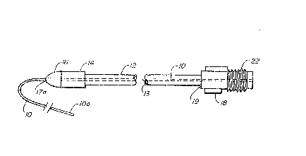
Fig. 1 is a fragmented longitudinal view of the preferred
long-texm epidural catheter of this invention illustra~ing the
assembly of the component members: an epidural catheter
inserted through a flexible reinforcement sleeve having an
advancement tip and a cuff, the sleeve being pre-attached to an
adapter for connecting the catheter to a liquid drug source.
Fig. 2 is a longitudinal sectional view of the epidural
catheter illustrating an internal lumen diameter in the
catheter;
Fig. 3 is an enlarged cross-sectional view of the epidura
catheter taken along lines 2-2 of Fig. 2
Fig. 4 is a longitudinal sectional view of the flexible
reinforce~ent sleeve showing the inner and outer diameters
thereof;
Fig. 5 is a sectional view of the cuff shown in Fig. l;
Fig. 6 is a cross-sectional view of the cuff taken along
lines 9-4 of Fig. 5, illustrating its circular configuration;
Fig. 7 is a longitudinal sectional view of a typical
molded adapter component for securing the proximal ends of the
ca~heter and sleeve;
Fig. 8 is a longitudinal sectional view of the advancement
tip illustrating its contoured body and extended shaft for
attachment to the reinforcement sleeve
Fig. 9 is a longitudinal sectional view of the flexible
reinforcement sleeve depicting the relationship of the
pre-attached advancement tip, the cuff and adapter;
Fig. 10 is an e~ploded longitudinal view of the invention
illustrating the assembly of the advancement tip, cuff, and the
adapter to the sleeve, and the insertion of the catheter
through the sleeve;
patO5123
J~
Fig. ll is an end view of the preferred embodiment, taken
~long the line 3.3 of Fig. 9 illustrating that the advancement
tip member and the cuff member are of the same diameter to
facilitate movement of the cuff through subcutaneous tlssue;
Fig. 12 is a fragmentary e~ploded elevational view of a
prior art two-piece catheter adapter contemplated for use in
connecting the epidural cathster of this invention to a liguid
drug source.
Fig. 13 is an enlarged fragmented longitudinal view of the
proximal end of the catheter showing the ports for
administering the narcotic in~o the body; and
Fig. 14 is a cross-sectional view of the proxlmal end of
the catheter taken along lines 5-5 of Fig. 13.
patO5123 8
4 ~
~ESCRI~TION OF TH~ PREFERRED EMBODIMENTS
As was heretofore men~ioned, the present invention is
directed to an improved long-term tunneled epidural catheter
for use in relieving intractable or chronic pain originating
below the cranial nerves.
In accordance with this invention, the aforementioned
disadvantages in the prior art two-piece tunneled epidural
catheters is obviated in an elegant and highly efficacious
manner by providing a one-piece epidural catheter which is
tunnæled from the paravertebral entry poin~ where it is
introduced into the epidural space to an exit site on thc
flank, the exteriori~ed proximal portion of the catheter being
protected by a sleeve which is pre-attached at one end to a
catheter connector, the free end of the sleeve extending over
the catheter being inserted a short distance into the passage
within the exit site provided by the tunneling step, as will be
detailed with particularity hereinafter. The catheter
connector is a component along with a liquid source connector
component to provide an adapter for placing the catheter in
liquid communica~ion with the drug to be administered into the
epidural space.
The arrangement of elements of the novel long-term
epidural catheter system of the present invention may best be
understood by reference to=t~e accompanying drawings taken in
conjunction with the following detailed clescription.
As shown therein, the exteriorized portion of the
catheter system, i.e. that proximal portion extending through
the exit site, comprises epidural catheter 10, sleeve 12 and
catheter connector 18 to ~hich the sleeve is pre-attached.
Catheter 10 has a lumen 11 through which a liqllid drug
may be transmitted for introduction in~o the epidural space.
Sleeve 12 has distal and proximal ends 12a and 12b,
respectively, between which extends a channel or hollow chamber
13 the inner diameter of which is greater than the outer
diameter of the catheter 10 in order to accommodate
patO5123 9
2 ~
~,
positioning of the catheter within cham~er 13, as seen, for
example, in Figs. 1 and 10. An advancement tip 16 is providsd
adjacent the distal end 12a of the sleeve in order to
facilitate inser~ion of the sleeve beneath the skin at the exit
site, as will be discussed hereina~ter. As seen more clearly
in Figs. 8-10, tip 16 has a generally bullet-shaped leading end
16a tapering from a base 16c to optimi~e ease of penetration
and a shaft 16b extending within the distal end 12a of the
sleeve. As will be well understood, tip 16 is provided with a
channel 17 communicating with an opeDing 17a at the leading end
16a to permit the catheter 10 to extend through the tip and
then through chamber 13 of the sleeve. As seen, the base 16c
of the bullet-shaped leading end of the tip is of greater outer
diameter than that of the sleeve to minimize friction~l
resistance to entry of the sleeve within the passage beneath
the skin created by the tunneling.
A cuff 1~ of a suitable material such as ~acron having
leading and trailing ends 14a and 14b between which a hollow
bore 15 extends is positioned on sleeve 12 with its leadi~g end
14a abutting base 16c of the advancement tip, as best seen in
Fig. 1. Cuff 14 and base 16c of the advancement tip have
substantially the same diameter, whereby the skin tissue is
caused to be opened enough by the tip to allow the cuff to
follow easily into the tissue. When the cuff 14 is introduced
into the tissue, it functions to encourage tissue ingrowth.
Additionally, there is evidence that the cuff may tend ta
pr~vent infection by precluding passage of bacteria through the
tunneled passageway and then ~own the external sur~ac~ of the
catheter.
As previously alluded to, the proximal or trailing end 12b
of the shaft is pre-attached to the leading or distal end 19 of
connector 18. connector 18 has an openlng 20 at its leading
end extending lnto the hollow bore 21 within the connector for
securing the catheter to the connector. Connector 18 also is
shown to have external threads 22 at its proximal or trailing
end adapted to mate with internal threads of a f luid source
connector ~not shown) in order to provide a fluid passageway
extending from the fluid source into the epidural space f or
drug admi~istration.
The particular adapter employed per se comprises no,part
patO5123 10
2~7~
~f,
of this invention. For purposes of further illustration, it
may for 'nstance comprise an adapter of the type descri~ed and
claimed in U.s.P. 4,187,848 issued to ~lenn N. Taylor and shown
in Fig. 12.
As seen, the adapter consists of two separate body mem~ers
which, for ease of reference, are designated as catheter
connec~or 100 (corresponding to connector 18 in the previous
figures) and a syringe connector 120. Both connectors have a
longitudlnally e~tending passageway for pumping narcotic from
the syringe into the catheter.
Catheter connector 100 has an opening 140 at its d~stal
end 160. A compressible plug 180 of elastomeric materlal
having a lonyitudinally extending channel 200 is seate~ within
a correspondingly shaped bore within connector 100 in a
relatively unco~pressed condition with channel 200 aligned with
opening 140 to receive the proximal end of a catheter (not
shown), as heretofore discussed.
Syringe connector 120 has a tapered port 220 at its
proximal end 240 to receive the tip of a syringe (not shown~.
The proximal end 240 has luer lock flanges 250 and a female
luer slip 260 adapted to receive the luer tip of the syringe so
that the syringe may be releasably locked to connector 120.
When employed in the practice of this invention to administer
morphine, a filter unit ~not shown) at the proximal end 240 is
also re~uired, as will be apparent tQ one skilled i~ the art.
Connector 100 has external thread~ 280 adjacent its
proximal end which mate with internal threads 300 adjacent the
distal end of syringe connector 120.
When the respective connectors are secured together, e.g.
by rotating wings 320 on catheter con,nector lOO, a compression
collar 340 having an opening 360 (in liquid communication with
channel 200 in external plug 180) compres~es plug 180 to
decrease the external dimensions (gap) of channel 200 to secure
the catheter end in the adapter.
A preferred adapter for placing the catheter in liquid
communication with the drug source is a one-piece adapter of
i
patOS123 11
~7.~9
Lhe type described and claimed in the copending application of
James R. ~ross, Serial No. ~00,859 filed August 30, 1989 ~nd
now U.S. Patent No.
It will of course be understood that routine modifications
may have to be made in the design of the adapter shown in Fig.
12 and/or that of the aforementioned application Serial No.
400,859 in order to accommodate pre-attachment of the sleeve on
the distal end of the adapter or the removable morphine
filter/injection cap assembly at the proximal end. In any
case, the adapter to be employed per se comprises no part of
the present invention and any modiiications in the illustra~ive
adapters sugyested above or the selection of alternative
adapters will be a matter of choice within the expected
judgement of the skilled worker in the light of the foregoing
disclosure.
Catheter 10, which should in yeneral be radiopaque for
visibility on X-ray to confirm catheter placement, may be made
of any of the materials heretofore employed in epidural
catheter manufacture, e.g. a synthetic polymeric amide of the
nylon family, "Teflon" ~trademark of DuPont ~or
polytetrafluoroethylene), polyurethane, silicone, etc., in
which case it may typically possess an outer diamter of on the
order of 1.3 mm (18 gauge~, like the aforementioned "Du Pen"
Long Term Epidural Catheter made of silicone rubber for
introduction with the aid of a stylet through a 14-gauge
Hustead needle. -_
However, in accordance with one embodiment of this
invention, the catheter may be made of a polymeric composition
which is characterized as being sufficiently stiff in the dry
state during insertion to eliminate the need for a stylet, but
which softens and swells on hydration in the body, which
material will be on the order of 19-20 gauge and may be
introduced with a smaller (17 gauge) needle.
Compositions of this general description are known in the
art for preparing medical products such as body implants,
tubular cannulas and the like which soiten and swell when
inserted into a living body and/or upon contact with an aqueous
med~um. ~y way of example of the state of the art pertaining
patO5123 12
~7~
there~o, mention may be made of the compositions disclosed in
U.s. Patents No. ~,883,699 and 4,911,691 issued to Anluk et al
and 4,728,322 and 4,846,812 issued to Walker et al.
As stated in the aforamentioned U.S.P. 4,911,691, for
instance, such compositions may compri~e a multiple phase
polymer composition comprising:
(a) a first phase which comprises a substantially
non-hyrophilic polymeric component; and
(b) a second phase which comprises a hydrophilic
polymeric component;
the composition (i) being capable of ab~orbin~ water to an
extent that it softens with a softening ratio of at least about
2:1 and/or swells with a swelling ratio of at least about
1.3:1; and tii) when substantially completely hydrated, having
an energy to break of at least about 700 N-cm~c~3 and a 2.5%
Secant modulus of less than about 7,000 N/cm2.
Preferably the non-hydrophilic polymeric component forms a
continuous phase. The hydrophilic polymeric component can form
a co-continuous phase with, or a dispersed phase in, the
non-hydrophilic polymer phase.
The non-hydrophilic polymeric component comprises a
poLymer which does not substantially absorb or attract water.
Preferably, the non-hydrophilic polymer is capable of absorbing
in an amount of no more than about 30%, more preferably no more
than about 1~%, and most preferably no more than about 10%, by
w~ight, based on the weight of the non-hydrophilic polymer.
The non-hydrophilic polymer can be for ex~nple, a
polyurethane such an aliphatlc polyurethane, a polyether
polyurethane, a polyester polyurethane; and ethylene copolymer
such as ethylene-vinyl acetate copolymer or ethylene-ethyl
acrylate copolymer; a polyamide, in particular a polyamide of
low crystallinity; aliphatic polyesters1 or the like. A
particularly preferred non-hydrophilic polymer is a
polyurethane, especially an aliphatic polyurethane.
The hydrophilic polymer pre~erably is a polymer that
absorbs at least about 50~ water, more preferably about 100~,
or example, at least about 150% by weight based on the weight
of the hydrophilic polymer. The hydrophilic polymer preferably
forms a hydro~el on absorption of water.
~ ~ ~ 5
",
The hydrophilic pol~nsr is preferably polyvinyl alcohol,
poly(ethylcne oxide), polypropylene oxide, poly(ethylene
glycol) polypropylene ~lycol, polytetramethylene o~ide,
polyvinyl pyrolidene, ~olyacrylamide, poly(hydroxy ethyl
acrylate), poly(hydroxyethyl methacrylate), or the llke.
Generally, the ratio of non-hydrophilic polymeric
component to hydrophilic polymeric component is 0.65:1 to 9:1.
Preferably the ratio of the polymeric component is 1:1 to 9:1.
The polymeric components are selected to provide a
multlple phase system. Generally, the polymeric components
each have a molecular weight of at least about 3,000 pre~erably
at least about 5,000 and most preferably at least about 10,000.
By way of further illustration, the composition selected
for use in the practice of this invention may have an inner
diameter of 0.019 inch in the dry state and 0.027 inch when
hydrated; and an outer diameter of 0.036 inch ~20 gauge~ in the
dry qtate and 0.050 inch (18 gauge) when hydrated.
Catheters prepared from swellable materials such as those
described above provide certain advantages in the preparation
of epidural or subarachnoid catheters.
Such materials possess a stiffness comparable to Teflon
when iD the dry state, thereby permittincl insertion without the
need of a wire stylet. This in turn permits one to employ a
smaller OD catheter, e.g. a 20 gauge which can be introduced
with a 17 gauge needle. ~owever, once in the body, the
composition hydrates and softens comparable to silicone.
The swelling which also occurs in a controlled reproducible
manner when hydrated enlarges the catheter, e.g. to approximate
the dimensions of the needle used to insert the catheter. In
this manner, the swellable catheter will seal the puncture made
by the needle, thereby sealing the ligamentum flavem and
tending to eliminate any drug leakage out o~ the epidural
space. The enlargement of the catheter after implantation may
also serve to increase retsntion of the catheter in the tissue,
thereby making it possible to retain the catheter in place
without the need for any sutuxing. Moreover, the enlarged
patO5123 14
lumen size allows for higher flow rates and, consequently,
improved dru~ delivery.
The reinforcement sleeve 12 should be made of a material
~hich is characterized as being tough, durable and possessing
good flexibility for optimum patient comfort. Preferably, it
should also be clear for visualizing the epidural catheter
within and possess low elongation so that the epidural catheter
retained inside cannot be pulled out of the epidural space. As
an example of a suitable material of th1s ger.eral descriptlon,
mention may be made of commercially available nylon-braided
polyurethane. It may, for example, be on the order of 18
inches in length and have an inner diameter of on the order of
O.071 inch and an outer diameter of around 0.142 inch (11
French).
Tip 16 may, for examplej be made of stainless steel or a
rigid plastic, the former being preferred. The outer diameter
at its widest point (base 16c) may be slightly less than twice
that of the sleeve, e.g. on the order of 0.245 inch. As will
be appreciated, the outer diameter of shaft 16b should be such
that it fits within the sleeve (as previously described) and
the outer diameter of channel 17 must of course accommodate
passage of the catheter.
Cuff 14 will be of no greater diameter than that of tip 16
and preferably will be of the same diameter. It may, for
in~tance, be on the order of 0.375 inch in length. greferably,
it is retained in place on the sleeve by being bonded thereto.
As heretofore mentioned, it may be made of a material such as
Dacron felt which encourages tissue ingrowth.
It will of course be appreciated that the foregoing
description of the materials and dimensions of the component
parts of the epidural catheter of this invention are by way of
illustration only and the scope of the invention is accordingly
not limited thereto. Various other materials and sizes may be
readily suggested to the skilled worker within the limits
required for introducing the catheter with a needle into the
epidural space.
patO5123 15
2 ~
The following description illustrates the preparation of
the epidural catheter system of this invention for
administration to a patien~.
The epidural catheter is first threaded through a needle
into the epidural space in per se known manner. With the
needle still in plac~ to avoid inadvertent damage to the
catheter, a small incision is made with a scalpel extending
cranially and caudally approximately 0.5 - t.o cm. All tissue
is dissected away from the needle to allow the catheter to fall
freely into the incision as the tunneler is later advanced.
The epidural needle is then removed. If a wire stylet is used
Eor insertion of the catheter, it is also removed.
With the aid of a tunneler, the catheter is then tunneled
to the desired exit site, e.g. on the patient ' s flank.
Tunnelers for use in this procedure are per se known in
the art. In general, they fall into two basic categories~
a solid tunneler of metal or plastic in which one end of the
catheter to be tunneled is slipped over the trailing end of the
tunneler (the end opposed from the leading end having the
cutting tip) and then dragged through the passageway created by
the tunneler; or (2) a hollow tunneler open at the trail~ng
end and having an opening in the cutting tip of sufficient
diameter to permit passage of the catheter therethrough, in
which case after the tunnel is made and with the tunneler still
in place, the catheter may then be threaded through thç opening
in the tip and out the trailing end of the hollow tunneler.
While either type of these malleable tunnelers is quite
satisfactory most of the time, each does nevertheless possess
inherent properties which may adversely a~fect the tunneling
step.
Since the solid tunneler functions by dragging the
catheter behind it through the passageway created by tunneling,
it follows that the catheter is dragged through the debris of
host origin caused by the tunneler. This may, in turn, cause
certain problems requiring the tunneling and, in some instances
the insertion o~ the epidural catheter itself to be repeated.
First, kinking of the catheter may be caused. Secondly, any
patO5123 16
~ ~ ~ t~
Y~
undue or sudden resistance in the advancPment of the catheter
behind the tunneler may cause the catheter to slip o~f the
trailing end of the tunneler. Finally, if the epidural
catheter is the component to be tunneled (as will be the case
wi~h the catheter system of this invention) any such resistance
may cause the distal end of the epidural catheter to become
dislodged from its position within the epidural space. Such
dislodgement may or may not require the catheter ts be removed
and re-introduced into the epidural space, depending upon the
extent of the d1slodgement.
The secon~ type of tunneling device heretofore used,
namely the hollow tunneler having an opening in the cutting
tip, does not suffer from the inherent dangers noted above.
However, it may instead cause different problems.
Since the cutting tip at the leading end of the tunneler
has an opening permitting passage of the catheter therethrough,
there is a tendency for flesh, blood and~or other debris from
the tunneling to enter the hollow tunneler through this opening
at the leading end. This in turn may at least partially clog
up the passageway within the tunneler, notably at the leading
end, thereby impairing threading the catheter therethrough and
possibly causing kinking within the tunneler. Rdditionally,
some of this debris of host origin may enter the leading end of
the catheter, thus providing an environment for infection due
to bacterial contamination.
Accordingly, the preferred tunneler for use in the
practice of this invention is that described and claimed in
Applicant's concurrently filed copending application, Serial
~o. (P.F. 1696).
As disclosed therein, the t~nneler consists o~ a malleable
hollow shaft having a solid cutting tip releasably secured to
one end thereo~, e.g. by threading.
After tunneling and advancement of the tunneler through
the exit site, the tip is then removed for passage of the
catheter through the tunneler and then through the exit site.
patO5123 17
~ ~ $ ~
",
Irrespective of the type of tunneler employed, after
lntroducing the catheter into the epidural space as described
about, the tunneler, being malleable, is then manually shaped
to match the contour of the flank. The skin at the
paravertebral incislon is lifted and the shaped tunneler is
introduced subcutaneously and then guided laterally toward the
contemplated exit site on the flank.
~ When the tip of the tunneler has reached the desired exit
point laterally, the tunneler is turned away from the pati~nt,
thereby forcing the cutting tip up against the skin . A scalpel
i5 then used to cut down to expose the tip, after which the
tunneler is advanced through the thus provided exit site.
When employing the novel tunneler descxibed and claimed in
the aforementioned copending application Serlal No. (P.F.
1696), following advancement of the leading end of the tunneler
throu~h the skin, the tunneler tip is removed and the catheter
passed through the chamber or lumen within hollow sha~t and out
through end. The shaft is then removed through the exit site.
In any case, after the proximal end section of the
catheter is exteriorized thxough the exit site, the protective
sleeve 12 is advanced over the free proximal end of the
catheter extending above the skin and down to the exit site.
The skin is then lifted with forceps and the sleeve advanced
through the exi~ site into the passage provided by the tunneler
until the cuff 14 is approximately two inches beneath the skin.
The epidural catheter may then be trimmed to fit within
the adapter 18 preattached to the sleeve and the adapter then
moved to the closed position to secure the catheter.
Both the paramedial incision and ventral exit site~ are
then closed with suitable sutures and sterile dressings
applied. After attaching a removable morphlne filter/injection
cap assembly, a saline solution may be injected to confirm the
catheter integrity.
The long-term epidural catheter is then ready to co~mence
introducing narcotic into the epidural space on an as-needed
dosage for pain management.
patO5123 18
J,
From the foregoing description it will thus be seen that
the novel one-piece epidural catheter of this invention
obviates all of the above-noted disadvantayes of the prior
two-piece systems in a simplified and elegant manner and
additionally provides a protective covering on that portion of
the catheter which is above the skin.
Since certain changes may be ~ade without departing from
the scope of the invention herein contemplated, it is to be
expressly understood that the foregoing description, including
the drawing, i9 by way of illustration and not be way of
limitation and the invention ls limited only as indicated in
the appended claims.
., ., . .,, . ----, .
patO5123 19