Note: Descriptions are shown in the official language in which they were submitted.
LASER VIDEC) ENDOSCOPE
Reference T Related App~
This invention relate~ in general to a small diameter
endoscope used for medical purposes and more particularly to one
in which illumination, viewing and laser operating unctions are
performed within a singls relatively small diameter endoscope.
The endoscope of this invention i5 designed particularly
for use in certain ophthalmological operations and thus the
disclosure herein will relate to such an embodi~nt~
It is known to apply laser energy, and othPr types of
energy, both directly and indirectly to variou~ parts of the eye
in order to ef~ect surgery. For example, it i~ ~nown to laser
the peripheral retina for treatment o~ retina detachment. It is
also know~, in appropria~e circumstances, to directly laser the
ciliary processes as one of th~ treat~ents ~or glaucoma.
~ecause of the difficulty o~ applying laser energy directly to
the ciliary processe~, the standard technique for disa~ling
ciliary proceRses has been a cryogenic technique. This
cryogenic technigue involves applylng a free~ing probe on the
external surface.o~ the eye overlying the ciliary process on
~6~3r31~
the inside of the eye. The ciliary processes are then frozen
and thawed. This destroys the ciliary processes and thus
reduces the aqueous output that builds up pressure. This
process is fairly brutal in its immediate effect on the eye.
Vision can be frequently lost. I~ is extremely difficult to
titrate. There is a risk o~ shrinkage and atrophy of the eye
due to overtreatment.
It is clearly preferable to apply a destructive element,
such as a laser, directly to the ciliary processes. This has
been done only where it is accompanied by a vitrectomy and
lensQctomy operation. That is only desirable or feasible in a
very small num~er of cases.
It is a specific purpose of this invention to provide an
intraocular endoscope that will be useful in photocoagulating
any internal ar~a of the eye including, most importantly, the
pars plana region, the cil~ary processes and th~ posterior
aspect of the iris.
A further and related purpose o~ this invention is to
allow more complete photocoagulation of the peripheral retina in
the treatment of complicated retina detachment or proli~erative
retinopathies such as in diabetes mellitus.
It is a further and a related purpose of this invention
to provide the above functions in a product which is relati~ely
easy ~or a surgeon to use so that the operations involved can be
2~
precisely determined and can be more complet~ than is presently
feasible.
Another related purpose of the invention is to providQ
an endoscope product that performs the above functions at a cost
which makes it feasible for appropriate ocular surgery to b~
undertaken on a relatively widespread basis by a rela~ively
large number o~ ophthalmoloqists.
: - 3 -
~` ' 2
Blief ~escription
In brie~, this invention involves a fiber optic
endoscope having a probe supported by a handpiece. The hand
piece is connected through a relatively long flexible lead to a
laser energy source, ~ source of illumination and an optical eye
piece. The flexible lead, hand piece and probe all contain a
laser optical ~iber, an optical fiber image ~uide and an optical
fiber illumination zone. The image guide ibsrs and the laser
fiber are surrounded by the set o~ illumination fibers.
The laser fiber is a monoPilament fiber that provides
the required pulses o~ laser energy to effect operation. In one
embodiment, it ha~ a diameter of approximately O.2 m~. The
image guide is a set of high resolution fused quartz image
~ibers that provide a 3,000 pixel image, each pixel having a
three micron diameter. The image guide has a diamet~r of 0.25
mm. An objectiva lens having a depth of ~ield ~rom down to
about one mm is bonded to the distal end of the image guide~
This image guide and laser fiber ar~ embedded within a
set o~ fibers whi~h carry illumination toward the distal end of
the probe.
Light transmitted down the illumination ~ibers emerges
at the ~istal end o~ the probe to provide illu~ination at the
area of operation. The image of at least part of the area
illuminated is transmitted back through the set o~ fibers that
constitute th~ image guide to b. viewed by the suxgeo~ at an
.
- 4 -
eyepiece or by video or still photography. With ~he image in
view and the pr~be in position, the surgeon can then control the
transmission of las~r energy, typically pulses o~ laser energy,
through the monofilament laser fiber to the zone of the
operation.
Bnef Description Of The Fig~res
FIG. 1 is a mechanical schematic longitudinal view of an
embodiment to this invention.
FIG. 2 is a cross-sectional view at the tip of the probe
illustrating the rela~ive deployment of the monofilament laser
fiber 22, the 3,000 pixel image guide 24 and the multi-~iber
illumination zone 26.
Y~
DescriptionQf ~ Pre_rred Embodimen~
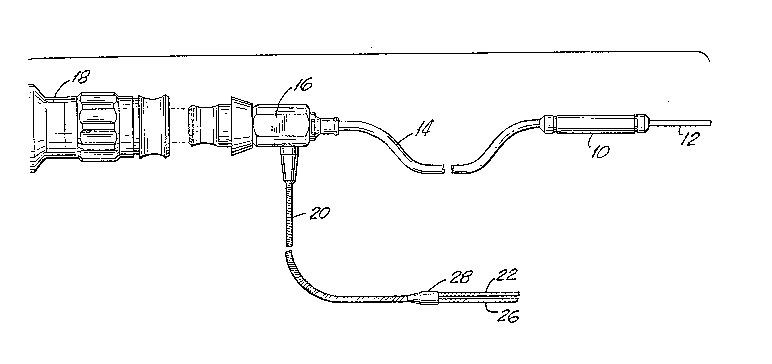
As shown in the FIGs., one embodiment of the endoscope
of this invention has a hand piece 10 and a probe 12, which are
connected through a ~irst flexible cable 14 to a connector 16
An eyepiece 18 is optically coupled to the connectGr 16 for
viewing purposes. A second flexible cable 20 extends out of the
~ide o the connector 16.
Within the probe 12, the hand piece 10 and the firs~
flexible cable 14 there is deployed three separate sets of
optical fibers that perform three separate functions. These are
shown in the cross-sectional view of FIG. 2. This cross-
sectional view is one taken at the tip of the probe 12. Within
the probe 12 there is a monofilament laser fiber 22, an image
guide 24 and an illumination zone 26~ The laser fiber 22 is a
monofilament optical ~iber that delivers the laser energy at the
tip of the probe 12 for performing operations. The image guide
24 is a set of high resolution fused quartz image fibers that
provide a 3,000 pixel image, each pixel having a 3 micron
diameter. The illumination zone 26 is composed sf a large
number of fibers which carry illumination toward the distal end
of the probe 12. All o~ these optical fiber elements are guart.
~ibers.
In operation, light i~ transmitted down the illumination
zone 22 to emerge at the distal end o~ the probe 12 to provide
~;
2 ~
illumination at ~he area of operation. The image of at least
part of the area illuminated is transmitted back through the
image guide 24 to be viewed by the surgeon at the eyepiece 18.
With the i~age in view and the proba 12 in position, the surgeon
can then control the transmission of laser ener~y (usually
pulses of laser energy) through the laser fiber 22 to the zone
of the operation.
Because of the small diameter of the probe 12 (under one
mm) this endoscope can be used for operations in areas
(particularly for eye operation~) where a combined viewing and
operating endoscope was not hithertv possible.
At the juncture 16, the imaging set of op~ical fibers 16
is separated from the other two sets of optical fi~ers so that
only the laser fiber 22 and illu~ination fibers 26 extend down
through the tube 20. As indicated at the juncture 28, these two
sets of ~ibers 22 and 26 are further separated to be
appropriately connected to a source of laser energy for the
optical fiber set 22 and to a source of light for the se~ of
~iber that constitute the illumination zone 26.
: It should be noted that the image provided by the image
guide 24 can be applied to an eye piece 18 or can be displayed
by a video or can be applied to cr~ate a still photograph.
Indeed, it is anticipated that a video display might be
pre~erable to ~acilitate the surgeon's positioning in order tQ
manipulate the probe 12 properly.
; ~ 7 ~
In one embodiment that ha~ been tested, the probe 12 has
an outer diameter of 950 microns with a steel side wall 12w o~ f
75 microns and thus an inner diameter of 800 microns.
In that embodiment, the laser fiber 22 is a monofilament
fiber with an active diameter of 200 microns. The cladding and
protective buffer layer to prevent mechanical abrasion of the
cladding brings the diameter to 250 microns.
In that embodiment, the illumination zone 26 has 500
optical fibers, each fiber having a diameter o~ 30 microns
including cladding. The optical fibers in the illumination zone
26 are strung randomly down.the length of the instrument and are
potted into place only at the tip of the operating probe 12.
In that embodiment, the image guide 24 has 3,000 quartz
~ibers, each only 3 microns in diameter including cladding. The
fibers are fused to provide a single convenient to handle guide
with 3,000 pixels. The guide wall is a thin black protective
PVC sleeve 45 microns thic~. This image guide 24 has a 250
micron diameter which with the PVC sleeve becomes 340 microns.
In that embodiment, the probe 12 is 30 mm long, the hand
piece 10 in 40 mm long and the cable 14 is 410 mm long~
In one preferred embodiment, which has been tested, an
objective lens is bonded to the distal end of ~he image guide 24
fibers and provides a depth of field ~rom one mm to infinity
with a field o~ view of 70 degrees, A depth of field that can
go down to as little as one mm is important to aid the surgeon
- 8 -
to position the end of the laser guide 22 as olose as one mm
from the tissue on which the laser energy is to be delivered.
It is important to have the distal end of the laser guide and
the distal end o~ th~ image guide at the same plane. The reason
relates to a combination of the fact that Sa) with a single
probe there is no stereopsis, (b) the need to deliver the laser
energy as close as possible to the tissue b~ing worked on, and
(c) the importance of avoiding tissue puncture or contact.
The lens is a triple lens o~ a known type. It is bonded
to the image guide 24 prior to assembling the image guide 24 in
the illumination zone so that the distal surface of the lens (~
mm which is one em~odiment) is flush with the distal end of the
probe.
Very minute working diskances have to be traversed by
means of the operator's hand moYements. These minute working
distances axe in part required by the fact that the laser energy
should be delivered only to a speci~i¢ small zone of tissue
which is to be operated on. This requires that the distal end
of the laser guide be brought as close as possible to the
tissue. A distance of one mm is desirable. But it is essential
that the surgeon be able to view the tissue being operated on at
the one mm distance in order to avoid having the probe contact,
damage or puncture the tissue.
In known types of operations where an operating
microscope is employed, an image is provided with ~ degree of
O g _
.~ .
~ ~ ~ 8 ~ ~ ~
stereopsis which aids the surgeon in moving toward the tissu~.
But with a probe that incorporates both imaging and laser
delivery, stereopsis is not available and it is essential that
the laser guide not extend past the image guide in order to make
sure that tissue damage is avoided.
Having a wide field of view, such a~ 70 degrees, is
useful to enable the surgeon to locate the various tissue zone
areas which have to be operated on and to which energy has to be
delivered.
As is known in the art, the particular laser guide
optical material is selected as a function of the frequency of
the laser l~ght pulses which are to be transmitted by the guid~.
In the embodiment tested, the laser used is a diode laser
composed of gallium-aluminum-arsenide semi-conducting crystals
to provide a wave length of about 810 nano meters (in the range
of 7~0 nm to 850 nm)
The joints 28 and 16 where the different components 22,
24 and 26 of the interior o~ the probe 12 are brought together
are fabricated as junctions by standard techniques known in the
art including the use of heat shrink tubing at the connector 28.
The embodiment ha~ the straight probe 12 shown~
~ppli~ant b~lieve~ there ~lay be advantage in a slight curvature
to the probe 12 so that the tip of the probe is displaced two to
three mm ~rom the axis. This might provide adYantage in use of
more readily clearing the lens of ~he eye.
-- 10 --
2 E!!Çi8~
It should be noted that the combination of the
illumination zone, the image guide and laser ~uide within a
single probe provides a particularly compact probe ~or
performing these three functions, which because it is compact
and circular in cross section requires a minimal incision o~ the
cornea while meeting the objectives of this invention including
an ability to access areas such as the ciliary processes which
otherwise require the use of multiple instruments.