Note: Descriptions are shown in the official language in which they were submitted.
WO 91/11956 PCT/SE91/00106
207~ps~
A Monitor which F~aiyses Puls Freouenc~- by Fhotopleth~-smo-
~raphic bieasLremert ann a Neasur_ne Nethoc trerefor
Technical Field
The present invention relates to monitoring apparatus
provided ~~ith an optical sensor and functioning to
analyze pulse frequencies by measuring the blood circu-
lation of a measuring object, such as a part of the
human body or an animal body using photoplethysmography
(PPG-measuring), said apparatus being of the kind set
forth in the preamble of Claim 1. The invention also
relates to a nethod of taking such measurements.
Photoplethysmography (hereinafter abbreviated to PPG)
has been kno~m to the art for more than 50 years and is
applied technically for measuring peripheral blood
circulation. The nethod is primarily used for Deasuring
heart frequencies and
blood circulation when perforaing surgery. The method
has many advantages, since, anong other things, it is
non-imrasive and does not subject the patient to any
appreciable trauma. Furthermore, the method requires no
highly expensive or complicated equipment in order to be
put into effect.
When light from a suitable light source impinges or.
the skin, the light is dampened or attenuated according
to the nature of the tissue on which the light impinges.
This light attenuation is assumed to be constant. The
light also passes through a number of blood vessels and
is also attenuated by the blood present. The light is
attenuated as a result of a number of complex processes,
such as absorption, reflection and different forms of
WO 91/11956 PC'T/SE91/00106
2
scattering. The PPG-technique is based on the assumption
that the more blood that is present in the volume inves-
tigated, the more the light is a~terua~ed. This results
in two signal components of interest, namely a DC-compo-
nent which corresponds to the total amount of blood in
the investigated volume and an AC-component which signi-
fies pulsation of the blood flow.
In order to study the aforedescribed phenomenon, it is
necessary to use a light source, a light detector,
amplifying electronics and a display unit, for instance
an oscilloscope or a printer.
The light source and the detector may be placed on a
respective side of the object on which blood circulation
is to be measured, and the detector consequently mea-
sures the light transmitted. This technique is, at
times, referred to as transmission-photoplethysmography
and can only be applied to a few skin surfaces, such as
fingers, ear lobes and toes.
A more general method is to place both the light source
and the detector in one and the same probe and measure
the light reflected. This technique is known as reflec-
tion * and is the dominating technique. It has long been
understood that the pulsating component or AC-component
of the PPG-signal is contingent on changes in blood
volume during each heart beat. The greater the volume of
blood, the less light will impinge on the detector.
It is obvious, however, that this is not the whole
truth. Tests have been carried out in which blood has
been allowed to pulsate in rigid glass tubes, where
changes in volume are impossible and where solely the
flow rate pulsates. A pulsating PPG-signal is also
obtained in this case, which can be explained by the
WO 91 / 11956 PCT/S E91 /00106
2o7~os4
3
detection of changes in orientation of the erythrocytes,
which varies during each heart beat.
In summary, there are at least two reasons for the AC-
component, namely a change in blood volume and the
orientation of the erythrocytes.
A typical PPG-signal has, in the time plane, the form of
a blood-pressure curve having the same periodicity as
the heart beats. The signal also includes a number of
low frequencies.
The present invention is based on the realization that
the lower frequencies occur as a result of changes in
blood flow caused by the sympathetic nerve system and by
respiration, this realization being based on the known
fact that the intrathoracic pressure is lowered when
breathing-in, or inspiring. This subpressure is utilized
to "suck" the venous blood into the atrium and ventri-
cle.
The invention departs from this starting point and
assumes that this subpressure causes variations in blood
flow in venous plexus and also that it should be possi-
ble to detect this variation with the aid of the PPG-
technique, particularly by using a technique which
enables measurements to be made in the venous plexus.
Nerve signals in the sympathetic nerve system also
influence the blood flow. The smooth muscle around the
vessels pulsates at a frequency which lies close to the
respiration frequency. This pulsation is normally re-
ferred to as Traube-Hering's waves, after the scien-
tists' Traube-Hering. Waves of a still lower frequency
are also found, these waves normally being referred to
as Mayer's waves.
WO 91/11956 PCT/SE91/00106
The blood also pulsates through the so-called arterio-
venous anastomises, so as to control body temperature.
This normally occurs at a frequency of about 0.3 Hz snd
is designated Burton waves.
Disclosure of the Invention
The present invention is based on the aforesaid realiza-
tion and on measurements, or assays, carried out with
the aid of apparatus that has been constructed in accor-
dance with the theory on which the present invention is
based.
The inventive monitoring apparatus intended for measur-
ing blood circulation is of the kind set forth in the
preamble of Claim 1 and has the characterizing features
set forth in the characterizing clause of said Claim.
The exhaustive experimentation, which forms the basis of
the invention and which is briefly described herebelow,
has shown that respiration is the totally dominating
low-frequency component of the PPG-signal.
It was found from the series of experiments performed
that the extracted signal can be encountered substan-
tially irrespective of where in the body the probe is
placed. This leads to the conclusion that the extracted
PPG-signal constitutes a measurement of variations in
blood pressure caused by respiration, and therewith also
variations in the flow of blood through the object under
examination.
It is expected that the inventive monitoring apparatus
will find universal use within human care establish-
ments. In the case of patients in intensive care wards
or under anaesthetic or under postoperative conditions,
WO 91/11956 PCT/SE91/00106
it is important to monitor heart and respiration fre-
quencies. When these two physiolog~.:al variables are
known, the doctcr or nursing syster will have a good
picture of the patient's general condition. Monitoring
5 of these variables under anaesthetic conditions can
facilitate the assessment of the depth of anaesthesia.
Above all, the invention avoids those serious disadvan-
tages that are associated with respiratory frequency
l0 monitoring methods and apparatus hitherto used, all of
which are normally unreliable, besides being both stren-
uous and complex.
It is generally more important to monitor respiration
parameters in the case of infant care than in the case
of adults under intensive care. In this regard, the
inventive monitoring apparatus is superior to the tech-
nique which has been used most frequently hitherto,
namely the use of impedance plethysmography with the aid
of ECG-electrodes placed on the surface of the skin,
among other things because such electrodes (normally
three) take-up a relatively large area of the thorax.
When requiring to make X-ray examinations, it is neces-
sary to remove the electrodes, since they are not trans-
parent to X-rays.
Furthermore, light disturbances from peripheral electri-
cal apparatus are induced in ECG-cables with associated
input amplifiers. The signal cables are coupled both
inductively and capacitively. When carying out surgery,
it is impossible to carry out ECG-recordings and to
measure heart frequencies over prolonged periods of
time, due to the surgical application of diathermy.
ECG-electrodes and the paste used together therewith
WO 91/11956 PCT/SE91/00106
~~'~ ~0~~
E
cause irritation of the skin, particularly when monitor-
ing is effected over a prolonged period and particularly
in the case of infants whose ~kir~ ~s very ~te:~:~~-r and
sensitive. The electrodes and associated leads or cables
also limit the ability of the child to move.
The present invention provides important direct advan-
tages in relation to the aforesaid, and also affords
indirect advantages with respect to methods of measuring
respiration frequency.
For example, the inventive monitoring apparatus can be
applied and handled with ease: it avoids the aforesaid
problems associated with prolonged use of skin elec-
trodes; it is free from disturbances during surgery in
which diathermy is applied: and affords a wide degree of
freedom with regard to positioning of the sensor. For
example, a sensor-provided probe can be placed on a
finger or on a toe, at a distance from the thorax region
where another investigation is being made. Furthermore,
the sensor element can be made very small, such as not
to interfere with X-ray examinations to any appreciable
extent.
An additional, very important advantage afforded by the
inventive monitoring apparatus is that it can be inte-
grated with a number of different medical instruments of
the kind where heart frequency and respiration frequency
are important parameters, for instance pulsoxymetry and
defibrillators.
In order to enable measurements to be taken directly on
patients, it is necessary to equip the monitoring appa-
ratus With one or more filters. This will result, how-
ever, in difficulty in selecting limit frequencies,
since the signal can exhibit pronounced variations.
WO 91 / 11956 PCT/SE91 /00106
20~~064 - -
Accordingly, one preferred embodiment of the invention
is characterized in that the apparatus includes one or
more filters, preferably digital filter , which have
means for setting limit frequencies and the degree of
amplification. Filters of this kind are suitably incor-
porated in the apparatus, which will also preferably
include means for electronically detecting the frequency
content of the signal for selection and setting of limit
frequencies.
l0
According to another embodiment of the monitoring appa-
ratus, the filters are adaptive and are constructed to
adapt to the prevailing heart and/or respiration fre-
quency, so as to optimize the filter properties.
Furthermore, the monitoring apparatus will preferably be
provided with means for DC-compensating the PPG-signal,
so as to balance-out the low frequency components of
said signal automatically, without experiencing harmful
energy losses.
In the case of one embodiment of the invention which is
particularly beneficial in practical application, the
monitoring apparatus includes a probe which is intended
to be placed on a suitable part of the body, for example
a finger, and which includes means for delivering light
to said body part and means for capturing light which
passes through said body part or which is reflected
therein, for the purpose of passing this light to the
detector unit. This monitoring apparatus is character-
ized by optical fibres connected to the probe and func-
tioning to conduct light from a light source to the skin
and from the skin to the detector unit respectively.
By conducting, in said apparatus, the light through
optical fibres to and from the skin, there is obtained a
WO 91/11956 PCT/SE91/00106
s
system which is highly resistant to electromagnetic
disturbances or interference, a feature which is ex-
tremely important within the sphere of medical treat-
ment. This particular feature enables the heart frequen-
cy and respiration frequency to be recorded during
surgery in which diathermy is applied. An apparatus of
this kind which is insensitive to disturbances during
surgery represents a very important step forwards in the
art.
The inventive monitoring apparatus preferably includes a
microprocessor which is programmed to calculate the
Fourier transform, and/or to separate signals concerning
the heart and respiration frequencies of the object by
digital filtration, and/or to eliminate disturbances,
emanating, for example, from stray light of frequency
50 Hz.
A further possibility afforded by the use of micropro-
cessor technology is that of combining the measuring
process with Sa02-measuring with pulsoxymetry.
The present invention also relates to a method of carry-
ing out photoplethysmographic measuring processes, this
method being characterized mainly by the characteristic
features set forth in Claim 9.
disclosure of the Experiments Performed
With the intention of confirming the aforedescribed
theory scientifically, namely the theory that it is also
possible to separate from a PPG-signal whose dominating
component forms a measurement of the heart frequency of
the object being examined, a signal component which
discloses the respiration frequency of said object, a
simple photoplethysmograph was constructed. Four differ-
WO 91 / 11956 PCT/SE91 /00106
207064
9
ent measuring probes were mounted on the photo-
plethysmograph, all of which probes used a light-
emitting diode as a light source. One probe utilized the
wavelength 875 nm, two utilized the wavelength 940 nm
and one utilized the wavelength 950 nm. All probes
measured reflected light. The photoplethysmograph oper-
ated either within the frequency range of 0.2-10 Hz or
the frequency range 0.2-20 Hz.
The photoplethysmograph was used to measure the blood
circulation of dogs, cats, adult males aged 35 years and
infants in incubators. In order to show both the respi-
ration frequency and heart frequency in the photo-
plethysmograph signal, the heart frequency and respira-
tion frequency of the adults and the infants were re-
corded separately with the aid of other methods. In the
case of the animals used in this experiment, solely the
respiration frequency was recorded separately. These
measurements were used as reference signals in the
measuring-data analysis. All measurements were recorded
on a measurement tape-recorder.
The measurement data was analyzed partly in the time
plane, where the two components in the photoplethysmo-
graph signal were filtered out, and partly in the fre-
quency plane, wherein the power spectrum was calculat-
ed?. A cross-correlation function for the photoplethys-
mograph signal and the reference signals was also calcu-
lated. The following conclusions can be drawn from these
analyses:
The apparatus functions well on adults. The heart fre-
quency and respiration frequency can be separated by
means of filter techniques. The heart frequency is the
dominant signal component. The two components are
clearly evident in the power spectrum and the cross-
WO 91/11956 PCT/SE91/00106
17f~06~
correlation function shows correlation with the
reference signals.
In the case of infants in respirators, respiration is
5 the totally dominant component. It is slightly more
difficult to filter-out the two components in the case
of infants than in the case of adults. Although the two
frequencies are evident in the power spectrum, respira-
tion dominates the spectrum totally. The cross-correla-
10 tion function shows correlation with the reference
signals.
The respiration frequency is the dominant signal compo-
nent in the case of animals. Although a high frequency
component can be filtered-out, it cannot be guaranteed
that this component is the heart frequency. The power
spectrum has a broad band with many peaks or spikes
whose origin cannot readily be established. The cross-
correlation function was constructed solely for respira-
tion, where correlation can be shown.
A more detailed account of the experiments carried out
is given below, with reference to the accompany draw-
ings, in which
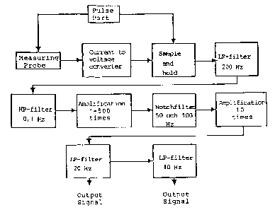
Figure 1 is a block schematic which illustrates the
principle construction of the measuring electronics
used:
Figures 2 and 3 are schematic views of measuring probes
used in the experiment series;
Figures 4 and 5 are circuit diagrams with associated
component signs; and
Figure 6 illustrates the principle of transmission
WO 91/11956 PCT/SE91/00106
11
measuring with the aid of fibre optics and with the aid
of a probe fitted to one finger of the object.
Infrared light-emitting diodes were used as the light
source. Light-emitting diodes are small, mechanically
insensitive and have a light intensity which is propor-
tional to the diode current. The AC-component of the
PPG-signal is weak and must be greatly amplified. The
light intensity should be high, in order to obtain a
high signal level. This is achieved by pulsating the
light-emitting diode with a high current. This enables a
much higher diode current to be used than in the case of
continuous light. The use of pulsated light results in
time-discrete measurement of a time-continuous signal.
According to the sampling theorem, it is necessary for
the pulse frequency to be twice as large as the
frequency content of the signal, in order to recreate
the continuous signal. This frequency content should be
beneath 20 Hz. The pulse frequency selected was 1 kHz,
which fulfils the sampling theorem more than well. The
diode illuminating time was 40 ~s, which constitutes a
fraction of the period time 1 ms. Since the illuminating
time is so short in relation to the dark time, it is
possible to use a very high diode current without de-
stroying the light-emitting diode.
For the purpose of obtaining a continuous measurement
response, the measuring values are maintained constant
between each new measuring process, with the aid of a
sample-and-hold circuit. In order to guarantee that a
measurement value is obtained when the diode emits light
at full intensity, the sample-and-hold circuit is closed
(and therewith holds the measurement value) before
extinguishing the light-emitting diode.
WO 91/11956 ~ PCT/SE91/00106
12
The signal from the sample-and-hold circuit is equalized
by passing said signal through a low-pass filter.
The principle construction of the measuring electronics
will be seen from Figure 1. References are made to the
circuit diagram shown in Figure 4.
The Measuring Probe
Four different measuring probes, referenced A-D, were
constructed during the experimental period. All of these
measuring probes included a detector in the form of a
light-emitting diode model S-4C from United Detector
Technology.
Three different light-emitting diodes were used, namely:
Probe Wavelength Light Power
A Philips CQY 58 875 nm 0.5 mW
B, C Teiefunken TSUS 5400 950 nm 15 mW
(corresponds to
Philips CQY 99)
D Hewlett-Packard HEMT 1001 940 nm 2.5 mW
It should be observed in this respect that the light was
comparitively broad-band light and that the wavelength
values refer to maximum intensity. The power value
denotes the power radiated totally in a hemisphere and
is estimated from existing data sheets in the case of
HEMT 1001. It should also be mentioned that the light-
emitting diodes spread light differently.
Figure 1 illustrates a measuring probe comprising an
acrylic tube 4 in which a light-emitting diode 1 and a
detector in the form of a light-emitting diode 2 are
embodied or cast with expoxy resin 5. The tube 4 has a
WO 91/11956 PCT/SE91/00106
13
diameter of 8 mm and is surrounded by a tube 3 of diame-
ter 12 mm.
The probe illustrated in Figure 3 differs from the probe
illustrated in Figure 2, in that the detector 2 is
angled in relation to the detector surface.
The following light-emitting diodes were used with the
different probes:
Probe uses light-emitting diode CQy 58
A
Probes and C uses light-emitting diode HEMT 1001
B
Probe uses light-emitting diode TSUS 5400
D
Probes A and D were constructed in the same way as the
probe B shown in Figure 2.
The cables used between probes and electronic devices
were very thin and flexible, screened four-conductor
cables sold by Telko under the trade name Pick-uptrad
PU 402.
The pulse electronics used had two functions, firstly to
drive the light-emitting diodes and secondly to generate
and deliver control signals to the sample-and-hold
circuit. The fundamental component of the pulse elec-
tronics is a bistable flip-flop which generates a square
wave having a frequency of 1 kHz. This flip-flop is
constructed around two Nand-gates (IC lA and B in the
circuit diagramme of Figure 4), and a buffer circuit (IC
2). The clock frequency is proportional to the product
R2 and C1. The capacitor C24 is required, to lead away
disturbing high frequencies.
WO 91/11956 PCT/SE91/00106
~'J~~'~ 14
The control pulses are generated by a monostable flip-
flop (IC3), which is triggered on positive flanks by
flanks of the square wave.
Those times at~.which the monostable flip-flop is "high"
is determined by the product R3 and C2 for the light-
emitting diode, and by the product R4 and C3 for the
sample-and-hold circuit.
Current is supplied to the light-emitting diode through
a transistor stage built around a Darlington transistor
(T1 in Figure 4). A Darlington transistor is actually
two transistors connected in series and has the positive
property of having a very high current-amplifying
factor.
The voltage drop across collector and emitter reached
about 1.4 V.
The light-emitting diode control pulse was connected to
the base of the transistor via a resistor (R5), which
was dimensioned so that the transistor would bottom at
high pulse values and throttle at low pulse values.
The resistor R41 was coupled in parallel with the col-
lector resistor R6 by means of a switch on the front
panel of the apparatus used, such as to obtain a high
collector current and a high light intensity. The col-
lector current was 130 mA in position "low" and 180 mA
in position "high".
The Current-Voltage Convert r
The light-emitting diode in the measuring probe was
biased electrically with +2.5 V in the reverse direc-
tion. This voltage was produced by IC11, which is a
WO 91/11956 PCT/SE91/00106
precision regulator and which held the voltage stable. A
linear detector response was guaranteed in this way. The
light-emitting diode now delivered a reverse current
which was proportional to the detected light intensity.
5 This current was converted to a voltage, by a current-
voltage converter constructed around an operational
amplifier (IC4).
It shall be noted that the current-voltage converter was
to an inverting circuit.
The Sample-and-Hold Circuit (,~C51
The circuit functioned to hold the time-discrete mea-
15 surement values constant between each new measuring
process. Sample-and-hold circuits, however, are encum-
bered with the disadvantage that disturbances in the
form of spikes from the control logic leak through to
the measurement value. This is particularly pronounced
in respect of the weak AC-component. In order to reduce
this disturbance, the amplitude of the control signal
was scaled down to about 2 V over the resistors R39 and
R40. An external holding capacitor C15 on 1.0 ~.F was
connected to the circuit. This capacitor also assisted
in damping the disturbance spikes.
Low-Pass Filter 200 Hz
The purpose of the first low-pass filter was to elimi-
pate the disturbance spikes deriving from the sample-
and-hold circuit. The filter was an active Tjebychev
filter of the fourth order, followed by a passive RC-
link. The filter had been designed to permit 0.5 dB
ripple in the pass band and to have a cut-off frequency
of 200 Hz. The filter was built-up around two
cascade-coupled operational amplifiers (IC 6A and B) and
WO 91/11956 PCT/SE91/00106
._. _
16
the RC-link R16 and C9.
~e Hiah-Pass Filter 0 1 Hz
The high-pass filter eliminated the DC-component, there-
by enabling amplification of the AC-component. The
filter was an active Tjebychev filter of the second
order, constructed around an operational amplifier
(IC7). The filter was designed to permit 0.5 dB ripple
in the pass band and to have a cut-off frequency of
0.1 Hz.
Amplification 1-500 Times
The amplifier was a non-inverting amplifier constructed
around a offset-compensated operational amplifier
(IC10). The amplification was varied with the aid of a
potentiometer positioned on the front panel.
Notch Filter 50 and 100 Hz
The weak AC-component was greatly disturbed by the net
frequency 50 Hz, and also by disturbances from lamps and
fluorescent tubes at 100 Hz. These disturbances were
eliminated in two cascade-coupled notch filters. The
notch filters were constructed around two operational
amplifiers (IC 8A for 50 Hz and IC 8B for 100 Hz). The
filters could be adjusted in the frequency direction?,
with the aid of potentiometers R29 for the 50 Hz-filter
and R30 for the 100 Hz-filter, such as to filter-off
precisely the desired frequency.
Amplif~cat~on 10 Times
The signal was amplified a further 10 times in a non-
inverting amplifier constructed around an operational
WO 91/11956 PCT/SE91/00106
1~207~06~
amplifier (IC9A).
Low-Pass Filter 20 Hz
The signal passed through an active Tjebychev filter of
the third order, having a cut-off frequency of 20 Hz.
This construction permitted a 0.3 dB ripple in the pass
band. The filter was constructed around an operational
amplifier (IC9B). The output signal was coupled to a
BNC-switch labelled "Output 20 Hz" located on the front
panel.
Low-Pass Filter 10 Hz
The signal finally passed through a low-pass filter of
the same kind as that described in the aforegoing, with
the cut-off frequency of 10 Hz. The signal was then
coupled to a BNC-switch labelled "Output 10 Hz" locatea
on the front panel.
The Net Part
The measuring electronics comprised a digital pulse
part, and an analogue amplifying and filtering part. one
problem which readily occurs when mixing digital tech-
nique with analogue technique is that disturbances occur
in the form of spikes from the digital side to the
analogue side. This disturbance can be reduced by using
a separate supply voltage on the two parts. Accordingly,
the built-in power unit was constructed around a trans-
former which had two secondary windings, each producing
a secondary voltage of 12 V. This alternating voltage
was rectified to ~5 V With the aid of rectifying
bridges, smoothing and disturbance-eliminating capaci-
tors and integrated regulators. Particular mention can
be made to the fact that the light-emitting diode of the
WO 91 / 11956 PCT/S E91 /00106
I8
~~~.J
p~~e was powered by a current of between 100 and 200
mA. Consequently, there was used a more powerful regula-
tor capable of delivering more current to the digital t5
V-side (cf the circuit diagramme shown in Figure 5).
The Lictht I_n_tP_r,_eity of the Probes
The following measuring process was carried out in order
to obtain an estimation of the mutual intensity rela-
tionship between the various probes.
The probes were connected and a photometer was held
directly against the probes. The luminous intensity of
the probes was observed from the photometer. It shall be
noted that this is an integrated measurement value and
not the intensity when the diodes are illuminated. The
measurement values are shown in the following table.
Intensity
r be " ow" "High"
A 25.1 ~tW -
B 15.2 ~1W 19.0 ~tW
C 18.4 ~tW 22.7 ~tW
2 5 D 3 2 .1 l,i,W 3 8 . 7 ~,W
It Was found that probe D could not be used to take
measurements in practice, since the system became self-
oscillating. This problem was not rectified. Probes B
and C were found to provide a better result than probe
A, probably because the light emitted had a longer
wavelength (875 nm for probe A, 950 nm for probes B and
C and 940 nm for probe D).
Measurements and Results
The auto-measuring process was effected by recording the
WO 91/11956 PCT/SE91/00106
2~7~06~
PPG-signal on tape. The respiration frequency was also
taped at the same time. When measuring blood circulation
on human beings, the heart frequency was also measured,
but with other methods. These signals were used as
reference signals for the two components of the PPG-
signal.
Measurements were taken on three different groups of
objects, namely animals, infants in incubators, and
adult males aged 35-years.
A narrow selection of the measurements taken are pre-
sented below. This selection is neither a random selec-
tion or a particularly representative selection. It is
rather an example of those measurements which were
considered to be of interest in evaluating the tech-
nique. An attempt to form a conclusion from these mea-
suring processes is made below.
Measuring Eauipment
In addition to the aforedescribed PPG-equipment, the
respiration frequency of animals and adult human beings
was measured with the aid of Strain Gauge equipment,
which had the form of a strain sensor consisting of a
thin rubber hose filled with mercury and connected to a
measuring bridge. The heart frequency of the adult human
beings was measured with the aid of laser-doppler equip-
ment. when laser-doppler equipment was used, a minor
investigation was also carried out in order to ascertain
whether or not the respiration frequency could be traced
in the laser-doppler signal. The heart frequency was not
measured separately on the animals used in the experi-
ment. In the case of infants, the heart frequency was
measured with ECG and respiration was measured with an
impedance plethysmograph.
WO 91/11956 PCT/SE91/00106
The measured signals were coupled directly to a measure-
ment tape-recorder and thereafter to an oscilloscope,
where they could be monitored during the actual measur-
ing process.
5
Processing Measurement Data
No measurement data was processed during the actual
10 measuring processes. All data processing was, instead,
carried out on the taped signals. The measurement data
was processed in three different ways, namely by filtra-
tion, Fourier analysis and cross-correlation.
15 Filtration
The PPG-signal was coupled to a system of active fil-
ters, where different types of filter, limit frequencies
and amplifications could be set. This enabled the dif-
20 ferent components to be filtered-out and compared with
respective reference signals. The two signals were
presented simultaneously on a printer.
Fourier A_naly~is
The signals were processed on an Ericsson PC, using the
signal processing programme ASYSTANT. The power spec-
trum of the PPG-signals and the reference signals was
calculated and comparisons subsequently made in the
frequency plane.
Cross-Correlat;nn
The cross-correlation function is a good way of ascer-
taining whether or not the periodicity of one signal is
found in another signal. For example, it can be ascer-
WO 91/11956 PCT/SE91/00106
21
207064
tained whether support is found for the assumption that
the respiration period is found in the PPG-signal.
The cross-correlation function C(k) is calculated by the
computer as:
N-1-;k;
C(k) - 1/N E X(n) Y(n+~ki)
n = o
where N is the number of measuring points, k is the
shift between the signals, X(n) is the one signal and
Y(n) is the other signal. It should be noted that the
computer works with sampled signals.
The cross-correlation function is thus a form of convo-
lution function where the one signal can be said to
"slide" over the other. Assume, for example, that one
signal is the PPG-signal and the other the respiration
signal measured with a Strain Gauge. If the respiration
frequency is found in the PPG-signal, there is obtained
a periodic cross-correlation function which has the same
periodicity as the respiration function. Furthermore, in
this case, the functions are symmetrical and, when there
is no time-shift between the signals, their maximum
amplitude for k will be equal to 0, since the signals
will then be superimposed and all values will be
contributing.
Measurements on Adult Males
When taking these measurements, the PPG-probe holder was
taped firmly to the skin of the patient. The Strain
WO 91/11956 ~ PCT/SE91/00106
22
Gauge sensor was fastened over the thorax. The laser-
dobbler probe was also secured with double-adhesive
tape. The measurement object rested on a bed during the
whole of the measuring process. Several measuring pro-
s cesses were carried out. The results obtained when
measuring the blood circulation on a finger, the thorax
and the forehead are given below.
Measuring Blood Circulation on a Finger
The finger tip is an extremely good region on which to
measure blood circulation using PPG-technology. The
signal is strong and has pronounced peaks or spikes for
each heartbeat. The question asked prior to effecting
this measuring process was whether or not it would be
possible to detect respiration frequency. The PPG-probe
C and low light intensity were used in this measuring
process.
Studies in the time plane showed that the PPG-signal
exhibited clear heart-signal peaks. A periodic low-
frequency variation was also observed. The variation in
amplitude between heartbeats was about 1 V, the maximum
variation reaching to about 3 V. When counting the
spikes or peaks in the two resultant diagrams, there is
obtained a pulse of 54 beats per minute in both cases.
In a subsequent series of tests, the PPG-signal was
filtered in a high-pass filter having a limit frequency
of 0.5 Hz. The low-frequency variation was then
filtered-out.
The PPG-signal was then passed through a low-pass filter
having a limit frequency of 0.5 Hz. The respiration
signal was used as a reference signal. Good agreement
was found between the signals. The amplitude variations
WO 91/11956 PCT/SE91/0(1106
2075064
23
in the PPG-signal were about 3 V. When counting the
peaks or spikes, a respiration frequency of 15 breaths
per minute was obtained.
In a subsequent test series, the PPG-signal was passed
through a low-pass filter having a limit frequency of
0.5 Hz and the test objects were asked to hold their
breath. Variations in the PPG-signal were observed to
fall and became smaller than 1 V.
Since the test objects were at rest throughout the whole
of the measuring process, it can be assumed that the
signal was stationary over a long period of time. Conse-
quently, frequency studies were carried out at measuring
intervals of slightly longer than 50 seconds (1024 occa-
sions). When studying the power spectrum of the PPG-
signal and the power spectrum of the respiration signal
and the laser-doppler signal, it was found that the PPG-
signal contained two frequency peaks which coincided
with the respiration peak and the heart-frequency peak.
Cross-Correlatic~
The cross-correlation function between the different
measuring methods was calculated at intervals of 25
seconds. It was evident from this that the PPG-signal
was well correlated with both the respiration and laser-
doppler measurement values. The laser-doppler signal, on
the other hand, was not particularly correlated with the
respiration signal. The laser-doppler technique is thus
not suited for measuring respiration frequency.
Conclusion
WO 91/11956 PCT/SE91/00106
~r~.
24
The finger tip is a splendid location in which to per-
form the PPG-technique. Both the respiration frequency
and the heart frequency could be measured with the
equipment used.
Thorax Measurement
A similar test series was carried out with the probe
placed in the third intercostal space. Probe A with low
light intensity was used in this instance.
The tests showed that the thorax is also a feasible
region on which to take measurements in accordance with
this technique. It is probable that thorax movements
were reflected in the signal.
forehead Measurement
The last measuring process carried out on adult human
beings comprised a series of tests with the probe placed
on the forehead of the object. Probe C with low inten-
sity was used in this case. It was established that the
forehead is not a good region on which to take measure-
ments with the aid of PPG-technology, since the response
is very weak. On the other hand, the forehead is a
region which exhibits low-frequency variation of blood
flow in the head, designated vasomotion. It is possible
to detect this variation with laser-doppler technique.
The investigation was intended to show whether or not
this variation is discernible with PPG-technology or
whether it is respiration that is measured.
WO 91/11956 PCT/SE91/00106
m~_ 2~7~0~4
A frequency study of the measuring results, and primari-
ly of the cross-correlation function, showed clearly
5 that the PPG-signal contained both respiration frequency
and heart frequency. This is not seen clearly in the
time-plane, however, which is possibly because the
forehead is not a good region on which to carry out the
measuring process. The laser-doppler signal also con-
l0 tained low-frequency components, as evident from the
frequency spectrum. This is probably due to vasomotion.
It is also possible that this variation is also found in
the PPG-signal.
15 Measuring the Blood Circulation of
Infants in Incubators
When measuring the blood circulation of infants, it was
found that the respiration component was clearly domi-
20 nating and not the heart frequency as in the case of the
adult human beings. This can be explained by the fact
that the thorax-configuration of infants produces a more
pronounced thoracical subpressure when infants inspire.
Furthermore, the heart frequency of infants is much
25 higher than that of adult human beings, up to 200 beats
per minute. The respiration frequency is also higher,
namely up to 100 breaths per minute. The infants were
monitored continuously with respect to heart frequency,
which was registered by ECG, and respiration, which was
registered by impedance plethysmography. The same elec-
trodes were used for both measuring processes. The two
measuring signals were taped simultaneously with the
PPG-signal. The blood circulation of three different
infants was measured in total.
WO 91/11956 ~ r~ ~ ~ PCT/SE91/00106
26
Conclusion
The series of tests carried out showed that the tech-
nique also functions with infants, although respiration
frequency is the dominant signal component in this case.
It is far more difficult to filter out both of the
components, since it is necessary to heavily suppress
the strong respiration frequency. The limit frequency
chosen for the high-pass filter lay close to the heart
frequency, in order to be able to suppress the respira-
tion frequency. The reason why the heart frequency was
very weak and needed to be further amplified can be
explained by the fact that the measuring process was
carried out on the thorax and that thorax movement
obviously contributed to the respiration component.
Additional Measuring Processes
A similar series of measuring processes was carried out
on infants who lay on their stomachs with the probe
fastened to the spines of respective infants and on the
soles of the feet of said infants. The results showed
that the respiration frequency was the dominant compo-
nent even when measuring blood circulation at locations
so widely spaced as the thorax and the soles of the
feet. The amplitude of the PPG-signal obtained when
measuring blood circulation in the soles of the feet was
lower, however, than the amplitude obtained in the other
measuring processes.
Measurinct Blood Circulation on Animals
The blood circulation of ten dogs and two cats was
measured. All animals were either given a general aes-
thetic or were locally anaesthetized before performing
the different surgery entailed. The stomachs of many of
WO 91 / 11956 PGT/SE91 /00106
2~ zo7~os4
the animals were shaved, in which case blood circulation
was measured on the shaved part of the skin. The probe
holder was attached to a rubber belt placed tightly
around the animal concerned. Respiration was recorded
with the aid of a Strain Gauge device. Heart frequency
was not measured separately.
It was more difficult to analyze the measuring data
obtained with animals than the measuring data obtained
with human beings. The signal quality was poorer. As
with the case when measuring the blood circulation of
infants, it was found thz~~ the respiration frequency was
the dominant component of the signal. The measuring
results obtained with the various dogs differed radical-
ly. It was difficult to establish a pattern in the
results obtained. Difficulties were also experienced in
filtering-out the two components. The heart frequency
was especially difficult to find. A heart-frequency
reference signal was often desired. Neither did the
frequency analysis provide a clear result. The spectrum
contained many peaks whose origins were difficult to
establish. A great deal of this uncertainty is probably
due to harmonics. Neither do dogs always have a uniform
heart frequency, since the heart frequency of dogs is
influenced by respiration, among other things. As a
result, the signal is not stationary for any length of
time, which makes frequency analysis difficult.
Evaluation of the Measuring Results
The series of tests carried out indicate that it is
quite possible to detect the respiration frequency and
heart frequency of adult human beings and infants with
the aid of equipment constructed in accordance with the
present invention. With regard to measuring the blood
circulation of animals, the results are more dubious.
WO 91 / 11956 PCT/SE91 /0(1106
28
Several of the measuring processes, however, showed
positive tendencies and it is probably also possible to
measure the blood circulation of animals, although it is
necessary to improve signal quality in this case.
An Attemt~t to Ouantifv the easurin4 Resul~rs
The following table summerizes the measuring processes
carried out and constitutes an attempt to quantify the
quality of the measurements on the processed signals and
calculated signal parameters. This assessment is object-
ive and the values presented in the table have the
following vague significance:
3 A very good and clear signal or signal parameter,
which should suffice for some form of electronic
detection process.
2 A relatively clear signal or signal parameter to
the eye of a human being, although not suitable for
eletronic detection unless processed.
1 A very unclear signal or signal parameter, although
capable of being discerned by the trained eye. Not
suitable for electronic detection.
0 The component sought cannot be discerned in the
signal or the signal parameter.
- No measuring process was carried out.
The mean value of the assessment made for each measuring
process is given in the table. This measurement is an
extremely rough measurement and should only be used to
obtain a general idea of the assessments carried out.
WO 91/11956 2 PCT/SE91/00106
9
z~aDle
1
Unprocessed Filtered Frequency oss-corr. Mean
Cr value
signal signal analysis
Heart. Resp.Heart.Resp. Heart. Resp. eart. Resp
H
Adult
Finger 3 1 3 3 3 3 3 3 2.8
Thorax 2 1 3 3 3 3 2 2 2.5
Forehead 3 0 - 2 3 3 3 3 2.4
Infants
Thorax 0 3 3 3 3 3 2 3 2.5
Spine 0 2 2 3 2 2 1 3 1.9
foot sole 0 2 3 3 3 2 2 2 2.1
Animals
Spaniel
Stomach 0 3 2 - 0 2 - 2 1.3
Rear leg 0 3 2 - 0 3 - 3 1.8
Terrier
Stomach 1 1 2 3 0 3 - 3 1.9
Spine 1 0 1 0 0 0 - 0 0.3
Cat
Stomach 0 3 2 - 0 3 - 3 1.8
Back paw 0 0 2 2 0 3 - 3 1.4
By unprocessed ignalis meant a signal having
s low-pass
filtered
the limit frequency or 10 .
5 Hz
f
WO 91/11 ~~'3~~ PCT/SE91/00106
Apparatus for Measurinct Blood Circulation
Directs y o~~ a Ob 'sect
On the basis of the test series briefly described above,
5 there has been developed an apparatus which enables
blood circulation to be measured directly on a object
such as a human being or an animal. This apparatus,
which is of the kind defined in Claim 1, enables the
heart frequency and respiration frequency of a patient,
10 or object, to be monitored continuously on a display or
by means of a printer.
The probe used with the apparatus includes a light
source, a detector unit and an electronic amplifier unit
15 and may be of the kind described above. The apparatus
may also include the aforedescribed filter units neces-
sary to separate from the detected PPG-signal that
signal component which indicates the respiration fre-
quency of the object.
The presentation unit may vary in accordance with re-
quirements, and may be a display or printer, for
instance.
The filter used is preferably a digital filter.
The filter or filters is/are provided with means for
adjusting limit frequencies and degrees of amplifica-
tion. The filter or filters is/are preferably adapted to
detect the frequency content of the PPG-signal elec-
tronically for selection and setting of suitable limit
frequencies.
In order to obtain a reliable monitoring apparatus, the
apparatus should also include a microprocessor which is
programmed to calculate the Fourier transform. A
WO 91/11956 2 0 ~~ ~ ~ ~ PCT/SE91/00106
31
microprocessor of this kind may also be programmed to
separate signals relating to the heart frequency and
respiration frequency of the object by digital filtra-
tion. The microprocessor may also be programmed to
eliminate disturbances, for example disturbances emanat
ing from stray light having a frequency of 50 Hz.
Figure 6 illustrates a modified embodiment in which a
probe 10 coacts with fibre optics and is placed on a
to finger 12. When light is passed through the optical
fibres 11, there is obtained a system which is resistant
to electromagnetic disturbances, a facility which is
extremely important within health care organizations.
The apparatus illustrated in Figure 6 enables heart
and respiration frequencies to be recorded while per-
forming surgery with diathermy, which was not previously
possible.
The presentation unit used with the inventive monitoring
apparatus shall be constructed to correspond to the
monitoring situation concerned. The unit should include
the possibility of storing data over long periods of
time, coupled with the possibility of quickly analyzing
both the respiration frequency and heart frequency. The
unit should also include conventional alarm functions,
for example bradycardia and tachycardia alarm functions
respectively, and also a respiration-state alarm func-
tion. The unit should also provide the possiblity of
documenting other media, i.e. printed media for medical
journal or case history entries.
The monitoring unit may also include other, purposeful
functions, for example a function which enables automa-
tic adjustment of different signal amplitudes of the
respiration and heart activities of different patients
WO 91/11956 ~ PCT/SE91/00106
32
and at different medical states or conditions. The
alarm-signalling part of the unit which includes the
relevant alarm limits will preferably be capable of
modification in accordance with prevailing circum-
stances.
It will be evident from the aforegoing that the inven-
tive monitoring apparatus which enables both the respi-
ration frequency and heart frequency to be measured
simultaneously in a disturbance-free and galvanically-
insulated fashion has a number of interesting applica-
tions in a plurality of situations where present-day
methods and apparatus become disturbed or are dangerous
to use. Examples of such applications include monitoring
of a patient during surgery in which diathermy is
applied, monitoring in high electric and magnetic
fields, for instance during 1~-investigations, and
working-physiological investigations in industrial
environments. The inventive monitoring apparatus can
also be used advantageously, for example, in combination
with other methods, such as pulsoxymetry, where it is
desirable to record heart frequency, respiration fre-
quency and oxygen-gas saturation, and also defibrilla-
tion with the aid of a probe.
The inventive monitoring apparatus can also be used in
special investigations or examinations, for example in a
magnetic camera while crushing kidney stones, and also
in irradiation processes where other methods become
disturbed.
The monitoring apparatus can also be used in veterinary
medicine, particularly for assessing the depth of anaes-
thesia during surgery.
WO 91 / 11956 PGT/SE91 /00106
Other applications in which present-day methods are
difficult to apply because of electromagnetic or
acoustic disturbances include, for e~:4mple, working-
physiological evaluations, studies in heavy industry,
the physiological reactions of fighter pilots under
flying conditions, etc.