Note: Descriptions are shown in the official language in which they were submitted.
"r
c
an
REAGENT COMPOSITIONS AND THEIR USE IN SPHERING CELLS
BACKGROUND OF THE INVENTION
1. Field of the Invention
The present invention relates to reagent compositions and
their use in cell_analysis, and more particularly to reagent
compositions and their use in preparing whole blood samples for
improved electrooptical determination of the volume and/or
protein concentration, protein content and/or nucleic acid
content of large numbers of cells by light scatter and
absorption and/or fluorescence flow cytometry techniques.
2. Description of the Prior Art
At the birth of the field of cytophotometry, the science of
the quantitative determination of the properties of individual
cells by measuring their interactions with beams of light
(usually through various kinds of microscopes), Caspersson
(Caspersson T., Skand. Arch. Physiol., 1936, 73 Suppl. 8.
1-151) pointed out that by focusing a point source of light at
the center of a spherical cell, one was assured that all optical
paths through the cell would be precisely the same and equal to
the cell diameter. In principle, this would permit accurate
measurement of the concentration of a light-absorbing species
MS-1717
,, ,
~~~'~'~9p
of molecules from the measured optical density of the cell
(Pollister, A:W. and Ornstein, L., "The Photometric Chemica l
Analysis of Cell", in Mellors, R.C.,,Ed., ~n :~ytical Cvtoloav,
2nd Ed., McGraw-Hill, New York, 1959, 431-518). Yet about 50
years passed before it was demonstrated that, by controlled
conversion of non-spherical cells into perfect spheres, the
analysis of flow cytometric data of light scattered by cells
could be vastly improved (see Kim and Ornstein, and Tycko
discussed below). The further improvement in such controlled
sphering of this invention now substantially extends the utility
of controlled sphering of cells to its use for the sensitive
measurement of minute quantities of absorbing molecules in cells
by absorption/scatter flow cytometry as well as for the accura a
simultaneous measurement of cell volume and cell total protein
concentration (or equivalently, refractive index, density or
total cell solids) by the Tycko method.
In all the higher animals, blood consists of an aqueous
fluid part (the plasma) in which are suspended corpuscles of
various kinds: the red blood cells (erythrocytes), the white
blood cells (Leukocytes) and the blood platelets. Plasma has a
composition comprising~roughly 90% water, 9% protein, 0.9%
salts and traces of other materials such as sugar, urea, uric
acid and the like.
-2-
MS-1717
_ . ~~~~'~9fl
The cells or corpuscles of the peripheral blood (i.e. the
blood outside the bone marrow) are divided into two main groups:
erythrocytes, whose primary object is to transport ozygen and
leukocytes. whose-primary functions relate to the immune system
and the destruction of materials foreign to the body. In addi-
tion to these two main groups, the blood also contains the so-
called blood platelets which are important in hemostasis.
The final stages of erythrocyte maturation occur after their
release from the bone marrow while these cells are circulating
in the peripheral blood. These young red cells, or "reticulo-
cytes", have lost their nucleus,.and thus, their ability to
divide or to synthesize ribonucleic acid (RNA). Although these
functions have.ceased, reticulocytes are still metabolically
active and for a while are capable of synthesizing protein,
taking up iron for the synthesis of heme, and carrying out the
necessary metabolic reactions required to maintain an energy-
rich state. These cells are usually most easily distinguished
from mature erythrocytes by ezposing them to solutions of
cationic dyes which react with the anionic RNA in the reticulo-
cytes and precipitate into a fine or coarse stained "reticulum"
within the reticulocytes, which gives the reticulocytes their
name.
-3-
MS-1717
,,. ~ , ~ ,
Although reticulocytes normally comprise about 0.5 to 2
percent of the total red blood cell population, this percentage
can change dramatically under abnormal conditions. For example,
reticulocyte counbs have been used for many years as a diag-
nostic aid in studying blood dyscrasias and as an index of red
blood cell regeneration following hemorrhage, as well as for
monitoring early toxicity in chemotherapy of certain malignant
diseases. -
Nucleic acids (RNA and DNA) are polyanions which can be
stained with practically any cationic dye. The RNA in reticulo-
cytes can be stained with only a few cationic dyes [including
Brilliant Cresyl Blue (BCG)~ New Methylene Blue (NMB), Auramine
O (Au0), Acridine Orange (AO), Thiaz.ole~Orange (TO) and Pyronine
Y (PY)]. Among these dyes, only a sub-set can be made to pene-
trate the cells (and therefore stain) rapidly. The sub-set
includes NMB and AO. The rate of, and degree of staining of
reticulocytes depends upon the eatracellular concentration of
the dye. the rate. of penetration of the dye through the reticu-
locyte membrane, and the strength of the specific binding
constant between the cationic dye and the reticulocyte RNA. The
latter two properties are different, and not easily predictable,
for each dye,lso that trial and error are necessary to discover
useful reticulocyte stains. Not all cationic substances are
-4-
MS-1717
capable of penetrating intact red cell (and reticulocyte) mem-
branes, and the nature of the anions which necessarily accompany
the cations, can effect whether or not the cationic substance
penetrates rapidly, slowly or not at all. Hydrophobic molecules
generally penetrate red cell membranes faster than hydrophilic
molecules. and small molecules generally penetrate membranes
faster than large molecules. Only a sub-set of salts or buffers
rni$ed with those cationic dyes which can stain reticulocytes
permit rapid staining; that is the "right" dye with the "wrong"
buffer can take "forever" to stain reticulocytes. Again, trial
and error are necessary to discover useful formulations of
reticulocyte staining miztures. Thus, despite various "rules"
which can be used as guides, it is not yet possible to predict,
a priori, whether, and under which conditions any particular
cationic dye may rapidly penetrate and stain reticulocytes.
The fundamental concept of flow cytometry is essentially
the passing of cells, one at a time, through a specific sensing
region. Typically, by means of hydrodynamic focusing, single
cells are passed through the sensing zone, which consists of a
focused light source and a detection system for the measurement
of scattered, absorbed or fluorescent light.
The effect a particle has on the light it intercepts can be
detected in a number of ways. In general, the particle has a
-5-
MS-1717
., ' '.
refractive indez which is different than that of the medium in
which it is suspended. It will therefore scatter light with
which it is illuminated through a range of angles, and with
varying intensities, that depend upon that refractive index
difference, the particle's size, its shape and any internal
variations in refractive index and structure as well as upon
the wavelength of the illuminating light. (For homogeneous
spheres, Mie Scatt-Bring Theory provides a complete description
of the distribution and intensities of scattered light.) A
particle may also absorb some of the incident light. In the
latter case, a portion of the absorbed light may be reemitted
as fluorescence, typically at a longer wavelength than the
wavelength of the absorbed light.
These and other effects can be measured with light
detectors arranged to measure different angular intervals of
scattered light, of unscattered light and of fluorescent light.
When particles are as small as cells, typically less than
15 micrometers in diameter, the numbers of photons in the
illuminating beam affected by their passage at high speed
(typically hundreds to thousands of widely-spaced cells per
second), and especially compared to the number of photons per
second falling on the illuminated part of the suspension
stream, [and compared to the background illumination of an
-6-
MS-1717
,' . ,
absorption detector (and even a fluorescence detector)] can be
very small. Therefore. the limits of sensitivity of detection
of small particular differences between particles depends
critically on thewphoton fluz (which depends at least on the
intrinsic "brightness" of the light source) and how large the
perturbations of the photon fluz are that are produced by other
small and large differences between particles.
The main sources of interfering noise in absorption, scatter
and fluorescence flow cytometry signals can be quite different
for each kind of signal. To a first order approzimation, the
magnitudes of fluorescence signals from stained or unstained
cells are almost uninfluenced by shape or orientation of the
cells from which the signals arise; whereas scatter and absorp-
tion signals are very strongly influenced by shape and orienta-
tion. As an eztreme ezample, the native biconcave shape of
human erythrocytes has a profound effect on the absorption and
scatter signals they generate; effects larger than the small
absorption signals of typical classically stained reticulocytes
(see FIG. 8). This is the main reason why, prior to the
present invention, absorption flow cytometry methods have not
been useful for~reticulocyte counting or generally for the
measurement of low concentrations of absorbing molecules in
cells. On the other hand, weakly fluorescence materials in
cells or (for example, unbound fluorescent dyes) in their
MS-1717
surrounding medium has virtually no effect on absorption or
scatter signals.
Several semi-automated methods are available which can be
used for counting the percentage of reticulocytes in an anti-
coagulated sample of whole blood. In each of the existing
methods, a diluent containing an organic cationic dye, such as
A0, Au0 or TO, is-used to stain the RNA within the reticulo-
cytes. The dye penetrates the cell membrane. binds to the RNA
and usually precipitates a "reticulum' within each reticulocyte.
The amount of signal from stained RNA is roughly proportional
to the RNA content. After proper staining, a fluorescence flow
cytometer, equipped with the proper e$citation light source
(typical.ly an argon ion laser emitting at 488 nm), and emission
detection system, can be used to determine the percentage of
reticulocytes in the effluent.
Illustrative methods for differentiating reticulocytes in
whole blood samples using fluorescent dyes and flow cytometric
methods are disclosed in the patent literature.
For example, U.S. Patent No. 3,684,377 to Adams and
Kamentsky discloses a dye composition for differential blood
analysis including an aqueous solution of acridine orange
having a pH factor and osmolality within normal physiological
_8-
MS-1717
2071790
ranges for human blood. The dye composition can be used for
counting reticulocytes by measuring the presence or absence of
a fluorescence signal with an erythrocyte scatter signal.
U.S. Patent No. 3,883,247 to Adams discloses a similar
method to that of Adams and Kamentsky using a dye composition
including acridine orange having a concentration of between
6 and 10 5 grams per ml.
U.S. Patent No. 4,336,029 to Natale discloses a reagent
composition comprising an aqueous solution of the dye AO,
citrate ion and paraformaldehyde at a pH of about 7.4 and an
isotonic osmolality. The concentrations of the various
ingredients were selected to maximize dye uptake of the
reticulocytes and platelets, and provided for dye uptake to be
achieved within 2-5 minutes of mining the blood sample and
reagent composition. An automated method for detection of
platelets and reticulocytes utilizing the Natale reagent is
disclosed in U.S. Patent No. 4,325,706 to Gershman, et al.
In the reagent disclosed in U.S. Patent No. 4,707,451 to
*
Sage, Jr., reticulocytes are stained with thioflavin T or
chrysaniline. A whole blood sample was found to be effectively
stained by mining a 25~u1 aliquot of the dye in an isotonic
*Trade-mark
_g_
MS-1717
~~~~~~0
saline solution (0.2 mg/ml~ with 10,t~1 of anticoagulated whole
blood with the mizture incubated for about 7 minutes.
U.S. Patent Nar. 4,883,867 to Lee, et al. discloses a dye
composition for staining RNA or DNA. The staining composition
includes TO as the preferred dye compound. The reticulocytes
are stained in a minimum time of 30 minutes.
A reagent for reticulocyte counting with flow cytometric
techniques is described in U.S. Patent No. 4,971,917 to Kuroda
which contains a carbonate salt to reduce the non-specific
staining of the mature erythrocytes by the dye, e.g. AuO, to
prevent the mature erythrocytes from being erroneously counted
as reticulocytes when analyzed by fluorescence flow cytometry.
U.S. Patent No. 4,981,803 describes a reagent for reticulo-
cyte counting which comprises two solutions, namely a stock
solution for staining in which a dye Au0 is dissolved in a
non-aqueous solvent and.a buffer solution which satisfies the
optimum staining conditions.
Another reticulocyte staining reagent for fluorescence flow
cytometric techniques including AuO is disclosed in U.S. Patent
No. 4,985,176 to Kuroda, et al. This reference teaches an
-10-
MS-1717
2077790
incubation time of the reagent and sample of anywhere
between 30 seconds and 20 minutes.
As noted above, only a small sub-set of cationic
dyes selectively stain reticulocytes, and only a
smaller sub-set of these penetrate reticulocytes
rapidly. The cationic dye compounds of the present
invention stain the reticulocytes in less than 5
minutes so that reticulocyte analysis by flow
cytometry can be performed shortly after the blood
sample and the reagent composition are mixed together,
thus making the present invention readily adaptable
for automated procedures.
Quaternized AO derivatives for quantitating
reticulocytes are described in copending Canadian
Application No. 2,024,166 by Fan and Discher entitled
"Compounds and Reagent Compositions and Their Use in
the Quantitative Determination of Reticulocytes in
Whole Blood" . The Fan, et al . reagent contains 10'6
gram per ml of an AO derivative in a buffer solution
including paraformaldehyde and potassium oxalate.
This reagent composition stains reticulocytes to
enable the quantitative fluorescence flow cytometric
analysis of reticulocytes in a blood sample. Neither
this reagent nor any of the above-mentioned reagents
contain a sphering agent to prevent orientational
noise problems as discussed below, and
JJ:VS - 11 -
y, . , , ,
none permit simultaneous determination of other diagnostically
significant parameters such as volume and hemog~.obin concentra-
tion of the reticulocytes and erythrocytes on a cell-by-cell
basis.
Shapiro and Stevens disclose the use of Ozazine 750 for the
determination of DNA content by flow cytometry in ~~ow Cvtometrv
of DNA Content Usi-na Ozazine 750 or Related Laser Dves With 633
nm Ezcit t~ ion, Cytometry, Vol. 7, pp. 107-110 (1986). The cells
are stained by 10 yM to 30~uM of Ogazine 750, and are fined by
the addition of ethanol for the DNA determination. Shapiro and
Stevens claim that Ogazine 750 does not appear to stain RNA.
Moreover, such protocols with Ogazine 750 do not permit reticu-
locyte counting or simultaneous determination of other
diagnostically significant red blood cell parameters such as
volume and hemoglobin concentration on a cell-by-cell basis.
As mentioned above, a disadvantage of reticulocyte quantita-
tion through the use of an absorption or scattered light flow
cytometer is the inability to differentiate between
orientational noise and reticulocyte signals. Human and many
other mammalian red blood cells have the shape of biconcave
disks. The amount of light scattered by such asymmetric red
blood cells varies with the orientation of the cell.
Accordingly, two identical red blood cells will generate very
-12-
MS-1717
different scattered light and absorption signals as they pass
through the sensing zone unless their orientations in the zone
are identical. The result is that the distribution of
magnitudes of scatter and absorption signals for normal red
cells is very broad and bimodal (see FIGS. 8A and 8B). Two red
blood cells which are identical, ezcept for the presence in one
of a small amount of stained reticulum, generally produce large
signal differences on scattered light and absorption detectors
because of their different orientations. When this occurs, the
very small difference the stained reticulum might generate is
buried in the orientational noise.
U.S. Patent Nos. 4,575,490 and 4,412,004 to Kim and Ornstein
teach a method for the elimination of orientational noise in the
measurement of the volume of red blood cells in a flow cyto-
meter. Their method involves isovolumetric sphering of
unstained red blood cells to eliminate any orientational diffe-
rences between the cells to permit more precise and accurate
measurement of cell volume. Each red blood cell is converted
from a biconcave shape to a perfect sphere by a surfactant
sphering agent. A "buffering' protein and/or an aldehyde fining
agent are used with the sphering agent to prevent lysis of the
erythrocytes. The anionic surfactants described by Kim and
Ornstein cannot be used with reticulocyte stains because they
-13-
MS-1717
207790
have been found to react rapidly with and precipitate the
cationic dyes used to stain and precipitate the reticulum.
U.S. Patent No'. 4,735,504 to Tycko discloses the red blood
cell channel of the TECFiNICON*H'1 system, a flow cytometer
which provides a fully automated method and means for deter-
mining the individual and mean erythrocyte volumes (MCV), and
individual and mean corpuscular hemoglobin concentrations (MCFiC)
of the erythrocytes in an anticoagulated whole blood sample.
In this method, the red blood cells in a two microliter aliguot
~of a whole blood sample are first diluted, and then
isovolumetrically sphered using the Kim and Ornstein method
just described. After a twenty second incubation period, these
cells are passed, essentially one at a time, through the
illuminated measurement zone within the red cell channel of the
analyzer. The magnitude of the light scattered by these cells
into two separate angular intervals is measured. The choice of
light source and detection angles are critical in this
application. When the light source. is a helium neon laser,
which emits light at 633 nm, the two scattered light collection
angle intervals are two to three degrees (20-3°) and five
to fifteen (5°-15°) degrees. Once the level of the
scattered Iight in each interval is known for a cell, the volume
and hemoglobin concentration for that cell are determined by
comparison with values predicted by Mie scattering theory. The
*Trade-mark
-14-
MS-1717
volume (V) and hemoglobin concentration (HC) for each cell are
stored in memory, and the MCV and MCHC are calculated at the
completion of the sample measurement.cycle by techniques known
in the art as discussed in Tycko. The V and HC distribution
cytogram and the V and HC histograms are produced using these
calculations.
Neither of the above methods distinguishes between reticulo-
cytes and non-reticulocytes, and the methods as previously
described and practiced cannot be used to determine separately,
the diagnostically significant. parameters of the reticulocytes
and erythrocytes such as volume and hemoglobin concentration on
a cell-by-cell basis.
Another difficulty in monitoring reticulocyte counts with a
flow cytometer is difficulty in differentiating between reticu-
locyte detection signals, mature red blood cell signals, and
system noise. The stained strands of RNA are numerous in young
reticulocytes, and generate signals of relative large magnitude
when detected by a flow cytometer. However, more mature cells
contain less stained RNA, and generate smaller signals which may
be masked by the noise of the flow cytometer measuring system.
There exists a need for methods and reagents useful for
identifying reticulocytes and simultaneously measuring
-15-
MS-1717
separately the volume, hemoglobin concentration and hemoglobin
content of reticulocytes and erythrocytes in a whole blood
sample by light scatter and absorption or fluorescence flow
cytometry techniques.
We started with the premise that we wanted to use a cationic
dye in a variant of well-known art to stain the reticulum. We
were also interested in developing flow cytometric methods which
could utilize fluorescence and/or absorption to detect reticulo-
cytes. In addition, in the case of absorption, we wanted to use
the sphering of red calls to eliminate orientational noise (see
FIGs. SC and 8D). (Note, that if one is not concerned about
also simultaneously recovering and measuring precisely the
original cell volume, it is not necessary for the sphering to
be isovolumetric or complete to eliminate most orientational
noise.) We also hoped, by using isovolumetric sphering and the
aforenoted methods of Tycko. that for fluorescence and absorp-
tion methods, we would be able to simultaneously measure
reticulocyte and mature red cell volume and hemoglobin on a
cell-by-cell basis using a reagent which also selectively
stained reticulocytes. (Note, if the sphering is complete, not
isovolumetric. but some known factor X of isotonicity where X
varies from about 0.5 to 2 [from 0.15 to 0.60 Osm] using
Tycko's method with a correction by 1/X for volume and a
-16-
MS-1717
i r
2071190
a
correction by X for protein [e. g. hemoglobin]
concentration, original valves can be calculated.)
These inventions are the subject of co-pending
Canadian Applications Serial Nos. 2,077,788 amd
2,077,789 both entitled "Reagent Compositions and
Their Use in the Identification and Characterization
of Reticulocytes in Whole Blood", filed concurrently
herewith and assigned to the assignees of the present
invention.
To utilize Tycko's method, a light source which
emits monochromatic light in a region where hemoglobin
is very transparent is required; typically a light
source like a red helium neon (HeNe) laser, or a laser
with even longer wavelength. This means that if that
wavelength is also to be used for the absorption
measurement, the dye must be a blue dye with a strong
absorption of red light.
We explored non-ionic, cationic and zwitterionic
surfactants for compability with cationic dyes, and as
red cell sphering agents as would be suggested by the
teaching of Kim and Ornstein. As in the Kim and
Ornstein method, we used a protein (typically bovine
serum albumin) to "buffer" the concentration of the
surfactants to slow down red cell lysis. A number of
JJ:vs - 17 -
.,_.
2011790
such surf actants (e. g. Triton X100 and Laurylpropylamidobetaine)
worked satisfactorily. We then inadvertantly discovered that
Laurylpropylamidobetaine and some other zwitterionic surf actants
(e. g. DAPS and TD~rPS) did not require protein buffering to delay
red cell lysis, and are ideal alternate sphering agents for all
kinds of blood cells for the methods of Kim and Ornstein.
Because they do not require protein buffering, they permit a
stable and simples reagent to be manufactured. (The fining
steps of Kim and Ornstein are no longer obligatory; alternately,
the problems of bacterial growth in protein-containing reagents
is also avoided.)
~~MARV nF THE INVENTION
Accordingly. it is a principal object. of the present inven-
tion to provide an improved reagent composition and method for
sphering cells in a blood sample .to reduce or eliminate
orientational noise.
Another object of the present invention is to provide a
reagent composition and method as above for the sphering of red
blood cells.
Still another object of the present invention is to provide
a reagent composition and method as above for the simultaneous
-18-
MS-1717
207779)
sphering of red blood cells and reticulocytes and staining of
reticulocytes.
A further object of the present invention is to provide a
reagent composition and method as above for simultaneously
determining the volume, hemoglobin concentration and hemoglobin
content of reticulocytes and erythrocytes in a whole blood
sample by absorption and scattered light flow cytometry.
Still yet another object of the present invention is to
provide a reagent composition and method as above for
simultaneously discriminating between and counting each of the
red blood cells and the reticulocytes within a blood sample,
and determining the volume, hemoglobin content, hemoglobin
concentration, mean erythrocyte volume, and mean corpuscular
hemoglobin concentration of each cell type determined from
measurements on a cell-by-cell basis.
In accordance with one embodiment of the present invention,
a reagent composition includes an organic cationic dye for
staining the reticulocytes and a buffer solution for maintaining
pH of about 6 to about 9. The dye may be the blue absorption
dye Ozazine*750 (available from Ezciton, Inc. of Dayton, Ohio)
having the structure:
-.19-
MS-1717
. ,.
N
\ \
\ \ + ~H
N C104
C2 Hs
or the blue absorption dye New Methylene Blue having the
structure: -
\ CH3
H
S ~ N' t C I'
H5C2 C2 f-15
The buffer system of the reagent composition includes
suitable buffers to maintain the pH of the reagent composition
between about 6 and about 9. The solution may include one or
more of the following constituents at the concentration noted
with the final osmolality adjusted with KC1 or NaCl to from
about 250 m Osm to about 330 m Osm:
Constituent concentration (mM)
K/Na HC03 5- 50
Mg C12 0- 88
KC1 4-104
Na3P04 0- 1.5
CaCl2 0- 0.6
-20-
MS-1717
Preferably, the solution is formulated to maintain the pH
of the reagent composition at between about 7 to about 8. and
may include one or more of the following constituents in the
concentration ranges given, and maintains an osmolality of from
about 280 m Osm to about 300 m Osm:
Sonstituent Concentration tmM)
Tris/TEA 0-150
K20z/EDTA 0-121
KC1/NaCl 0-155
It has been found that the reagent composition should
contain certain anions and cations to facilitate the dye
penetration through the red cell membrane. Such anions may
include bicarbonate, chloride. borate, barbital, ozalate (Oz)
or ethylenediaminetetraacetic acid .(EDTA~). But not all anions
have been found effective in promoting dye penetration across
the cell membranes. For ezample, when one or more of the
following anions: malate, tartarate, phosphate, were included
in the reagent compositions as the only major anions, little,
if any, distinction could be made between reticulocytes and
erythrocytes. Possible cations include potassium, sodium,
trishydrozymethylamino (iris), or triethanolamine (TEA).
The reagent composition may be used to identify reticulo-
cytes in a whole blood sample using the technique of scatter/
-21-
MS-1717
'~'~ ~° 9 ~
absorption flow cytometry. The method in its broadest applica-
tion includes mixing an aliquot of whole blood with one.of the
above reagent compositions. After a suitable incubation period,
the sample/reagent' mizture is then passed. one cell at a time,
through a specific sensing region of the flow cytometer., By
means of hydrodynamic focusing, single cells are passed through
the sensing zone, where they are illuminated by a focused light
source having a suitable illumination wavelength. At least one
scattered light signal and at least one absorption signal are
measured for the cells on a cell-by-cell basis. From these
measurements, the reticulocytes can be distinguished from the
erythrocytes.
In accordance with the preferred embodiment of the present
invention, the above reagent composition further includes a
zwitterionic surfactant to isovolumetrically sphere the red
blood cells and reticulocytes. The zwitterionic sphering agent
is preferably an alkyl amido betaine or an alkyl betaine such
as lauramidopropylbetaine (LAB), cocoamidopropylbetaine (CAPB)
and cocoamidosulfobetaine (CASB). Other preferred sphering
agents are N-tetradecyl-N, N-dimethyl-3-ammonio-1-
propanesulfonate (TDAPS) and N-dodecyl-N, N-dimethyl-3-ammonio-
1-propanesulfonate (DDAPS). TDAPS and DDAPS are most preferred
sphering agents because they give the most stable sample
preparation. .
-22-
MS-1717
~'~'~9a
To effectively isovolumetrically sphere the reticulocytes
and red blood cells within a blood sample, the concentration of
the sphering agent in the reagent composition is from about 3.9
~g/ml to about 14~ ~g/ml. The sphering agent is preferably
present in an amount of from about l2~txg/ml to about 87.5 ~g/ml
of LAB; f rom about 3 . 9 ~urg/ml to about 11. 8 ~ug/ml of TDAPS; from
about 49 . 3 ~ug/ml to about 148 ~trg/ml of DDAPS; f rom about 8 . 8
~ttg/ml to about 17:5 ~tg/ml of CAPB; or from about 12. 5 ,urg/ml to
about 15 ~utg/ml of CASE.
We have found that, in the presence of the buffer systems
described above, the concentration of New Methylene Blue in the
reagent composition required for staining RNA is in the range
of from about 10 to 100 ~g/ml.
We have found, for e$ample, that in the presence of the
buffer systems described above, the concentration of Ozazine
750 in the reagent composition required for RNA staining is
low, i.e. in the range of from about 2 ~g/ml to about 15 jtg/ml,
and the buffer enhanced penetration results in the dye staining
RNA in the reticulocytes in less than 5 minutes. Such a low
concentration of dye minimizes non-reticulocyte staining of
mature erythrocytes which leads to a good signal separation
from the noise background. Such rapid staining makes the
reagent composition highly compatible with automated methods.
-23-
MS-1717
~"""', ' ' ' '
When this whole blood/reagent composition mizture is passed
through the sensing region of a flow cytometer, the light
scattered and absorbed by each cell is measured, the erythro-
cytes can be distinguished from reticulocytes and the volume and
hemoglobin concentration of each reticulocyte or erythrocyte can
be determined. The number of reticulocytes and erythrocytes,
and the hemoglobin content, mean cell volume, mean corpuscular
hemoglobin concentration, and mean cell hemoglobin of the
reticulocytes or erythrocytes are calculated from the measured
cell-by-cell volume and hemoglobin concentration.
The invention accordingly comprises the compositions and
methods hereinafter described, the scope of the invention being
indicated in the claims.
BRIEF DESCRIPTION F THE DRAWINGS
The above and other objects and significant advantages of
the present invention are believed made clear by the following
detailed description thereof taken in conjunction with the
accompanying drawings wherein:
FIGS. 1A, 1B and 1C are schematic representations of the
illumination optics, detection optics and detection signal
processing system, respectively, of a scatter/absorption flow
-24-
MS-1717
'' ~ .
cytometer for practicing the principles of the present
invention;
FIGS. 2A(1) acrd 2B(1) are cytograms of red light scatter vs.
red absorption, and FIGs. 2A(2) and 2B(2) are cytograms of red
light low angle scatter vs. red light high angle scatter for a
whole blood sample containing partially sphered red cells and
reticulocytes stained with Oxazine 750 and New Methylene Blue,
respectively, in accordance with Example 1;
FIGs. 3A(1) and 3B(1) are cytograms of red light scatter vs.
red absorption, and FIGS. 3A(2) and 3B(2) are cytograms of red
light low angle scatter vs. red light high angle scatter for a
whole blood sample containing completely sphered red cells and
reticulocytes stained with Oxazine 750, and. New Methylene Blue,
respectively. in accordance with Example 2;
FIGs. 4A and 4B show the correlation between the MCV and
MCHC data for reticulocytes stained with Oxazine 750 dye in
accordance with Ezample 4;
FIGS. 5A(1) and 5B(1) are cytograms of red light scatter vs.
red absorption, and FIGs. 5A(2) and 5B(2) are cytograms of red
light low angle scatter vs. red light high angle scatter for a
whole blood sample containing completely sphered red cells and
-25-
MS-1717
r~, . . .
2~'~"~'~9D
reticulocytes stained with Osazine 750 and New Methylene Blue,
respectively, with pseudo-absorption correction in accordance
with Ezample 3~
FIG. 6 is a comparison of the percentage of reticulocytes
detected in a whole blood sample using the Osazine 750 contain-
ing reagent of the present invention and the NCCLS reference
method in accordance with Ezample 3:
FIG. 7 is a cytogram of HC vs. V with reticulocytes identi-
fied by "+" and red cells as "~": and
FIGs. 8A and 88 are cytograms of red light scatter vs. red
absorption, and red light high angle scatter vs. red light low
angle scatter, respectively, for unstained, unsphered red blood
cells: 8C and 8D are similar cytograms for unstained, sphered
red blood cells; FIG,8C has been corrected for pseudo-absorption.
DE.$~RIPTION OF THE PREFERRFT~ EMBODIMENTS
Referring to FISs. 1A and 18, there are shown stylized,
functional and structural representations of portions of a flow
cytometric apparatus which may be utilized in practicing the
principles of the present invention. In fact, the apparatus
depicts a particular system which is a modification of a system
-26-
MS-1717
commercially available under the trade designation TECHNICON
H'l, sold by the assignee hereof.
The apparatus-incorporates the principles of f low cytometry
for cell analysis, and includes the capacity for sensing the
light scattering and light absorption responses of cells to
specific types of illumination. Only those components of
primary interest with respect to the invention are shown. Thus,
the drawings do not illustrate all of the mechanical and elec-
trical elements, i.e. motors, solenoids, pumps, valves, sensors,
required for driving and controlling the various components of
the apparatus. All of these elements may have any known,
conventional form, which can readily be realized by one of
normal skill in the art having knowledge of the information
hereinafter given with regard to the desired mode of operation
of the various components in a flow cytometric apparatus
according'to the invention for treating the samples in the
manner intended.
Described in its most general terms. a sheath-stream flow-
cell and supporting hydraulics deliver prepared cells to the
point of measurement. The cells are confined to a cylindrical
volume which is central to the square-cross-section flow channel
of the flowcell. The flowcell construction is identical to that
used in the TECHNICON H'1 system. The hydraulic system is
-27-
MS-1717
fl~~~~~
r .
quite simple, consisting of only two peristaltic pumps and their
associated tubing. The sheath pump and tube deliver the sheath
at a rate of 1.6 a 10 7 m3/sec; the sample is delivered at
a rate of 3.5 z 10-10 m3/sec: the flow channel within the
flowcell is 250~um by 250,,um. The resulting cylindrical sample
stream flowing azially within the sheath stream has a diameter
of 7 ~m and a velocity of 2.5 m/s.
The primary objective is to provide an optical system which
will support an absorption measurement, in addition to the two
red cell scatter channels provided by the TECHNICON H'1
system. The optical system of the scatter/absorption flow
cytometer can be divided generally.into two subsystems: a) the
illumination optics (FIG. 1A); and b) the detection optics
(FIG. 1B).
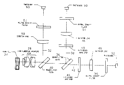
Referring first to FIG. 1A, the illumination optical system
is generally identified by the reference numeral 10, and incor-
porates a helium-neon laser 12 that emits a 2 mw beam of light
at 633 nm. The beam is folded by two reflecting mirrors 14 and
16 that provide adjustment of the laser beam position. The
adjustment enables the beam azis to coincide with the physical
optical azis of the illumination optics. The beam is then
shaped by the pair of cylinder lenses 18 and 20 into a 192 z 77
~tm elliptically shaped beam (at the 1/e2). The 192 ~m
-28-
MS-1717
~'~'~"~94
dimension is formed by the 150 mm focal length cylinder lens 18,
and it illuminates the long azis of the A-1 aperture 22 (which
is parallel to the plane of the page, in FIG. 1A). The 77,um
dimension is formed by the 60 mm focal length cylinder lens 20,
and it illuminates the short azis of the A-1 aperture. The A-1
aperture is 653 z 89 pm. The illumination doublet 23 produces
an elliptically shaped Gaussian intensity distribution of 37.4
x 12.6~um in the flowcell 24. The minor azis of the ellipse is
parallel to the direction of flow, which is vertical, i.e. in
the direction of arrow 26.
Cells that pass through the measuring volume scatter and
absorb the incident radiation. The light scattered and absorbed
is captured and measured in the detection optics illustrated
schematically in FIG. 18. The unscattered light and the light
that is scattered up to 19.5° is collected by the high
numerical aperture (Hi-NA) lens 28 and collimated. The beam is
divided into two parts by the 30/70 (30% reflection, 70%
transmission) beamsplitter 30. The beam 32 is reflected onto a
photodiode, and is used for the absorption measurement, while
the transmitted beam 34 is further split by the 20/80 (20%
reflection, 80% transmission) beam-splitter 36 to make the two
scatter channels. The reflected scatter channel 38 has a
5-15o darkstop 40, while the transmitted channel 42 has a
2-3o darkstop 44. The light passing through each of these
-29-
MS-1717
darkstops 40, 44 is then focused down through lenses 46 and 48
onto photodiodes 50 and 52, respectively. Neutral density
filters 54, 56 and 57 are then used to reduce the light levels
at each photodiode to a level that is appropriate for the
standard detectors and preamplifiers.
The beam 32 is focused through lens 58 onto a detector/
preamplifier 60. The preamplifier output is proportional to
the optical power transmitted through the system. It collects
unscattered light and light that is scattered into angles of up
to about 19.50. Within this angular interval, about 98% of
the light scattered by sphered erythrocytes and reticulocytes
is collected.
The absorption channel of the commercially available
TECHNICON H'1 instrument is not optimized for measuring cellu-
lar absorption. The absorption signals are of the same level as
the noise on the absorption preamplifier. A mathematical model
of the absorption detection process was developed. This model
predicted that the signal would improve dramatically with a
decrease in the area of illumination by the laser in the flow-
cell. The size of the slit was reduced from a nominal 150 z 20
microns (on the TECHNICON H'1 system) to a nominal 40 z 20
microns to increase the signal to noise ratio by a factor of
3.75.
-30-
MS-1717
2071790
The signal (pulse height) from the absorption preamplifier
is between 20 and 50 millivolts. This is much smaller than the
signal processing electronics requires. A second gain stage was
added to the absorption preamplifier with a gain of about 25.
This brought the pulse heights up to about 1 volt.
The gain of the preamplifier circuit and optical density of
the neutral density filter in each scatter channel were chosen
to produce mean.pulse signal levels of about 2 volts at the
output of each channel when Technicon (TCN) Optical Test
Material (OTM. TCH T03-1?04) was.assayed. OTM consists of
sphered and hard fized red blood cells. This material is
commercially available from the assignee hereof, and is adapted
for use on the TECHNICON H'1 system. This then allows fine
adjustment of the overall gain in each-channel using a variable
gain amplifier in the post detection signal processing hardware.
A functional block diagram of the post detection signal
processing system..is shown in FIG. 1C. The system consists of
pre-amplifier 62, a variable gain amplifier 64, pulse height
analyzer 66, analog-to-digital converter 68, and data acquisi-
tion hardware (computer) ?0 and software.
The electronic systems for the system consist largely of
the 4Cyte*system available from Howard Shapiro, M.D., P.C.,
-31-
MS-1?1?
201770
Cambridge, MA, (4Cyte Model FE Front End and 4Cyte Model I
Interface Card). The pulse-height analyzer, analog-to-digital
converter and data acquisition software are all components of
the 4Cyte system. These components produce held pulses repre-
senting the pulse heights for up to four input signals. and
allow setting of the 'valid" pulse height threshold level. The
4Cyte interface card is used in conjunction with the 4Cyte
software for analog-to-digital conversion of up to four input
signals, and the capture of those values in the RAM memory of
the host computer. The digitized signals are stored in list
mode. There are five eight-bit bytes of information for each
cell, one for each of the four parameters measured, and one for
flagging. The host computer for these ezgeriments was an IHM
PC/XT clone equipped with a color monitor ar<d a math
coprocessor. Data reduction can be performed on any IBM
compatible computer.
The following ezamples set forth reagent compositions and
methods incorporating the same for the identification of
reticulocytes and characterization of reticulocytes and red
blood cells using absorption flow cytometry techniques.
Standard commercially available reagent grade materials were
used whenever possible. It will be understood that the formula-
tions and the procedures which follow are provided for purpose
of illustration only. and that other ingredients, proportions
-32-
MS-1717
~_
and procedures can be employed in accordance with the disclo-
sures of this invention.
Example 1: Scatter and Absorption Measurements for Distinguish-
ing Reticulocytes and Erythrocytes Within a Blood
Sample Using a Reagent Composition and Method of
the Present Invention
Ozazine 750 dye was stored in a 1 mg/ml N, N-dimethylfor-
mamide stock solution. A working reagent was created by adding
the dye stock to a buffer solution containing the following
components at the concentrations noted:
O$azine 750 . ~ 6 ug/ml
Calcium Chloride 0.3 mM
Potassium Chloride 4.0 mM
Magnesium Chloride 88.0 mM
Sodium Phosphate (Tribasic) 0.5 mM
Sodium Bicarbonate 20.0 mM
The final osmolality and pH of the working reagent used in
this study were 272 mmol/kg and 8.1, respectively.
Samples were hand-mined in a manner which simulated the
automated TECHNICON H'1 system red cell sample processing
-33-
MS-1717
,.
scheme. Glass test tubes were filled with 5 milliliters of the
working reagent. Five microliters of a blood sample were then
pipetted into the reagent while the reagent was undergoing
agitation on a vortex miter. The 1:1000 dilution of blood was
then fed into the sample line of the flow cytometric apparatus.
In approzimately two minutes the sample passed through the flow
cell, and was then ezposed to a helium-neon laser source for
red cell and reticulocyte analysis. Each sample was measured
in duplicate if the sample volume permitted. Microscopic
ezamination revealed that most red cells and reticulocytes in
this mizture were partially sphered.
At the completion of the analysis, the raw data was dis-
played in the form of a Red Scatter v. Red Absorption cytograrn,
FIG. 2A. Distinct cell populations were clearly observed based
on their particular scatter and absorption signals. The ery-
throcyte population falls within Region A between the vertical
azis and vertical line X. These cells show high scatter signals
and low cell absorption signals. The major portion of the
reticulocyte population falls within the region to the right of
X, Region B. These cells are distinguishable from the mature
erythrocytes due to the higher absorption signals from their
Ozazir~e 750 stained RNA. The platelet population lies within
Region C below line Y, and the coincidence region lies within
-34-
MS-1717
Region D above line Z. The platelets have relatively low
scatter signals when compared to the reticulocytes.
Based on the absorption separation between mature erythro-
cytes and reticulocytes, the reticulocyte count of a patient
sample may be determined by creating electronic "windows" which
define the ranges of scattered light and absorption which
identify reticulocytes and erythrocytes. The number of reticu-
locytes and mature erythrocytes falling within each "window"
are determined so that the percentage of the reticulocytes and
erythrocytes present in the total cell population is then
calculated. In FIG. 2A(1), the reticulocyte "window" is~
determined by Region B, and the mature erythrocyte "window" by
Region A. Note, in FIG. 2A(2) and in all following scatter/
scatter cytograms, the non-linear grid overlaps indicate the
loci of constant volume and constant refractive indez for
perfect spheres according to the above-noted method of Tycko.
The reference percentage of reticulocytes in each sample was
determined using the manual microscopic procedure recommended by
the National Committee for Clinical Laboratory Standards
(NCCLS). In this procedure, a small volume of the sample was
prepared. and the percentage of reticulocytes in the sample was
counted with the aid of a microscope. The microscope was
equipped with a 100X oil immersion objective and a lOX ocular.
-35-
MS-1717
A minimum of 1000 cells were counted for each sample. A Miller
disc was inserted in the ocular of the microscope to improve
counting precision. Any red cell containing two or more parti-
cles of blue material after staining was labeled a reticulocyte.
The reticulocyte count of a patient sample was measured as
2.3% by this flow cytometric technique. The same blood sample
was also analyzed by the NCCLS method. The result was a
reticulocyte count of 1.7%.
A second experiment was conducted to discriminate between
reticulocyte and erythrocyte populations when cells were stained
with New Methylene.Blue and measured by the scatter/absorption
'flow cytometer. The buffer formulation was the same as that for
the reagent composition containing Oxazine 750. The concentra- -
tion of New Methylene Blue dye in the working reagent. which
replaced the Oxazine 750, was 60 ~g/ml. The sample preparation
and analysis protocols as described above were followed.
However, microscopic eaamination revealed that the red cells
and reticulocytes were less sphered than in the Oxazine 750
mixture. The raw data from the analysis was displayed in the
form of a Red Scatter v. Red Absorption cytogram {FIG.~2B(1)).
Note, in comparison to FIG. 2A(1), both the absorption and
scatter signals are more spread out, presumably due to
orientational noise. Based on the absorption separation
-36-
MS-1717
2 ~'~ 7'7 9 ~
between erythrocytes and reticulocytes, the reticulocyte count
of the patient sample was measured as 2.2%. When analyzed by
the NCCLS method, a reticulocyte count of 1.7~C was obtained.
Eaamn~ 2: Scatter and Absorption Measurements for Distinguish-
ing Reticulocytes and Erythrocytes Within a Blood
Sample Using the Reagent Composition of Ezample 1
Containing a Zwitterionic Surfactant
A second set of ezperiments was conducted utilizing the
reagent compositions of Ezample l, but further including a
zwitterionic surfactant to isovolumentrically sphere the red
blood cells and reticulocytes.
For each experiment, a working reagent was created by adding
to the reagent composition the surfactant, lauramidopropyl
betaine so that the final concentration of the surfactant in the
reagent was 63 pg/ml.
The sample preparation as described above with regard to
Ezample 1 was followed.
When viewed through a microscope, the mature red cells and
reticulocytes in a prepared sample were found to be perfectly
-37-
MS-1717
~~~~~9~
sphered and the reticulocytes stained. Note the difference
between FIGs. 8A and 8H and 2 and 3 which demonstrates
increasing reduction in orientational noise with increasing
completeness of sphering.
FIG. 3A(1) demonstrates the higher degree of discrimination
between reticulocyte and erythrocyte populations when cells were
stained with the reagent composition containing the Ozazine 750
dye and above-noted surfactant. Note that in FIG. 8C, an
unstained control, Region B is devoid of cells.
The reticulocyte count of a patient sample was measured as
8.0% by this technique. The same blood sample was also
analyzed by the NCCLS method. The result was a reticulocyte
count of 9.1%.
FIG. 38(1) demonstrates the degree of discrimination between
reticulocyte and erythrocyte populations when cells were stained
with the reagent composition containing the New Methylene Blue
dye and above-noted surfactant.
The reticulocyte count of a patient sample was measured as
5.0% by this technique. The same blood sample was also
analyzed by the NCCLS method. The result was a reticulocyte
count of 9.1%.
-38-
MS-1717
Example 3: Correlation Study with the Reagent Composition and
Method of the Present Invention and the NCCLS
Reference Method Using Absorption Data Corrected
for Pseudo Abosrption
The detection optical subsystem collects both the scattered
and unscattered light from cells passing through the laser beam
in the flowcell. -Cells scatter light into all directions. The
relatively Hi-NA lens in the optical system, which is described
above, accepts the light that is scattered into a cone that is
centered on the optical axis with a half angle of up to 19.5
degrees. Thus, the light that is scattered into angles greater
than 19.5 degrees is lost. As a result, when attempting to
measure cellular absorption, completely non-absorbing cells
"appear" to absorb up to a few percent of the incident light
(pseudo-absorption). The measured absorption can be
represented as follows:
Absorption - . Pseudo- + Hemoglobin + Dye
Signal Absorption Absorption Absorption
The pseudo-absorption signal of a mature red blood cell is
typically of the same magnitude as the actual absorption signal
from a stained reticulocyte. This reduces the degree of separa-
tion of the stained reticulocytes from the unstained red blood
cells on the absorption cytogram. The signal to noise ratio of
-39-
MS-1717
2~'~~?~~
the absorption channel can be improved by correcting the signal
to remove the pseudo-absorption and hemoglobin absorption compo-
vents from each red cell and reticulocyte absorption signal.
The amount of pseudo-absorption and hemoglobin absorption can
be calculated for any given cell by using the well-known Mie
light scattering theory described in the aforenoted Tycko
patent. The scattering cross-section for the angular interval
19.5° to 180° plus-the hemoglobin absorption component,
S3, can be calculated as follows:
S3 - ~ a2 Qext S ( ~ ' ns' e3' ~'v~ HC)
where a is the radius of the sphered cell, is the excitation
(or illuminating) wavelength, ns.is the refractive index of
the sample stream and sheath, Qext is the extinction
efficiency of the cell, and for the case of pseudo-absorption,
3=0°, and 113=19.5°. S3 values have been tabulated
for all expected values of v and HC.
The pseudo-absorption correction is made as follows: The V
and HC must first be determined from the two scattering signals
from a cell from the scatter-scatter cytogram as described in
Tycko. S3 is then found in the look-up table entry for the
measured V and HC, and subtracted from the value measured by
the absorption channel. The result is the actual absorption
-40-
MS-1717
~~"~'~'~9~
due to staining of the cell. The measured absorption signal
can be adjusted using the following relation to leave only the
dye absorption for each cell:
w
Dye - Absorption - Hemoglobin - Pseudo
Absorption Signal Absorption Absorption
- Absorption Signal - 53
For all data,-the adjusted value is substituted for-the raw
data parameter prior to thresholding, and flagging. Any objects
whose red scatter parameters do not appear on the V-HC rnap are
ignored in the data analysis scheme. This data is then redis-
played with the red scatter v. absorption cytogram reflecting
the corrected values as shown in FIGs. 5A and 5B.
A study was conducted to compare the performance of Ozazine
750 when used in a reagent composition in the scatter/absorption
flow cytometer with the NCCLS manual method. Blood samples were
stained with the reagent composition. Reticulocytes in the same
set of blood samples were also counted using the NCCLS method.
The sample preparation and analysis protocols were the same
as those described with regard to Ezample 2, ezcept for the
additional pseudo-absorption correction.
-41-
MS-1717
~ ~'~'~'19 Q
When viewed through a microscope, the mature red cells and
reticulocytes in a prepared sample were found to be perfectly
sphered and the reticulocytes stained.
The percentage reticulocyte counts obtained f rorn these two
methods are compared in FIG. 6. At a concentration of 2,vg
Osazine 750/m1 in the reagent composition, close correlation
was shown to ezis~ between the measurements using the reagent
composition and flow cytometric apparatus of FIG. 1, and those
obtained by the NCCLS reference method. The correlation
coefficient for the measurements as obtained by orthogonal
regression analysis was 0.92.
Example 4; Correlation Study with the Absorption Flow Cytometer
and the TECHNICON H'1 Reference Method Using
Absorption Data Corrected for Pseudo-Absorption
After pseudo-absorption correction and appropriate gating,
the erythrocyte and reticulocyte indices. MCV and MCHC were
separately determined and compared between the values obtained
using the reagent composition of the present invention in the
scatter/absorption flow cytometer and the TECHNICON H'1
measurements. FIGs. 4A and 4B show the correlation data for
total red blood cell MCV and MCHC, respectively.
-42-
MS-1717
FIG. 7 shows reticulocytes marked as "+" in a HC vs. V
cytogram.
Some advantages of the present invention evident from the
foregoing description include a reagent composition and method
for preparing a whole blood sample for electrooptical simul-
taneous quantitation of the volume, hemoglobin content and
hemoglobin concentration of reticulocytes and erythrocytes by
absorption flow cytometric techniques.
In view of the above, it will be seen that several objects
of the invention are achieved, and other advantageous results
obtained.
As various changes can be made in the above constructions
and methods without departing from the scope of the invention,
it is intended that all matter contained in the above descrip-
tion, or shown on the accompanying drawings, shall be inter-
preted as illustrative, not in a limiting sense.
-43-
MS-1717