Note: Descriptions are shown in the official language in which they were submitted.
CA 02081559 2001-11-21
DETERMINATION OF ANALYTES IN BIOLOGICAL FLUIDS IN THE
PRESENCE OF SUBSTANCES INTERFERING WITH ASSAYS THEREFOR
Technical Field
s The present invention relates to the determination
of analytes in the presence of substances interfering with
assays for such analytes. The invention also relates to the
use of devices for implementing such determination. The
invention can be used for the detection, in a biological
to fluid sample, of analytes in targeted lipoprotein classes;
and targeted isozymes of creatine kinase, lactate
dehydrogenase, amylase, and alkaline or acid phosphatases;
and targeted immunoglobulins; as well as other analytes.
15 Background Art
A. General
Modern medical practice typically requires routine
clinical tests of sera and urine for biological analytes such
as cholesterol, enzymes (such as creatine kinase, lactate
2o dehydrogenase, acid phosphatase, alkaline phosphatase,
amylase, etc.), immunoglobulins, as well as other substances.
More specific (and, typically, more time-consuming)
diagnostic tests are also performed in addition to routine
tests. For example, the detection of certain isozymes of
2s acid phosphatase is used clinically as an indicator of
prostatic cancer as well as various leukemias. Levels of
certain isozymes of alkaline phosphatase detected in a blood
or serum sample serve as an indicator of bone and liver
metabolic activity. Levels of pancreatic specific amylase
3o are an indicator for pancreatitis. Serum levels of the MB
isozyme of creative kinase (CKMB), as well as levels of
isozymes of lactate dehydrogenase, are indicators of
myocardial infarction (Noel, S., et al. "Enzymes" In Clinical
WO 91/17441 PCT/US91/02919
2 -
Chemistry (Kaplan, L. and Pesce, A., eds. The C.V. Mosby
Company, St. Louis, MI). pp. 454-483 (1989)). Similarly,
the detection of cholesterol, in specific lipoprotein
classes, is used in the determination of coronary heart
disease risk. (Russel et al. "Lipids" in Clinical Chemistry
(Kaplan, L. andPesce , A., eds. The C.V. Mosby Company, St.
Louis, MI): pp. 968-1004 (1989)). These more specialized
tests are often directed°to a specific class of analyte that
is already the subject"of routine tests.
The efficacy of assays for analytes in a biological
fluid sample can be reduced due to the presence of
substances which interfere with the assay (Kaplan, L. and
Pesce, A., "Interferences in Chemical Analysis" in Clinical
Chemistry (Kaplan, L. and Pesce, A., eds., The C.V. Mosby
Company, St. Louis, MI): pp. 808-819 (1989)). For example,
compounds such as hemoglobin or bilirubin, which have a
strong visible absorbance, can interfere with a
spectrophotometric assay for an analyte. Kaplan and Pesce,
id.
Clinical testing, in the case of both routine and more
specialized tests, demands strict adherence to carefully
developed quality assurance and quality control procedures
in order to assure accuracy and to minimize variability of
test results. Concerns over variability and inaccuracy of
test results have in fact led to further regulation of
clinical laboratories by the Health Care Financing
Administration of the U.S. Department of Health and Human
Services. 53 Federal Register 29590-29632 (Friday, August
5 , 1988) (proposed amendments to 42 CFR part 74 et seq.); 55
Federal Register 9538-9610 (Wednesday, March 14, 1990)
(revision of laboratory regulations, final rule with request
for comments). These new regulations impose additional
burdens on clinical testing laboratories. Such laboratories
thus have a need for testing procedures that can be readily
verified for adherence to quality control standards. The
more specialized tests (as opposed to the routine tests) may
readily permit verification, but the inherently
WO 91/17441 PCT/US91/02919
,.. _ 3
Q ~ ~'
sophisticated nature of these tests requires mastery by the
laboratory technician of a set of testing'protocols entirely
a different from those used in connection with routine tests.
The result is that quality control of such specialized tests
0 5 typically requires more extensive laboratory procedures and
training of laboratory personnel.
An additional complication is posed in the
interpretation of test results, even assuming that there is
good quality control from one test run to another. For
example, because of the important diagnostic information
gained from cholesterol results and the need to eliminate
interlaboratory variability,
uniform cholesterol cutpoints based on national population
studies have been adopted. Additionally, a national
reference system for cholesterol has been developed so that
cholesterol measurements are standardized and values are
therefore traceable to the National Reference System for
Cholesterol. Due to the absence of accepted National
Reference Systems for triglycerides, lipoproteins, and
apolipoproteins, much remains to be done in the
elimination of interlaboratory variability associated with
these lipid related tests. .Presently, these tests and other
specialized tests for cholesterol may not be directly
related to the National Reference System for Cholesterol.
With this discussion as background, the remainder of
this Background Art section discusses cholesterol
determination, as an example of the state of the art in the
detection of analytes in biological fluids. The prior art
known to the inventors lacks an assay; for cholesterol in
specific lipoprotein classes, that is simultaneously (i)
easily interpretable from an epidemiological point of viek;
(ii) easily, quickly and inexpensively implemented, and
(iii) universally applicable to all routine clinical
chemistry testing systems. Indeed, the inventors are
unaware of any assays, for specific classes of an analyte
that are the subject of the routine tests described above,
that meets these two criteria.
WO 91/17441 PCT/US91/02919
B. Cholesterol
_Biochemical Background
Triglycerides and cholesterol are transported in the
blood via lipoprotein particles. Abnormalities in these
lipoproteins, either inherited, environmentally contributed,
or a combination of both, lead to a variety of disorders
including a predisposition to premature coronary heart
disease (CHD) and atherosclerosis (N.I.H. Publication Number
88-2925 (1988); and Schaefer and Levy, The New England
Journal of Medicine 312:1300-1310 (1985)). The underlying
cellular and genetic mechanisms of many of the disease
states have been intensively and elegantly explored in the
preceding 30 years (Brown and Goldstein, Science 232:34-47
(1986), and Lusis, J. Lipid Research 29:397-428 (1988)).
The chemistry, biosynthesis, function, metabolism, cell
biology, and molecular genetics of lipoprotein particles
have been extensively reviewed (Segrest, Jere P., and
Albers, John J., editors, 128 Methods in Enzymology (1986)
and Albers, John J., and Segrest, Jere P., editors, 129
Methods in Enzymology (1986)).
Lipoprotein particles are divided into four major
classes based on their density, composition, and
electrophoretic mobility: The classes are chylomicrons, very
low density lipoproteins (VLDL), low density lipoproteins
(LDL) and high density lipoproteins (HDL). LDL and HDL
particles may be further subdivided on the basis of density.
The lipoprotein particles are composed of triglycerides,
cholesterol, fatty acids esters of cholesterol, phospholipid
and protein. The varying ratios of protein to lipid, in
different lipoprotein classes, account for the physical
differences by which these particles can be fractionated by
density gradient centrifugation.
The protein components, known as apolipoproteins, are
responsible for a variety of cellular functions. Increased
levels of LDL cholesterol and decreased levels of HDL
cholesterol have been shown to be risk factors for CHD.
Consequently, clinical diagnostic assays for cholesterol
WO 91/17441 PCT/US91/02919
-
content in the major lipoprotein classes are performed
extensively and a large body of statistical data on the
normal ranges for these classes is available standardized by
the Centers for Disease Control (McNamara and Schaefer,
5 Clinica Chimica Acta 166:1-8 (1987)).
_Cholesterol Determination
In the clinical laboratory, the following assays are
performed routinely to characterize the lipid and
cholesterol profile of a plasma or serum sample: (l)
Triglycerides are determined using the enzyme lipase(s) plus
enzymes linked to a color indicator system, (ii) Total
cholesterol is determined enzymatically using cholesterol
esterase, cholesterol oxidase and other enzymes and reagents
which translate the oxidation of cholesterol into a
detectable color change, and (iii) HDL cholesterol is
determined enzymatically as for (ii) above in the
supernatant of a sample following selective precipitation of
the VLDL and LDL fractions using a mixture of polyanions,
e.g. sulfated polysaccharides, or phosphotungstate and
divalent cations (Burstein et. al., J. Lipid Research 11:583
(1970): and Mulder et. al. Clinica Chimica Acta 143:29-35
(1987)). VLDL and LDL cholesterol (VLDL~.C, LDL.C) are
measured indirectly using the Friedewald equation
(Friedewald et. al., Clin. Chem. 18:499-502 (1972)):
Total cholesterol = HDL.C + VLDL.C + LDL.C
LDL.C = Total.C - (YLDL.C + HDL.C)
LDL.C = Total.C - (Triglycerides/5 + HDL.C)
The equation assumes (l) that no chylomicrons are
~ present, for example, in a blood sample from a fasting
patient, and (ii) that there is a constant relationship
between cholesterol and triglycerides: This is known to be
untrue in hypertriglyceridemic conditions (Cohn et. al.,
Clinical Chemistry 34:2456-2459 (1988): and Rao et. al.,
Clinical Chemistry 34:2532-2534 (1988)).
WO 91/17441 PCT/US91/02919
- 6 -
Thus the above analytical procedures suffer from
several disadvantages. (i) The VLDL.C and LDL.C are not
measured directly but rather are estimated using a formula.
(ii) The Friedewald formula is known to be imprecise under
conditions of clinical relevance i.e. elevated triglyceride
levels (>400 mg/100 ml) (Cohn et. al., Clinical Chemistry
34:2456-2459 (1988); and Rao et. al., Clinical Chemistry
34:2532-2534 (1988)). (iii) HDL.C determination relies on
the selective precipitation of VLDL and LDL particles by a
l0 polyanion, or by phosphotungstate, plus divalent cations,
with subsequent total cholesterol measurement of the
separated supernatant. In general, cholesterol detection in
specific lipoprotein classes lacks a standardized reference
system.
Mulder et. al: (Clinical Chimica Acta 143:29-35 (1987))
report on the direct measurement of cholesterol in
redissolved LDL precipitates but such measurements are not
performed in routine diagnostic surveys.
More recent efforts toward separation and quantitation
of lipoprotein classes have utilized antibodies., either
polyclonal or monoclonal, directed against apolipoproteins
which are specific to distinct, clinically relevant
lipoprotein particles (Tikkanen et. al., J. Lipid Research
24:1494-1498 (1983): and Ordovas et. al. J. Lipid Research
28:1216-1224 (1987)).
In research laboratories a variety of immuno-based
analytical techniques have been employed to quantitate
lipoproteins, including radial immunodiffusion,
radioimmunoassay and electroimmunoassay, but these
techniques are too cumbersome to be employed in a clinical
diagnostic setting where large numbers of samples must be
handled rapidly. This disadvantage may be addressed by
using an enzyme-linked immunoabsorbant assay (ELISA), and
this is an area of active investigation (Ordovas et. al., J.
Lipid Research 28:1216-1224 (1987)).
However, there is a further disadvantage which some of
these immuno-based techniques, including ELISA suffer, and
WO 91/17441 PCT/US91/02919
- ~ -
that is that these procedures quantitate an epitope
associated with specific lipoprotein classes - they do not
measure cholesterol levels. The significance of this
situation is that there must be a very large number of
samples analyzed by an immuno-based procedure to establish
its correlation with cholesterol values (measured
enzymatically) and which are interpretable
epidemiologically. Thus, ELISA-based tests require a long
lead time to gain acceptance in the clinical diagnostic
industry.
Methods which utilize immobilized antibodies to measure
levels of substances in biological fluids are known.
Longenecker (U.S. Pat. No. 4302536 (1981)) reported the
determination of antigenic materials in biological fluids
and cells by colorimetric immunoassay with an adduct of
antibody and chromo-protein. Onishi and Ito (Eur. Pat. No.
327,918 (1989)) reported an immunoassay using the
homogeneous competitive reaction between a target and
labelled substance and a specific binder. Freytag and
Ishikawa (U. S: Pat. No. 4,657,853 (1987)) reported a high
sensitivity immunoassay using a polymeric enzyme-antibody
conjugate. Nippon (Jap: Pat. No. 59226864 (1984)) reported
an immunoassay in which levels of transforming growth factor
(TGF) in a liquid sample are detected using an immobilized
TGF antibody and an enzyme labelled TGF antibody. Gomez and
Wicks (U. S. Pat. No. 4353982 (1982)) report an immunoassay
for creative kinase in blood serum using iodine-125 labelled
antibody to precipitate immune complex mixtures.
Several groups have examined selective
immunoprecipitation of specific lipoprotein classes followed
by cholesterol quantitation in the lipoprotein class
remaining in solution. Heuck et al. reported the use of
antibodies to ApoB to precipitate LDL and VLDL followed by
measuring cholesterol levels in the HDL left in the
supernatant. Antibodies to apoAI and apoC were also used,
to precipitate HDL and VLDL, followed by determination of
cholesterol levels in the LDL left in the supernatant.
WO 91/17441 PCT/US91/02919
- 8 -
(Heuck et al. Clin. Chem. 31: 252-258 (1985)). Kerscher et
al. reported the use of antibodies to HDL to precipitate HDL
and VLDL, followed by centrifugation to separate the
precipitate, followed by analysis of cholesterol levels, or
other component levels, in the LDL in the supernatant
(Kerscher et al. U.S.P. 4,746,605 (1988): Fed. Rep. Germany
Patent No. P32 15 310 (1983): Kerscher et al. Clin. Bioch.
18:118-125 (1985)). Antibodies to both apoproteins and
whole lipoproteins, including immobilized antibodies, have
20 been used to immunoprecipitate lipoproteins followed by
determination of the cholesterol content of the lipoprotein
class remaining in solution (Ziegenhorn et al. Canadian
Patent No. 1 211 707 (1986)). This reference, however, does
not describe any specific structure or device on which the
antibodies are immobilized.
Summary of the Invention
The invention disclosed herein provides in one
embodiment a method for detecting an analyte in a biological
fluid sample in the presence of a substance interfering with
an assay for the analyte. This embodiment is implemented by
using antibodies to cause the selective immunoprecipitation
of at least one of the analyte or the interfering substance
and then conducting an assay for the analyte in at least one
of the immuno-reactants or the non-reactants. In another
embodiment, the invention provides a disposable reaction
device to implement the method.
The invention is applicable to the detection of a wide
variety of biological analytes, including but not limited to
cholesterol in a targeted lipoprotein class in the presence
of cholesterol in another class; as well as to targeted
isozymes of creatine kinase, lactate dehydrogenase, amylase,
alkaline or acid phosphatase in the presence of non-targeted
isozymes; and targeted immunaglobulins in the presence of
non-targeted immunoglobulins.
2081559
-8a-
According to a still further broad aspect of the present invention, there is
provided a method for measuring an amount of an analyte in a targeted class of
biological
molecule in a biological fluid in the presence of an interfering substance
from another
related class. The method comprises obtaining a disposable reaction device
having a
reaction chamber and a collection chamber. The reaction chamber is positioned
within the
collection chamber and contains stabilized antibodies for binding each
interfering
substance from the biological fluid. A biological fluid sample is added to the
reaction
chamber of the device for reacting with the stabilized antibodies and causes
selective
immunoreaction of each interfering substance with antibodies. The method
further
comprises the step of causing substantially all non-reactant material,
including the analyte
to be removed from the reaction device into the collection chamber through a
permeable
filter, the antibody bound interfering substance remaining on the filter in
the reaction
device. A clinical assay for the analyte on the non-reactant material
including the analyte
is then conducted.
According to a still further broad aspect of the present invention there is
provide a disposable reaction device for detecting an analyte in a biological
fluid in the
presence of a substance interfering with an assay for the analyte. The device
comprises a
reaction chamber into which biological fluid may be placed. Stabilized
antibodies to the
interfering substance are disposed within the reaction chamber. A permeable
filter is
provided for filtering components upon exit from the reaction chamber, the
filter being in
fluid communication with the reaction chamber. A collection chamber is
provided for
receiving non-reactant material including analyte. The reaction chamber is
disposed
within the collection chamber.
.~
W0 91/17441 PCT/US91/02919
- 9 -
Brief Description of Drawings
The foregoing features of the invention will be more
readily understood by reference to the following detailed
description taken with the accompanying drawings, in which:
Fig. 1 is a schematic illustration of the sequential
density gradient u1 racentrifugation eparation of the
lipoproteins used as antigens for obtaining antisera used in
Example 1.
Fig. 2 is a correlation of supernatant cholesterol
values between dextran sulfate and anti-ND-LDL sera
precipitation method over l9 human plasma samples, in
accordance with Example 1.
Fig. 3 is a graph showing between-run precision of the
immunoprecipitation method over eight human plasma samples,
in accordance with Example 1.
Fig. 4 is a graph showing the time course for the
precipitation of human plasma ApoB-containing lipoprotein
particles by freeze-dried anti-ND-LDL sera as determined by
supernatant cholesterol estimation, in accordance with
Example 1.
Fig. S is a graph similar to that of Fig. 4 but wherein
the precipitant is non freeze-dried antisera, in accordance
with Example 1.
Fig. 6 is a graph supernatant cholesterol and ApoB
levels in accordance with Example 1.
Fig. 7 is a vertical section of a reaction device in
accordance with the embodiment of the invention described in
Example 2.
Fig: 8 illustrates a variety of solid support matrices
for antibody immobilization in accordance with the
invention.
Fig. 9 is a vertical section of he inner reaction
chamber 74 of the reaction device 70 of Fig. 7.
Fig. l0 is a vertical section of the outer collection
chamber 72 of Fig. 7.
W091/1~~'' ' 2081559
PCT/US91 /02919
- 10 -
Fig. 11 is a perspective view of another embodiment of
a device in accordance with the invention as described in
Example 3.
Fig. 12 illustrates schematically detail of the
reaction device of Fig. 11.
Fig. l3 illustrates a variety of solid support matrices
for antibody immobilization in accordance with the
invention.
Fig. 14 is a vertical section of the collection-
reaction pipette 114 of Fig. 12.
Fig. 15 is a vertical section of the docking device 116
of Fig. I2.
Fig. 16 is a vertical section of the reaction device cf
Fig. 11 loaded in a work station.
Fig. 1? is a fully loaded sample preparation system
containing multiple units of the reaction device of Fig. 11
within a work station.
Fig. 18 is a cut away view of an embodiment of a
reaction device similar to that of Fig. 11, but with certain
enhancements.
Fig. 19 is a vertical section of the reaction pipette
202 of Fig. 18.
Fig. 20 is an exploded view of the filter unit 220 of
Fig. 18 with related components.
Detailed Description of Specific Embodiments
The invention provides, in one embodiment, a method for
5 the detection of an analyte in a biological fluid sample, in
the presence of a substance interfering with an assay for
such analyte, using immunoseparation technology. In the
invention, antibodies are used to cause selective
immunoreaction of the analyte or a substance interfering
10 with an assay for the analyte, and then the analyte is
detected by assay of one of either the immunoreactants or
the non-reactants. In one embodiment of the invention, a
reaction device is used to rapidly and inexpensively
implement the immunoreaction-separation. The antibodies
15 used in the immunoreaction may be freeze dried or used as a
2081559
- 11 -
preparation on a suitable carrier. A wide range of
suitable carriers and separation techniques for this
purpose are available. Thus the antibodies may be
found to the surface of an insoluble carrier whose
mass, density, surface area or charge will
facilitate separation of immuno-reactants from non-
reactants. Examples of high molecular weight
insoluble are microporous beads, latex particles,
magnetic particles, controlled pore glass, gel
matrices from cross linked dextran, cross linked
polysaccharides, or cross linked acrylamide,
microporous filters or membranes. Other suitable
insoluble carriers include a coiled strip or the
interior wall of the reaction device itself.
Antibodies may also be bound to soluble large Mt~1
polymers to effect a more readily precipitable
immune complex with the analyte. Examples of
suitable polymer carriers are polysaccharides,
proteins or polynucleotides. This approach can also
enhance the kinetics of the immunoseparation. The
immuno-precipitating antibodies may be either
polyclonal or monoclonal or mixtures of either or
both, provided they possess sufficient specificity,
lack of cross-reactivity, ability to quantitatively
absorb to a broad range of substrate concentrations,
and separability of the immunoreacted complexes.
Purification of an analyte a.n a biological
fluid sample using the invention permits the
enhancement of the efficiency of existing routine
diagnostic tests currently in clinical use. The
invention also provides a systematic way of making
these routine tests more specific; such specific
tests can replace specialized testing, procedures,
with the result that the invention assists in
achieving the quality control, cost reduction, and
availability of tests of specific analytes in a
class of analytes.
2081559
- 11a -
There are several advantages of the invention,
applied to cholesterol testing, over the prior art.
Embodiments of the invention may be used in
connection with the determination of cholesterol in
an immunoseparated lipoprotein class based on
conventional enzymatic
WO 91/1744 2 0 815 5 9 pL'f/US91/02919
_ 12 _ ,°~, ,
techniques. Because the invention permits utilization of
existing routine clinical tests (applied to the immuno-
separated lipoprotein class), the results of testing, in
accordance with a preferred embodiment of the invention, may
be directly related to llhe national reference system.
(Other immuno-based assays currently provided by embodiments '
of the invention "include apoliproprotein quantitation, for
which statistically significant clinical data, i.e., normal
ranges in the population, are not yet available.) Thus the
analytical data generated by practicing this invention may
be directly related to existing data bases, and may
foreshorten dramatically the lead time in bringing the
benefits of immunospecificity to the cholesterol diagnosis
industry. Furthermore, the invention allows for the design
of analytical regimens which will provide an internal
control on individual cholesterol measurements within the
regimen. For example, a plasma or serum sample may be first
analyzed for total cholesterol in the absence of any
separation of lipoprotein fractions. Subsequently, aliquots
of the original sample may be subjected to
immunoprecipitation by various specific antibody
preparations either sequentially or on separate aliquots of
the original sample. In each situation it is possible to
validate the testing regimen by summing the cholesterol
results of all fractionations and comparing this sum to the
unfractionated value. The use of antibodies in the
invention permits highly specific fractionation of
lipoprotein classes.
In the practice of the invention, mixtures of poly- and
monoclonal antibodies are feasible. The antibodies need not
be optimized for characteristics relevant to other immuno-
based techniques, e.g., good binding to plastic surfaces as
needed for ELISA procedures. In addition, assay in
accordance with this invention may be implemented with a
device containing stabilized antibodies, which allows rapid,
inexpensive, and efficient analysis. It is universally
applicable to all routine clinical testing systems.
WO 91/17441 PCT/US91/02919
- 13 - ~0~
The embodiments describe3 below are discussed
principally in the context of the detection of cholesterol
in specific lipoprotein classes. However, as shown below,
the invention is equally applicable to the detection of a
wide variety of other analytes.
A. Antigen Preparation
1 Narrow density lipoprotein fractions
Pooled human plasma in CPD, collected at the New
England Medical Center Blood Bank, was used for the
preparation of lipoprotein fractions for use as antigens.
The plasma, at an assumed density of 1.006 (g/ml), was
fractionated by sequential ultracentrifugation as follows:
8 x 38 ml plasma + 1 ml KBr at 1:006 density were
loaded into Beckman quickseal tubes and spun for 18-22 hr @
4~C, 45k rpm in a Ti60 rotor. Following centrifugation the
tubes were stored on ice and the 1.006 floating lipoprotein
fraction was sliced off the top of the tube, cutting close
to the lower interface. The 1.006 bottom fractions were
pooled and gravity filtered through a Whatman filter (grade
#1). The filtrate volume was measured and the density
adjusted to 1.03 solid RBr (vacuum oven-dried) according to
fonaula l:
(1) gKBR = Vi lDf - Di)
1 - (v. D f)
Vi = Initial volume:
Df = Final density
Di = Initial density
v - Partial specific volume of KBr
The second ultracentrifugation of plasma (now at 1.03)
density) was carried out under the same conditions as above
and the tubes, again maintained on ice post-centrifugation,
were sliced to remove the upper fraction. The 1.03 bottom
5 fractions were pooled, filtered as before and adjusted to a
density of 1.05g/ml according to formula 1. The third
ultracentrifugation run was performed under the same
conditions as above and the collected tubes were maintained
on ice prior to slicing off the top lipoprotein layer. This
10 top layer was sliced just at the lower interface; the 1.05
WO 91/17441 2 0 815 5 9 PCT/US91/02919
- 14 - ..
top layers were pooled and contain the LDL fraction referred
to as narrow density LDL (ND-LDL).
The 1.05 bottom fractions were pooled, filtered as
before and the density adjusted to 1.107 with KBr (formula
1). This materia::l underwent ultracentrifugation number 4
under the following conditions: 4°C, 50k rpm, 42-48hr in a
Ti60 rotor. The collected tubes were maintained as
described above and the upper lipoprotein fraction was
sliced off close to the lower interface.
The 1.107 bottom fractions were pooled, filtered and
the density adjusted to 1.19 (formula 1): this fraction
underwent ultracentrifugation number 5 under the same
conditions as run 4. The collected tubes were sliced close
to the lower interface of the top layer to yield a 1.19 top
fraction containing narrow density HDL (ND-HDL).
The fractionation of the lipoprotein particles through
sequential ultracentrifugation is summarized schematically
in Figure 1. The harvested narrow density fractions were
washed and refractioned by repeated ultracentrifugation.
the ND-LDL (1.05 top fraction) was respun at a density of
1.05 @ 4~C, 18-22hr, 45k rpm, in a Ti50 rotor; the top
fraction was collected, respun, collected and respun and
finally harvested and dialyzed vs. PBS. Typically, 20-25 ml
of refractionated ND-LDL is dialyzed into 3.0L PBS for 18 hr
at 4~C. The PBS formula is as follows:
lOx Stock = Soln. A - 80 g NaCl
2.0 g KCl
1.97 g CaCl2- 6H20
1.0 g MgCl2~ 6H20
0.01 g NaN~ in 1.0L
deionized HZO
- Soln. B - 11.5 g Na2HP04
2.0 g KH2P04
0.01 g NaN3 in 1.0L
deionized H20
Final PBS Soln.:100 ml Soln. A + 100 ml Soln. B
Bring to 900 ml with deionized HZO
Adjust to pH 7.4 with 1N NaOH
Adjust to 1.0L final volume
WO 91/17441 2 0 815 5 9 PCT/US91/02919
- 15 -
The ND-HDL fraction (1.19 top) was respun in an
ultracentrifuge at a density of 1.107 @ 4~C, 50k rpm, 48 hr
in a Ti60 rotors the top fraction was collected and respun
under the same conditions. Finally the upper fraction was
dialyzed vs. PBS: typically 10-15 ml',into 3.0L PBS @ 4~C for
18 hr.
The purity of the dialyzed narrow density fractions was
checked by electrophoresing an aliquot over a 4-22.5%
polyacrylamide gradient gel under denaturing conditions
to using the system described essentially by Laemmli (1970).
The protein content of each fraction was determined by the
standard methods of either Lowry or using the Biorad
reagents. The preparation of narrow density lipoprotein
fractions described above is an improvement on the method
described by Schumaker, V.N. and Poppione, O.L. (1986)
Methods in Enzymology, Vol. l28 pp. 155-170.
The yields of narrow density lipoprotein fractions
isolated in this manner are typically: from 266 m1 plasma,
31 ml of ND-LDL at 1.2 mg/ml protein and 12 ml ND-HDL at 2.5
mg/ml protein are obtained.
2 Purification of Agoproteins
Starting with purified ND-LDL, ApoB was isolated by
electrophoresis through a preparative 15% polyacrylamide gel
followed by exci ion of the separated ApoB band. Upon
completion of electrophoresis the gel was stained with
sodium acetate to visualize protein bands, as described in
E. Harlow and D. Lane, editors, Antibodies. A Laboratory
Manual (Cold Spring Harbor Press, 1988). The ApoB
containing gel region was excised using a scalpel or razor
and the polyacrylamide gel was homogenized by repeated
passages though progressively narrower gauge needles
according to the method described essentially in Antibodies,
ibid. The ApoB in homogenized acrylamide may be stored at @
4~C prior to immunization.
Starting with approximately 10 ml purified ND-HDL,
ApoAI and ApoAII were purified by chromatography over
Sephacryl S-200 essentially as described by Brewer, et al.
WO 91/17441 2 0 815 5 9 PCT/US91/02919
- 16 -
(1986), Methods in Enzymology 128:223-246. Fractions
containing separated ApoAI and All were quantitated for
protein by Biorad assay and electrophoresed through a
preparative 15% polyacrylamide gel under denaturing
conditions. Protein bands Were visualized, excised and
prepared for immunization as described above.
B. Immunization
1. Narrow density LDL fraction: One ml of a 1 mg
(protein)/ml solution mixed in an equal volume of Freund's
complete adjuvant was used to inject goats intramuscularly.
At roughly 2-4 week intervals the goats received booster
injections of first, 1 mg protein (equivalent) in incomplete
Freund's adjuvant, followed by 0.5 mg protein (equivalent)
in incomplete Freund's adjuvant.
Apoprotein B in homogenized polyacrylamide, prepared as
described above, was used to maintain high antibody titre
via injections of approximately 0.5 mg of Apo B in
incomplete Freund's adjuvant every 2-3 weeks.
2. Narrow density HDL fraction: 0.5 ml of a 5 mg
(protein)/ml solution mixed in an equal volume~of complete
Freund's adjuvant and was used for the primary goat
immunization. The first boost utilized 0.5 m1 of the 5
mg/ml solution in an equal volume of incomplete adjuvant and
the second boost used 50% of the above level of immunogen.
It is within the scope of the invention to use purified
ApoAI and ApoAII to immunize individual goats, and obtain
the corresponding antisera.
Similarly, it is within the scope of the invention to
use purified ApoCI-III to supplement the narrow density HDL
immunogen.
Furthermore, it is within the scope of the invention to
use purified ApoE both individually and as a supplement to
narrow density HDL to immunize goats, by standard
immunological techniques, and to obtain corresponding
antisera.
C. Antisera Characterization and Purification
WO 91/17441 ~ ~ ~ ~ PGT/US91/02919
,. _ 17 -
Approximately 2-10 ml of°serum were prepared from
initial test bleeds following booster injections. The
antisera was,characterized by Western blot (as described
below) against isolated VLDL, LDL, HDL, and whole human
plasma. By this method, the anti-narrow density LDL sera
Was shown to be free of material cross-reactive to any of
the apoproteins of purified HDL. The ND-LDL antisera showed
cross-reactivity only to ApoB in VLDL, LDL, and whole
plasma.
The anti-narrow density HDL sera showed reactivity to
ApoAI in HDL and whole plasma and reactivity to ApoCs and
ApoE in HDL, VLDL and whole plasma. A small amount of
cross-reactive material to ApoB was detected and can be
removed by immunoaffinity chromatography over an ND-HDL-
sepharose column. The column is prepared in the following
manner:
i) Ligand (HDL) Coupling
Weigh out 3g freeze dried CNBr-activated Sepharose-4B
(Pharmacia) powder. Resuspend powderin 11.0 ml imM HC1 in
a 50 ml centrifuge tube. Wash gel for 15 minutes with 1mM
HC1 on a scintered glass filter (slow drip using the vacuum;
use 200 ml/g powder). Dialyze the ligand (IiDL) in coupling
buffer (0.1M NaHC03, 0.5M NaCl, pH 8.3: 10-20 ml in 2.0L
Buffer, overnight at 4°C and mix with gel in a centrifuge
tube overnight on a rocker at 4°C (use 5 ml coupling
buffer/g powder).
ii) Glutaraldehyde Crosslinking
Sepharose-HDL beads are centrifuged at 2000 g for 15
minutes to settle beads or are passed through a scintered
glass filter to collect the beads, then the beads are
incubated for one hour with 4 vol of solution 2. The beads
are collected by filtration or sedimentation, the
supernatant is discarded, and the beads are incubated for 1
hour with 4 vol NaHC03 and glutaraldehyde. The unreacted
aldehyde groups are blocked by incubating coupled sepharose
in 4 vol. 1M Tris-HC1 (pH 7.8 overnight at 4~C).
WO 91/17441 ~ ~ PCT/LJS91/02919
- 18 -
The sepharose is then collected and washed with three
cycles of alternating pH, each cycle being: O.1M acetate
buffer pH 4.0, 0.5M NaCl followed by O.lM Tris pH 8.0, 0.5M
NaCl. The sepharose is then suspended and coupled with
Buffer A and incubated overnight at room temperature. The
sepharose is then collected and resuspended in Buffer A and
stored at 4~C.
Solutions:
I) NaHCO , 0.25 M, pH 8.8
2) 0.015 Glutaraldehyde in Solution 1.
3) 1 M Tris HC1, pH 7.8
4) Buffer A
lOmM KPOi
150mM NaCl
1mM EDTA
0.1% NP40
ND-HDL IgG was affinity purified over this column
essentially by the procedures described by McConathy, W.J.
et al. (1985), Cuatrecasas, P. (1970) and Kowal, R., &
Parsons, R.G. (1980).
Antisera was characterized further by examining
immunoreactivity against lipoprotein particles that had been
electrophoresed through an agarose gel under non-denaturing
conditions using the Corning Agarose Universal
Electrophoresis System6. Following electrophoresis, 10-20
ml of antisera (control and sample antisera were tested
separately) were loaded into the vertical wells in the gel
and incubated for approximately 18 hours at ambient
temperature in a sealed moist environment. The gel was
examined for the presence of opaque precipitin lines to
determine the specificity of the sample antisera versus
control antisera. The gel was equilibrated in PBS by 1-2
soakings in approximately 200 ml PBS at room temperature
with gentle agitation. The gel was the air-dried for 16-20
hours at ambient temperature and stained for protein by
Coomassie Blue.
The anti-ND-LDL sera showed cross-reactivity with the
Beta migratory lipoprotein region only: no reactivity was
WO 91/17441 PCT/US91/02919
- 19 -
detectable either to the alpha migrating region or to
albumin.
Antisera was delipidated and the IgG fractions further
purified using a combination of standard techniques
a 5 including ammonium sulfate precipitation,
ultracentrifugation, and affinity chromatography either over
a protein A or over the appropriate immunoaffinity material
(i.e.: ND-HDL or ND-LDL) as described above.
ND-LDL antisera purification was monitored by Western
blot by the following procedure. The lipoprotein particles
were electrophoresed through an SDS-PAG described above
followed by electrotransfer to nitrocellulose (S & S) using
the following conditions: 40 volts for 16-18 hours; the
transfer buffer is: 20mM Tris pH 8.3; 150mM glycine, 20%
methanol. ND-LDL antisera was reacted with the
nitrocellulose sheet and cross-reactivity was detected using
a secondary antibody conjugated with calf intestinal
alkaline phosphatase followed by .incubation with a
phosphatase substrate-chromophore complex: color development
was observed visually. These procedures are essentially as
described 'in Vogel, et al. (1979) and Mason, et al.
(1978).
Finally, the anti-ND-LDL sera was freeze dried using a
Virtis Freezemobile 24 freeze drier at -55~C to -57~C to 65
millitorr overnight.
E
Example 1:
Immunoprecipitation of Beta lipoproteins in human
plasma using anti-ND-LDL sera was examined and compared with
the prior art. 200 ~t1 of fresh human plasma was added to a
1-.5 ml polypropylene conical tube containing the equivalent
of 50 JJl of anti-ND-LDL sera prepared essentially as above.
The contents were mixed gently and incubated at ambient
temperature for 30 minutes followed by centrifugation for 15
minutes in a Beckman microfuge at setting 12. The
supernatant was withdrawn and gravity filtered through a
cotton wool plugged pipette tip.
WO 91/17441 ~ ~ PCT/US91/02919
- 20 -
The filtrate Was assayed for total cholesterol using an
Abbott ACA 200 chemistry analyzer and Abbott A-Gent
cholesterol reagents, although any cholesterol determination
method should give equivalent results. Fig. 2 depicts the
correlation of immunoprecipitated supernatant (FIDL)
cholesterol values (mg/dl) for 19 human plasma samples
compared with values obtained using dextran sulfate (50 kd
molecular weight) as a control Beta lipoprotein
precipitating agent, used essentially as described by
Warnick, et. al (1985). Fig. 3 indicates the between run
precision of the antibody immunaseparation method over 8
human plasma samples tested (in duplicate) on two separate
occasions. A separate batch of purified, freeze dried anti-
ND-LDL sera was used to examine the time course of the
immunoseparation reaction on a human plasma sample. Fig. 4
illustrates the time course results using the equivalent of
300 Ell of antisera in the reaction with 200 E!1 of plasma;
the dextran sulfate control supernatant value was 58.5
mg/dl. This same batch of antisera was titred for
immunoprecipitation performance on human plasma prior to
freeze drying and the results are indicated in Fig. 5; on .
this plasma the dextran sulfate control value was 42 mg/dl.
Apoprotein immunoassays were used to evaluate the
comparative specificity of lipoprotein particle separation
between the immunoseparation method and the dextran sulfate
method. Fig. 6 demonstrates the immunoprecipitation of
lipoprotein particles by anti-ND-LDL sera monitored by the
reduction in supernatant (filtrate) cholesterol values (as
described above) and by Apoprotein B immunoassay measured by
the method of Ordovas, et al. (1987). At the equivalent of
350 Ell anti-ND-LDL sera, the supernatant cholesterol value
was 32.9 mg/dl, the dextran sulfate control value was 30.0
mg/dl. By immunoassay, the following results were obtained:
at 350 EJ1 of antisera the supernatant ApoB level was
undetectable above the background control; the dextran
sulfate supernatant yielded 1.2 mg/d1 ApoB remaining in the
supernatant.
WO 91/17441 2 p g i 5 5 9 PCT/US91/02919
- 21 -
Example 2:
As an alternative to the separation method of example
1, which employs centrifugation of the reacted sample
followed by supernatant removal and subsequent filtration, a
specialized reaction device may be used in accordance with
the invention to achieve both the immunoreaction and
separation. Fig. 7 is a schematic illustration of one
embodiment of such a device. Figs. 8, 9 and 10 illustrate
individual components of this embodiment. The device has
three components: (i) an outer collection chamber 72
(separately shown in Fig. 10) which collects the filtrate at
the completion of sample preparation, (ii) an inner reaction
chamber 74 (separately shown in Fig. 9) which is supported
(for example by a flange 71 forming part of the reaction
chamber; resting on the threaded shoulder 73 of the
collection chamber 72) within the outer collection chamber
72 and which has a suitable filter material 78 built into
its base, and (iii) an immobilized antibody component 76
which may, for example, be antibody-coated, or beads or a
coiled strip, such as represented in Fig. 8), which is
contained within the reaction chamber 74. Any other
suitable means for immobilization of the antibodies may be
employed, such as coating them on the interior surface of
the reaction chamber 74. The reaction chamber 74 includes a
cap 75 that is hingedly attached to the flange 71, so that
the reaction chamber 74 may be conveniently capped prior to
centrifuging as described below.
The immunoreaction-separation is implemented by the
device by pipetting a serum or plasma-or blood sample into
the reaction chamber 74 (Fig. 7), the cap 75 on the reaction
chamber 74 is closed, the sample is mixed with the
immobilized antibody and incubated to allow the
immunoreaction to occur. At an appropriate time the device
is centrifuged, so that the filter retains crude debris and
3~ the solid antibody support material, and the non-reactants
pass into the'collection chamber as filtrate. The reaction
chamber may be discarded following centrifugation and the
WO 91/17441 2 0 8 15 5 9 PCT/US91/02919
- 22 -
collection chamber may be capped (using a screw cap gripping
the threaded shoulder 73 in Fig. 10) or the filtrate or an
aliquot thereof may be assayed for non-reactant analyte(s),
using routine assays.
Examgle 3:
An alternative embodiment of a reaction device in
accordance with the present invention is depicted as item
110 in Figs. 11 - 15. Fig. 11 shows a docking assembly 116
of the device 110 in which the reaction pipette 114 of the
device is docked. The docking assembly includes a filter
118 at its bottom. The reaction pipette 114 contains
prepacked aliquots of antibodies immobilized, for example,
on beads or a coiled strip (Fig. 13), or otherwise in the
manner as discussed above in connection with Fig. 8. The
reaction pipette 114 may be fabricated as a single piece of
plastic or other suitable material, and includes, as shown
in Fig. 12, an upper flexible bulb region 121, a reaction
region 122, a locking position 126, and a port 123.
Squeezing and releasing the bulb region 121 can supply
positive and negative pressure differentials to the reaction
region 122 so that fluid may be alternately drawn into and
expelled from the reaction region 122 via the port 123.
Although a flexible bulb is here shown as the source of
positive and negative pressure differentials, other suitable
sources may be employed. The reaction pipette 114 is
employed by aspirating a sample of serum or plasma or blood
by means of the bulb region 121 (Figs. 12 and 14). The
docking assembly 116 (which may be a modified pipette tip)
is used to hold the collection-reaction pipette 114 during
the immunoreaction (Figs l2 and 15). The docking assembly
116 contains an inner ring of resilient material on its
inner surface 151, shown in Fig. 15 as a band around the
upper lip of the assesbly, to engage the locking position
126 of the reaction pipette 114; however, the resilient
material may cover a larger portion of the inner surface
thereof and may extend the entire length of the inner
surface or a suitable proportion thereof. At the base of
WO 91/17441 2 0 8 ~ 5 5 9 P~/US91/02919
- 23
the docking assembly 116 (FiS. 15), suitable filter material
118 is positioned, during or post-manufacturing, to retain
cells and crude debris.
Operationally, the reaction pipette 114 is designed to
aspirate a volume of liquid sample, by controlled
compression of the flexible bulb portion 121 (Fig. 14); the
aspirated sample is thereby brought into contact with the
immobilized antibodies 112 within the reaction pipette 114;
the reaction pipette 114 is then placed into the docking
assembly 116 (Figs. l2 and 15), where locking portion 126
engages against the inner ring 151. Mixing takes place,
and the device 110 is then placed in the work station 160
(Fig. 16) for an appropriate incubation period.
At the completion of the incubation period, the device
110 may be removed from the work station 160 and the non-
immunoreacted contents of the collection reaction pipette
114 are expelled from the pipette by compression of the
flexible bulb region 121, through the filter 118, into a
separate sample container which may be brought into position
beneath the docking assembly 116.
Figure l7 illustrates a fully engaged sample
preparation system, including a plurality of devices 110
oriented in a work station 160, which may be used in a kit
format for the preparation of samples for analysis. The
work station 160 and the device 110, and/or components of
the device may be color coded to aid'user recognition of
alternative sample treatment~systems.
Alternatively, the work station 160 may be modified so
as-to contain individual docking chambers into which
individual reaction devices may be suspended during
incubation and which may collect the filtrate from the
expirated devices within the work station. Following sample
expellation, the devices may be discarded and the collected
filtrate may be analyzed.
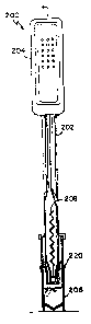
Fig. 18 shows an embodiment of a reaction device 200,
similar to that of Figs. 11-l7 but with certain
enhancements. The device 200 includes a reaction pipette
WO 91/17441 ~PCT/US91/02919
-
24 -
202, a filter assembly 220, and a sample cup 206. The
reaction pipette 202 (Fig. 19) contains aspiration bulb 204,
antibodies bound to a support matrix 208, a molded ring 210
which holds the immobilized antibodies in place in the
reaction pipette, and a fluid sample port 212 for entry and
exit of the biological fluid sample. The filter assembly
220, shown in an exploded view in Fig. 20, includes a wiper
224, a filter retainer 226, a filter 228, and a filter
carrier 230. Operation of this embodiment is similar to the
operation of the embodiment described above in connection
with Figs. 1l-17. The collected filtrate or immunoreactants
can then be assayed using routine tests.
The efficacy of the embodiments of the reaction device
illustrated above can be enhanced if one considers the
effective volume of the reaction chamber in relation to the
quantity of stabilized antibodies contained in the chamber.
In particular, the relationship should be such that there
are sufficient antibodies present to cause the target
analyte to be completely immunoreacted and then removed from
any anticipated biological fluid sample that may fill the
effective volume of the reaction chamber. In addition to
control of the effective volume of the reaction chamber by
limiting its physical size, the effective volume of the
reaction chamber may be limited in the embodiments of Figs.
11-20 by limiting the maximum volume that can be displaced
by squeezing the bulbs 121 or 204. The displacement can be
limited in turn by limiting the bulb size or imposing
physical constraints on the amount the bulb may be squeezed,
for example, by inserting a large solid object into the
bulb.
Although the foregoing discussion has been principally
directed toward devices containing immobilized antibodies,
it is only necessary that the antibodies be suitably
stabilized and contained within the reaction chamber at the
time of the immunoseparation reaction. Thus freeze dried
antibodies may be contained in a water-soluble or permeable
structure in the reaction chamber. Alternatively, the
WO 91/17441 PCT/US91/02919
,,, - 2 5
antibodies may be stored in « liquid suspension in a
container im fluid communication with the reaction chamber,
in such a way that they are put in contact with the
biological fluid sample when the device is used. The
container could be ruptured at the time of use, or the
reaction chamber can be designed to hold the suspension
sealed from the environment until the device is used.
Although the above discussion has been with respect
primarily to the detection of cholesterol in specific
to lipoprotein classes; the invention is also widely applicable
to the detection of a targeted analyte in a class of
analytes, such as targeted isozymes of an enzyme in the
presence of other isozymes and targeted immunoglobulins in
the presence of non-targeted immunoglobulins. For example,
the invention is applicable to targeted isozymes of creative
kinase, lactate dehydrogenase, amylase, and alkaline and
acid phosphatases. The invention may be implemented in a
manner similar to that described above in the case of
cholesterol testing, except that the antibodies used in the
reaction devices of Figs. 7 through 20 must be antibodies to
one of the targeted analyte or to the non-targeted analyte,
depending on how the assay is conducted following the
immunoseparation. The antibodies may be prepared using
methods known in the art.
It can be seen however that if the targeted analyte
is separated from the non-targeted analytes in the
applicable class of analytes and there exists a routine
_test for the class of analytes then following separation in
accordance with the invention the targeted analvte can be
assayed using the routine test for the class of analytes.
For example, one may use a routine test for amylase to
detect pancreatic specific amylase if the invention is
employed as a "front end" to the routine test. In other
words, embodiment of the invention may be employed to
separate pancreatic specific amylase from other isozymes of
amylase, achieving separation, for example, using antibodies
to all isozymes of amylase other than pancreatic specific
W0 91/17441 0 8 1 ~ 9 PCT/US91/02919
- 26 -
amylase. Thereafter the filtrate may be assayed using the
routine test to identify the level of pancreatic specific
amylase in the sample. A similar strategy may be used to
assay any targeted analyte in a class of analytes for which
a routine test exists.
The invention may also be used to remove substances
such as bilirubin and hemoglobin that can interfere with
spectrophotometric or other assays for an analyte. In such
instances, antibodies to bilirubin and hemoglobin may be
l0 employed to achieve their immunoseparation (using the
invention) from the sample prior to conduct of an assay in
accordance with prior art techniques.
It can be seen that the antibodies employed in the
invention need not be restricted to those for a particular
molecule, since any undesired substances may be immuno-
separated in accordance with the invention, as long as
undesired cross reactions are avoided.