Note: Descriptions are shown in the official language in which they were submitted.
20~1~~~
31688-O1
- 1 -
TITLE
MALLEABLE, BIOABSORBABLE, PLASTIC STAPLE; AND
METHOD AND APPARATUS FOR DEFORMING SUCH STAPLE
The present invention relates to surgical staples and
to a method and an apparatus, particularly an anvil for
a surgical stapling device, used to deform such staples
to secure adjacent layers of tissue together. More
specifically, this invention relates to the
configuration of malleable, bioabsorbable, plastic or
polymeric staples for suturing body organs and tissue,
and to a precision-formed anvil for deforming the
staples into that suturing configuration.
Gastrointestinal anastomosis-type devices drive and
bend the staples aligned in a row one after the other
in rapid sequence. Transverse anastomosis-type
devices, drive and bend all staples in a row
simultaneously.
One type of conventional staple 10, shown in Figure 1,
used with both gastrointestinal anastomosis and
transverse anastomosis-type surgical stapling devices
is made of a metal, like stainless steel or titanium,
that is substantially inert in the body. The
undeformed staple 10, or staple blank, is generally
20~1~~~
2 -
U-shaped and includes a back span 12 and two legs 14
depending perpendicularly from the back span in
parallel to one another. Each leg 14 has a sharp
chiseled end point 16 for cleanly piercing body organs
or tissue. The metal staple blank is bent by having
the legs engage and follow a conventional anvil to form
a B-shaped closed staple 18 as shown in Figure 2.
The anvil used to bend metal surgical staples is also
formed of a hardened metal and includes a staple-
bending face having a pair of coined or punched pockets
located to oppose each staple in the magazine of the
stapling device. The pockets are ordinarily elongated
arcuate depressions, co-linearly arranged in parallel
to the back span of a corresponding staple held in the
magazine. Thus the anvil closely resembles the anvil
of a conventional paper stapler.
When the staples 10 are driven from the magazine toward
the anvil, the staple legs 14 each engage one pocket so
that both legs are bent toward each other initially and
thereafter upwardly toward the back span 12. Thus, as
shown in Figure 2, the end points 16 may come to rest
against the underside of the back span 12.
Although metal staples inserted in the manner described
above provide an effective and relatively simple means
of suturing, one significant disadvantage is that they
remain in the patient's body permanently. While
generally not injurious to the body they may
nevertheless interfere with post-operative X-ray or
other diagnostic imaging of the patient.
This disadvantage can be overcome by using
bioabsorbable polymeric staples that are degradable in
the body after a short period of time. However,
conventional polymeric staples are not malleable and
2~~~.~54
- 3 -
thus cannot be easily bent into the B-shaped
configuration shown in Figure 2, to complete a suture.
Therefore, as shown in Figure 3, such conventional
bioabsorbable staples instead are made in two parts,
namely U-shaped polymeric staple body 20, the legs 22
of which are joined by a polymeric bar-like closure 24.
The closure has two end point-receiving holes 26 that
fit over the end points of the staple body 20 after
they have pierced the tissue to be sutured. The staple
body 20 and closure 24 are then forced toward each
other to complete the suture.
While this two-part staple will dissolve in the body
and, therefore, does not interfere with post-operative
procedures, it has the drawback of requiring a part in
addition to the basic staple blank and thus requires a
more complicated mechanical stapling device for
properly aligning the two parts and driving them
together.
More recently, a breakthrough has been made in the
bioabsorbable staple field. Specifically,
bioabsorbable or partially bioabsorbable surgical
staples have been developed using polymeric materials.
(Hereinafter the term "bioabsorbable" will be used
generically to describe surgical staples of the type
described in both of the applications mentioned above.)
These staples retain all of the beneficial attributes
of known bioabsorbable staples, but in addition are
malleable or plastically deformable like metal staples.
That is, these staples may be bent into complex shapes
that are then retained. Therefore, they may be made of
a single piece, not requiring independent staple body
and closure parts.
Nevertheless, it has been found that if these new
bioabsorbable staples are bent in the same way as are
CA 02081654 2002-11-21
74702-42
- 4 -
conventional metal staples, as shown in Figure 2, so that
the chiseled end points of the staple legs hit the back
span, the points may crush or break.
Therefore, further improvement in surgical staples
and devices for inserting them, taking advantage of the
attributes of the new polymeric materials described above,
are desirable.
The present invention enhances the benefits
obtained by using malleable, bioabsorbable, polymeric
staples in surgical stapling techniques. The present
invention provides a malleable, bioabsorbable, polymeric
staple deformed into a precise shape that securely joins
tissue sections together with minimal tissue injury and
damage to the staple itself. The present invention provides
a malleable, bioabsorbable, polymeric staple having a cross-
sectional shape that enhances tissue or clinching strength
once deformed. The present invention provides a high
precision anvil for surgical stapling devices that will
precisely and uniformly deform malleable, bioabsorbable,
polymeric staples, as well as other staples, into a desired
configuration. The present invention provides a method for
deforming the malleable, bioabsorbable, polymeric staple
into the desired shape. The present invention provides a
unique anvil that takes advantage of the
CA 02081654 2002-11-21
74702-42
- 5 -
beneficial properties of malleable, bioabsorbable,
polymeric staples of the type described above to in
turn provide an improved surgical stapling device.
These and other aspects are achieved by the malleable,
bioabsorbable, polymeric surgical staple of the present
invention, which in a preferred embodiment comprises a
back span, and first and second legs extending
generally in the same direction from opposite ends of
the back span, with the first and second legs having
first and second end points, respectively. The first
and second legs are deformed inwardly toward each other
and upwardly toward the underside of the back span such
that the end points of the respective legs extends past
opposite sides of the back span.
In another preferred embodiment, the present invention
comprises a method of deforming a malleable,
bioabsorbable, polymeric staple wherein in an initial
undeformed configuration the staple has a back span and
first and second legs each having an end point and each
extending in the same direction from opposite ends of
the back span substantially perpendicularly thereto.
The method includes the steps of initially deforming
the first and second legs inwardly toward each other,
and thereafter deforming the first and second legs
upwardly such that the first and second end points
extend past the back span of the staple on opposite
sides thereof.
The polymeric surgical staple of the present invention
also preferably has a noncircular oval or rectangular
cross-sectional shape that enhances its ability to
retain its deformed, tissue-joining configuration, as
will be described in detail below.
- 6 -
In accordance with yet another aspect, a preferred
embodiment of the invention is a surgical stapling
device anvil for forming a staple having, in an
undeformed state, a back span and first and second legs
extending in the same direction from opposite ends of
the back span substantially perpendicularly thereto.
The anvil comprises a supporting body having a
longitudinal axis and including a staple-receiving
face, that may confront the end points of the legs of
the staple. First and second pocket-like depressions
are formed in the supporting body and each begins with
an entry end located at the face, continues to a
depressed portion within the body below the face, and
terminates in an exit end at the face. The first and
second pocket-like depressions extend in non-collinear
relation with the entry end of each located
substantially on the longitudinal axis and the exit end
of each located on a side of the axis opposite the side
on which the exit end of the other depression is
located. The entry ends of the first and second
depressions are spaced by a distance substantially
equal to the distance between the depending legs of a
staple blank. Accordingly, a staple such as described
above driven toward this anvil will be deformed by
first bending the staple legs toward each other and
thereafter upwardly toward the back span. However, the
end points of the legs will be steered toward opposite
sides of the back span past the back span.
It will be appreciated, of course, that the surgical
stapling device anvil configured in accordance with the
present invention may be used with surgical staples of
any material. However, because it is specifically
designed for use with malleable, bioabsorbable,
polymeric staples that are non-metallic, it may be made
of plastic materials that are less expensive and in
CA 02081654 2002-11-21
74702-42
which the high precision pocket-like depressions may be more
easily formed than known hardened metal anvils.
In one embodiment, the invention provides a
deformed malleable, polymeric surgical staple, comprising: a
back span; and first and second legs extending in one
direction from opposite ends of said back span, each of said
first and second legs terminating in an end point; wherein
said first and second legs are permanently deformed inwardly
toward each other and toward said back span in a direction
generally opposed to the one direction with each of said end
points extending in the opposed direction toward at least
one side of said back span.
In a further embodiment, the invention provides a
deformable, malleable, polymeric surgical staple,
comprising: a back span; and first and second legs extending
in one direction from opposite ends of said back span,
wherein each of said first and second legs are permanently
deformed and terminate in an end point; wherein at least a
portion of said back span, said first leg, and said second
leg at which staple is deformable having a noncircular
cross-sectional shape.
In a still further embodiment, the invention
provides a method of deforming a malleable, polymeric
surgical staple having a back span, first and second legs
extending in one direction from opposite ends of the back
span and substantially perpendicularly thereto in an
undeformed state, the first and second legs each terminating
in an end point; said method comprising the steps of:
initially deforming the first and second legs inwardly
toward each other; thereafter deforming the first and second
legs toward the back span in a direction generally opposed
to the one direction to cause the end points respectively of
CA 02081654 2002-11-21
74702-42
- 7a -
the first and second legs to extend in the opposed direction
past the back span.
These and other aspects, features, and advantages
of the present invention will become apparent from the
following detailed description of the preferred embodiments
taken in conjunction with the drawings.
T1DTWTTT!'?C
Figure 1 is a front elevational view of a
conventional metal staple bank made, for example, of
stainless steel or titanium;
Figure 2 is a front elevational view of a
conventional staple in a deformed configuration;
Figure 3 is a front elevational view of a
conventional two-piece bioabsorbable polymeric staple;
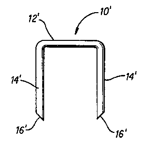
Figure 4 is a front elevational view of a
malleable, bioabsorbable, polymeric staple, which is not yet
deformed, in accordance with the present invention;
Figure 5 is a front elevational view of a
malleable, bioabsorbable, polymeric staple in accordance
with the present invention deformed to a shape for suturing
adjacent tissue sections together;
Figure 6 is a top plan view of the staple shown in
Figure 5 in its deformed state;
Figure 6A is a detailed cross-sectional view of
one particular embodiment of the staple shown in Figure 5;
2~~~~~4
_ g -
Figure 6B is a detailed cross-sectional view of another
embodiment of the staple shown in Figure 5;
Figure 7 is a schematic perspective view of a surgical
stapling device anvil formed in accordance with one
embodiment of the invention;
Figure 8 is a schematic top plan view of the surgical
stapling device anvil shown in Figure 7;
Figure 9 is a vertical cross-sectional view of the
surgical stapling device anvil shown in Figures 7 and
8, and taken on plane 9-9 in Figure 8.
Figure 10 is a schematic perspective view of a surgical
stapling device anvil formed in accordance with another
embodiment of the invention;
Figure 11 is a schematic top view of the anvil shown in
Figure 10;
Figure 12 is a vertical cross-sectional view of the
anvil shown in Figure 11 taken along plane 12-12;
Figure 13 is a vertical cross-sectional view of the
anvil shown in Figure 11 taken along plane 13-13;
Figure 14 a vertical is cross-sectional view of the
anvil shown in Figure 11 taken along plane 14-14;
Figure 15 is a vertical cross-sectional view similar to
Figure 14, showing a staple point being received and
steered in an anvil pocket toward a desired position;
Figure 16 is a vertical cross-sectional view similar to
Figure 15, showing the staple point properly aligned in
the anvil to be steered to the desired position;
2~8~ ~~~
_ g _
Figure 17 is a top plan view of the anvil of Figures 10
through 14 and of a staple fully deformed thereby;
Figure 18 is a schematic perspective view of a surgical
stapling device anvil formed in accordance with yet
another embodiment of the invention;
Figure 19 is a schematic top view of the anvil shown in
Figure 18;
Figure 20 is a schematic side elevational view of the
anvil shown in Figure 18; and
Figure 21 is a vertical cross-sectional view of the
anvil shown in Figures 18 to 20, taken along plane
21-21 in Figure 20.
As noted above, surgical staples in accordance with the
present invention are made of an inventive polymeric or
plastic material, some of the material being disclosed
in the prior art. Because they are made of this
material, these staples are plastically deformable or
malleable as well as bioabsorbable. The present
invention takes advantage of these unique properties to
provide a surgical staple having an improved deformed.
configuration, a method of deforming the staple to that
configuration, and a surgical stapling device anvil,
the use of which results in that configuration. Of
course other bioabsorbable or partially bioabsarbable
malleable polymeric staples later developed may be
adapted to the present invention.
More particularly, in its undeformed state shown in
Figure 4, the surgical staple or staple blank 10' in
accordance with the present invention is generally U-
shaped as are conventional staples shown in Figure 1.
Thus the improved staple 10' also includes a back span
2~~.~~~~
- to -
12', two legs 14', and an end point 16' formed at the
extreme of each leg 14'. The end points are sharply
chiseled to cleanly pierce the body organs or tissue to
be sutured. However, while the polymeric staple is
malleable, the end points may be brittle and can break
or crush if pressed against a hard surface.
Figures 5 and 6 show the plastic staple 10' in
accordance with the present invention in its deformed
state. As can be seen there, the legs 14' are bent
from their configuration perpendicular to the back span
12' into an arcuate shape with the end points 16'
extending toward opposite sides of the back span 12'.
Thus the brittle end points 16' do not encounter the
underside of the back span 12' during deformation and
breaking or crushing of them is mitigated. Preferably,
one end point 16' is guided toward one side of the back
span and the other end point is guided toward the other
side of the back span to further prevent the end points
from engaging each other. The end points may desirably
be closely adjacent opposite sides of the back span and
may extend beyond or past the backspan. The end points
can also be bent so that each extends in an opposite
direction across an axial plane A-A perpendicular to
the back span 12' of the staple.
While Figure 6 described above and Figure 17 to be
described below illustrate the surgical staple of the
present invention as having a generally circular cross-
section, it is preferred that that cross-section be
noncircular, for example, oval or rectangular, at least
in the regions where the staple is to be bent.
More particularly, it is known that the flexural
rigidity of a beam may be defined as E x I where E is
the modules of elasticity of the beam material and I is
the moment of inertia. I is determined by the cross-
2~~16~~
- 11
sectional shape of the beam. Therefore, beam stiffness
can be controlled by appropriately determining the
cross-sectional beam shape.
Using these principles, in one form the polymerical
staple of the present invention has an oval cross
sectional shape equal in cross-sectional area to a
metal staple having a conventional circular cross-
section, resulting in a 30 percent increase in beam
stiffness without a resulting increase in the size of
the tissue puncture area.
In greater detail, surgical polymeric staples that
would otherwise be circular might arbitrarily have a
diameter equal to about 0.018 inch or a cross-sectional
area A given as follows:
'~' ° '~ a2 - 2.545 x 10'~ inch2. (1)
4
The moment of inertia I of such a staple is given by:
4
I = Tr (64 ) (2)
- 5 .15 x 10-9
However, in accordance with one preferred embodiment of
the present invention the cross-sectional shape is
generally an oval as shown in Figure 6A. The cross-
sectional area is equal to the sum of the areas of
sections 1, 2 and 3, or:
a
( 4 ) + x(y-x) (3)
Setting the cross-sectional area of the oval staple to
be equal to the cross-sectional area of the round
staple for the reasons stated above, and empirically
setting x to be 0.015 inch, then y = 0.0202 inch.
~~81~~~~
- 12 -
The moment of inertia Ib of the staple of Figure 6A
about axis b - b, that is assuming the staple will be
bent up and down as shown in that figure, is given by
the sum of the moments of inertia about that axis of
sections 1, 2, and 3.
The moment of inertia of section 2 about axis b-b is
given by the known formula:
Ib (z) = xi3/ 12 ( 4 )
- (0.015) (0.0052)3/12
- 1.758 x 10'10
where i = y-x.
The moment of inertia of each of sections 1 and 3 about
axis b - b is given by:
Ib(1,3) = Ia '~' ~1,3)k2 ( 5 )
where Ia is given by the known formula:
I, = 0.007x4, (6)
A(1,3) equals the cross-sectional area of section 1 or 3;
and k equals the distance from axis a - a, which is the
neutral axis or center of mass of sections 1 or 3, to
axis b - b.
k is given by:
k =- ( 2 ) - J (7)
where j = 0.288x in accordance with the known formula.
2~~1~~4
- 13
Thus,
Ia = 0.007X4
- 3.544 x 10'1°
Ib(1,3) = Ia + w(1,3)k2
0.0202
- (3.544 x 10'1°) + (~rx2/8) [ ( 2 ) - (0.288) (.015)]2
- (3.544 x 10'1°) + (8.836 x 10⁻⁵) (5.78 x 10'3)2
- 3 . 31 x 10'9
Accordingly, Ib(1,2,3) for all three sections 1, 2, and 3 is
Ib(1,2,3) = Ib(I) -I' Ib(2) i' Ib(3) ( s )
- 3.31 x 10'9 + 1.758 x 10⁻¹° + 3.31 x 10⁻⁹
- 6.79 x 10'9
Comparing this value with that calculated using
equation (2) for a staple having a round cross-section
of equivalent area shows that the present invention
achieves more than 30 percent greater beam stiffness
than a staple having the circular cross-section of
equivalent area.
Using the principles described above, polymeric
surgical staples having other noncircular cross-
sectional shapes than ovals are also possible. For
example, another beneficial cross-sectional shape is
the rectangle as shown in Figure 6B. In this
embodiment, the moment of inertia I is given by
I = xy3/12 (9)
If x = 0.015 inch and y = 0.0202 inch, which are the
nominal dimensions of the oval staple described above,
then
I = 1.03 x 10'8
~~~16~4
- 14 -
This represents a 100 percent increase in beam
stiffness above that provided by the comparable
circular cross-section staple.
It is preferred that the aspect ratio of the minor
dimension x to the major dimension y of the noncircular
cross-section of the polymeric staple in accordance
with the present invention is about 0.75. In the case
described above, the aspect ratio is 0.74.
A precisely formed anvil in accordance with the present
invention is used to guide the polymeric staple
components with the accuracy necessary to locate the
end points 16' adjacent the back span 12' on opposite
sides thereof. The end points should be guided
sufficiently close to the back span so the stapled body
organ cannot work its way off of the end points.
One such type of anvil 24 used to deform the polymeric
staple in accordance with the present invention is
shown in Figures 7 through 9. That anvil 24 has a
supporting structure 26 having a staple-receiving face
28. The face and supporting structure can be either a
one or multi-piece construction.
The face 28 includes two pockets 30 for receiving and
guiding or steering the staple legs to the desired
configuration.
It will be appreciated that while the anvil shown in
Figures 7 through 9 includes but one pair of pockets
30, in the usual case an elongated row of pairs of such
pockets would be formed in a similarly elongated
support structure 26 so that a large number of surgical
staples can be driven simultaneously or in rapid
sequence.
2081054
- 15 -
Each pocket is defined by opposing first and second
walls 32 and 34 which slope downwardly and inwardly
toward each other to meet and form an endpoint guiding
path 36. The guiding paths 36 curve from respective
entry ends 38 to exit ends 40 in the face 28. As also
can be seen in Figures 7 and 8 the entry end 38 of each
path 36 is located substantially on the longitudinal
axis B-B of the anvil 24, but the paths also curve from
the entry ends in opposite directions so that the
respective exit ends 40 lie on opposite sides of the
longitudinal axis.
The anvil is arranged in the surgical stapling device
so that the longitudinal axis B-B is substantially
parallel to the back span of a staple to be driven
toward the anvil. Moreover, the entry ends 38 of the
respective paths 36 are spaced so as to receive the
respective end points 16' of the legs 14' of the staple
driven toward the anvil. Accordingly, when the staple
is so driven, the end points 16' each first encounter
the entry end 38 of one guide path 36. As driving of
the staple toward the anvil continues, the end points
16' are steered along the curved guide paths 36
ultimately to be pointed past opposite sides of the
staple back span 12' when driving is completed. The
anvil pockets 30 further are of suitable depth relative
to the length of the staple legs to achieve this result
arid so that the staple end points are finally located
on opposite sides of the axial plane A-A of the staple
as shown in Figures 5 and 6.
Thus it can be seen that the surgical stapling device
anvil in accordance with the present invention will
cause a malleable staple driven theretoward to assume
as unique desired deformed configuration. Moreover,
since this anvil is specifically designed to be used
with malleable, bioabsorbable staples, which are made
~~~1~~~
- 16 -
of polymeric material, it need not itself be made of a
hardened material like metal. This factor is important
because precisely shaped anvil pockets such as
described above are difficult to form in hardened metal
by other than very expensive machining techniques.
Indeed coining or punching techniques for forming anvil
pockets of conventional shape in known metal anvils are
not suitable for forming the precisely shaped anvil
pockets in accordance with the present invention. Thus
plastics can be used to make the inventive anvil using
precise yet inexpensive injection molding methods in
the production process. Still further, plastics from
which the anvil of the present invention may be made
are themselves less expensive than metals used in
conventional anvils. Therefore, the present invention
provides significant advances over the prior art.
It has been found that polymeric materials like
polycarbonate and liquid crystal polymer (LCP) may
suitably be used for the inventive anvil.
Figures 10 through 17 show a surgical stapling device
anvil in accordance with another embodiment of the
present invention. This second anvil 42 differs from
that shown in Figures 7 through 9 by providing a
supporting structure 44 having a staple-receiving face
46 formed with alternatively shaped pocket-like
depressions 48. More particularly, the face 46
includes two anvil pockets 48 each having a contoured
staple-forming groove 50. The staple-forming grooves
50 extend in a direction parallel to each other but
canted relative to the longitudinal axis C-C of the
anvil so the staple legs when deformed are again offset
to opposite sides of the back span of the staple.
Each pocket has a generally arcuate longitudinal
configuration, as shown in Figure 12, that extends from
- 17
a wide conical entry end 52 to a narrow exit end 54.
That is, the conical entry end 52 has a diameter at the
face 46 which is larger than the diameter of a staple
leg and point as can be seen in Figure 15, whereas the
width of the exit end 54 should be smaller than the
diameter of a staple leg or point as depicted in
Figures 15 and 16. The bottom of the conical entry end
52 leads smoothly to the floor 56 of the pocket, which
is defined by the staple guiding groove 50. At its
center near its lowest section and extending toward the
exit end, the groove 50 in each pocket is bounded on
its lateral sides by stepped sloping walls 58 and 60
that narrow the pocket in that region as shown in
Figures 11 and 13. As can be seen in Figure 13 the
surface 58 has a steeper slope than does the surface
60. Ultimately the groove 50 in each pocket terminates
at the face in its exit end 54 which is the narrowest
section of the pocket.
As can be seen in Figures 13 and 16, the floor 56 of
the groove 50 lies significantly below the lower
extreme of the surfaces 58. Thus the apex of the
chiseled staple point is prevented from engaging and
thereby digging into the floor 56.
Moreover, while the entry end 52 is relatively large,
at the surface 46 it quickly necks down to a relatively
narrow configuration. This arrangement minimizes
"tenting" or pushing of tissue into the anvil pocket by
a staple driven into the pocket.
Thus it will be understood that this anvil
configuration provides a relatively large target, the
conical entry end 52, for each end point 16' of a
staple leg 14' at the start of staple driving. The end
point can be received in the entry end off center as
shown in Figure 15, so certain variations from staple
- 18 -
to staple can be tolerated. However, as driving
continues the staple end point is quickly guided to the
groove 50 by the sloping walls 58 and 60 as shown in
Figure 16. Finally the end point is guided to exit
from the groove itself at the exit end 54 of the
pocket, without digging into the floor of the groove,
as driving is completed, thereby to form the staple
into the fully deformed configuration, as shown in
Figure 17.
Again, the anvil in accordance with this embodiment is
arranged in the stapling device so that its
longitudinal axis C-C is parallel to the back span of a
staple held to be driven theretoward. Driving of the
staple by the device toward the anvil may then proceed
in the same way as described with reference to the
embodiment shown in Figures 7 to 9.
Figures 18 to 21 show a surgical stapling device anvil
in accordance with yet another embodiment of the
present invention. This third anvil 80 includes a
supporting structure 82 having a staple receiving face
84 again formed with two pocket-like depressions 86
each having a contoured staple-forming groove 88. The
staple-forming grooves 88 extend in directions
generally longitudinally parallel to each other, but
again offset relative to a longitudinal axis D-D of the
anvil, as shown in Figure 19, so the staple legs when
deformed are offset to opposite sides of the back span
of the staple.
Each pocket-like depression 86 has an arculate
longitudinal configuration in side elevation as shown
in Figure 20. Further, as shown in Figure 21, each
depression 86 also has a generally C-shaped lateral
cross-section that has a portion 90 relatively deeply
recessed to define the staple-forming groove 88. Each
- 19 -
depression 86 then slopes gently at a wall 91 upwardly
toward the receiving face 84 to a shallow portion 92.
Finally, on each longitudinal side each depression
slopes steeply upwardly toward the receiving face 84 at
walls 94 and 96 from the deeply recessed portion 90 and
shallow portion 92 respectively.
Thus it can be seen that each depression provides a
large, wide target at opposing entry ends 98 so
opposing end points of a staple can be received in the
entry end 98, which is considered to be as wide as the
depression 86 at this point, off center relative to the
axis D-D. Nevertheless, the end point quickly is
guided by the steep slopes 94 or 96 of the depressions
86 and the gentle slopes 91 to the deeply recessed
portions 90 defining the staple-forming grooves 88. In
this way, the end points are guided to positions
oppositely laterally offset relative to the axis D-D to
exit from the grooves 88 at exit ends 100 defined by
inner terminations of the respective grooves 88,
thereby to form the staple into the fully deformed
configuration, again as shown in Figures 5 and 6.
As with the prior embodiments, the anvil in accordance
with this one is arranged in the stapling device, so
that its longitudinal axis D-D is parallel to the back
span of a staple held to the driven theretoward so that
deformation of the staple proceeds as described above.
As will be readily appreciated by those skilled in the
art, the present invention provides marked improvements
over known surgical staples and stapling device anvils.
It achieves all of the benefits of known bioabsorbable,
polymeric staples without the associate drawbacks.
Moreover, by taking advantage of the unique properties
of recently developed malleable, bioabsorbable,
polymeric staples, this invention provides a unique
- 20 -
deformed staple shape, as well as a unique surgical
stapling device anvil structure and method for
producing that shape.
Although a specific embodiment of the present invention
has been described above in detail, it will be
understood that this description is merely for purposes
of illustration. Various modifications of and
equivalent structures corresponding to the disclosed
aspects of the preferred embodiment in addition to
those described above may be made by those skilled in
the art without departing from the spirit of the
present invention which is defined in the following
claims, the scope of which is to be accorded the
broadest interpretation so as to encompass such
modifications and equivalent structures.