Note: Descriptions are shown in the official language in which they were submitted.
WO 91/18926 PC1'/SE91/00129
1
PROTEIN D - AN IgD-BINDING PROTEIN OF HAEMOPHILUS
INFLUENZAE
The present invention is related to a surface exposed
protein named protein D which is conserved in many strains
of Haemophilus influenzae or related Haemophilus species.
Protein D is an Ig receptor for human IgD.
Several immunoglobulin (Ig) binding bacterial cell
wall proteins have been isolated and/or cloned during the
last two decades. The best characterized of these are
protein A of Staphylococcus aureus and protein G of group
G beta-hemolytic streptococci. The classical Fc-binding
capacity of protein A involves IgG from humans and several
mammalian species but the binding is restricted to human
IgG subclasses 1, 2 and 4. Also other human classes of Ig
(G, A, M, E) have been shown to bind to protein A, a reac-
tivity that has been designed the alternative Ig binding
which is mediated by Fab structures and characterized by a
variable occurrence in the different Ig classes.
Protein G of group G streptococci binds all human IgG
subclasses and has also a wider binding spectrum for ani-
mal IgG than protein A. On the IgG molecule the Fc part is
mainly responsible for the interaction with protein G
although a low degree of interaction was also recorded fore
Fab fragments. IgM, IgA and IgD, however, show no binding
to protein G. Both protein A and protein G have acquired
many applications for immunoglobulin separation and detec-
tion. (EP 0 200 909, EP 0 131 142, WO 87/05631, US
3,800,798, US 3,995,018.)
Certain strains of group A streptococci are also
known to produce an IgG-binding protein which has been
purified or cloned. The Ig-binding protein from group A
streptococci is relatively specific for human IgG. Infor-
mation about bacterial molecules that selectively bind IgA
and IgM is more limited. However, IgA-binding proteins
have been isolated from both group A and group B strepto-
cocci, two frequent human pathogens. The IgA receptor of
WO 91/18926 PCT/SE91/00129
~oa~~~2
2
group A streptococci h~a been named protein Arp. Certain
strains of the anaerobic bacterium Clostridium perfringens
preferentially bind IgM but also IgA and IgG. This binding
is due to a cell surface protein (protein P). Recently a
bacterial protein, protein L, with unique binding proper-
ties for L-chains was isolated from Peptococcus magnus.
Protein L has been shown to bind IgG, IgA and IgM from
human and several mammalian species. Among gram-negative
bacteria, Ig receptors have been reported among veterinary
pathogens. Brucella abortus binds bovine IgM and Taylorel-
la equigenitalis, a venereal pathogen of horses, binds
equine IgG. Recently Haemophilus somnus was reported to
bind bovine IgG.
A decade ago Haemophilus influenzae and Moraxella
(Branhamella) catarrhalis were shown to have a high bind-
ing capacity for human IgD (Forsgren A. and Grubb A, J.
Immunol. 122:1468, 1979).
The present invention describes the solub:ilization
and purification of a H. influenzae surface protein res-
ponsible for the interaction with IgD. It also describes
the cloning, expression and nucleotide sequence of the
IgD-binding protein gene of the H.-influenzae in Esche-
-richia coli. In addition it describes the Ig-binding
properties of this molecule, named protein D, which were
found to be different compared with previously isolated
Ig-binding proteins. Protein D was found only to interact
with IgD and not with other human immunoglobulin classes.
Thus, protein D could be an important tool for studies,
separation and detection of IgD in a way similar to the
way in which protein A and protein G previously have been
used for IgG. Protein D could also be a valuable tool
alone and in combination with other molecules (for example
proteins and polysaccharides) in the stimulation of the
immune system through an interaction with B-lymphocytes.
Protein i is not identical with any previously described
protein rrom H. influenzae.
WO 91 / 18926 PCT/SE91 /00129
2fl~31'~2
3
H. influenzae is a common human parasite and pathogen
which colonizes the mucosa of the upper respiratory tract
and causes disease by local spread or invasion. An impor-
tant distinguishing feature between H, influenzae isolates
is whether or not they are encapsulated. Encapsulated H.
influenzae type b is a primary cause of bacterial menin-
gitis and other invasive infections in children under 4
years of age in Europe and the United States. Non-encapsu-
lated (non-typable) H. influenzae rarely cause invasive
infection in healthy children and adults but are a
frequent cause of otitis media in children and have been
implicated as a cause of sinusitis in both adults and
children. H. influenzae are also commonly isolated in
purulent secretions of patients with cystic fibrosis and
chronic bronchitis and have recently been recognized as an
important cause of pneumonia.
A vaccine composed of purified type b capsular poly-
saccharide has proven effective against H. influenzae type
b disease in children of 2 to 5 years of age. However,
since children under two years of age respond poorly to
this vaccine, conjugate vaccines with enhanced immunogeni-
city have been developed by covalently bonding the cap-
sular polysaccharide to certain proteins. However, the
polysaccharide vaccines, non-conjugated and... conjugated,
are of no value against nontypable H. influenzae disease.
Hence, other cell surface components and in particular
outer membrane proteins (OMPs) have been looked at as
potential vaccine candidates both against type b and
nontypable H. influenzae. (EP 0 281 673, EP 0 320 289.)
The outer membrane of H. influenzae is typical of
gram-negative bacteria and consists of phospholipids,
lipopolysacchaicide (LPS), and about 24 proteins. Four
different Haemophilus OMPs have been shown to be targets
for antibodies protective against experimental Haemophilus
disease. These include the P1 heat-modifiable major outer
membrane protein, the P2 porin protein, the P6 lipoprotein
and a surface protein with an apparent molecular weight"or
WO 91/1$926 ~~ PCT/SE91/00129
4
98,000 (98 K protein). Of these at least antibodies to P2
have been shown not to protect against challenge with
heterologous Haemophilus strains. (Loeb, M. R. Infect.
Immun. 55:2612, 1987; Munson Jr, R. S.~et al J. Clin.
Invest. 72:677, 1983; Munson Jr, R. S. and Granoff, D. N.
Infect. Immun. 49:544, 1985 and Kimura, A. et al, Infect.
Immun. 194:495, 1985).
Analysis of nontypable H. influenzae has shown that
there are marked differences in OMP composition amcng
strains (See e.g. Murphy et al. "A subtyping system for
nontypable Haemophilus influenzae based on outer membrane
proteins" J Infect Dis 147:838, 1983; Barenkamp et al.
"Outer membrane protein and biotype analysis of pathogenic
nontypable Haemophilus influenzae" Infect Immun 30:709,
1983).
If a surface exposed antigen (immunogen) which is
conserved in all strains of H. influenzae could be found
it would be an important tool in developing a method of
identifying H. influenzae in clinical specimens as well as
a vaccine against H. influenzae. The present invention
shows that protein D with an identical apparent molecular
weight (42,000), reacting with three different monoclonal
antibodies and human IgD, was found in all 116 H. in~luen-
zae strains (encapsulated and nonencapsulated) studied, as
well as in two other related Haemophilus species, namely
H. haemolyticus and H. aegypticus.
Thus, according to the invention there is provided a
surface exposed protein, which is conserved in many
strains of Haemophilus influenzae or related Haemophilus
species, having an apparent molecular weight of 42,000 and
a capacity of binding human IgD. The invention also com-
prises naturally occurring or artificially modified
variants of said protein, and also immunogenic or IgD-
-binding portions of said protein and variants. The pro-
tein is named protein D and has the amino acid sequence
depicted in Fig. 9.
_ CA 02083172 2000-07-06
a
There is also provided a plasmid or phage containing
a genetic code for protein D or the above defined
variants or portions.
Further there is provided a non-human host containing
5 the above plasmid or phage and capable of producing said'
protein or variants, or said portions thereof..The host is
chosen among bacteria, yeasts or plants. A presently pre-
ferred host is E. coli.
In a further aspect the invention provides for a DNA
segment comprising a DNA sequence which codes for protein
D, or said variants thereof, or for said portions. The DNA
sequence is shown ire Fig. 9.
In yet another aspect;~the invention provides for a
recombinant DNA molecule containing a nucleotide sequence
coding for protein D, or said variants or portions, which
nucleotide sequence could be fused to another gene.
A plasmid or a phage containing the fused nucleotide
defined above could also be constructed.
Further such a plasmid or phage could be inserted in
a non-human host, such as bacteria, yeasts or plants. At
present, E. coli is the preferred host.
The invention also comprises a fusion protein or
polypeptide in which protein D, or said variants or
portions, could be combined with another protein by the
use of a recombinant DNA molecule, defined above.
Furthermore, a fusion product in which protein D, or
said variants or portions, is covalently or by any other
means bound to a protein, carbohydrate or matrix (such as
gold, "Sephadex " particles, polymeric surfaces) could be
constructed.
The invention also comprises a vaccine containing
protein D, or said variants or portions. Other forms of
vaccines contain the same protein D or variants or por-
tions, combined with another vaccine, or combined with Ian
immunogenic portion of another molecule.
* Trademark
V!'O 91/1926 P~.T/SE91/00129
~?,Q~ ~~-~v
6
There is also provided a hybridoma cell capable of
producing a monoclonal antibody to an immunogenic portion
of protein D, or of naturally occurring or artificially
modified variants thereof.
Further there is provided a purified antibady which
is specific to an immunogenic portion of protein D or of
naturally ooccurring or artificially modified variants
thereof. This antibody is used in a method of detecting
the presence of Haemophilus influenzae or related Haemo-
philus species in a sample by contacting said sample with
the antibody in the presence of an indicator.
The invention also comprises a method of detecting
the presence of Haemophilus influenzae or related Haemo-
philus species in a sample by contacting said sample with
a DNA probe or primer constructed to correspond to the
nucleic acids which code for protein D, or for naturally
occurring or artificially modified variants thereof, or
for an immunogenic or IgD-binding portion of said protein
or variants.
Protein D, or said variants or portions, is also used
in a method of detecting IgD. In such a detecting method
the protein may be labelled or bound to a matrix.
- Finally, the invention comprises a method of sepa-
rating IgD using protein D, or said variants or portions,
optionally bound to a matrix.
MATERIALS AND METHODS
Bacteria
116 H. influenzae strains representing serotypes a-f
and nontypable and in addition bacterial strains represen-
ting 12 species related to H. influenzae were obtained
from different laboratories in Denmark, Sweden and the
U.S.A.
Culture cpnditions
All strains of Haemophilus, ,Ekinella and Acinobacil-
lus were grown on chocolate agar. H. ducreyi were grown in
microaerophilic atmosphere at 37°C and all other Haemo-
philus strains in an atmosphere containing 5~ C02. 30
CA 02083172 2000-07-06
7
isolates of H. influenzae were also grown overnight at
37°C in brain-heart infusion broth (Difco Lab., Inc.
Detroit, Mi.) supplemented with nicotinamide adenine di-
nucleotide and hemin (Sigma Chemical Co. St Louis, Mo.),
each at 10 Ng/ml.
Immunoglobulins and proteins
IgD myeloma proteins from four different patients
were purified as described (Forsgren, A. and Grubb, A., J.
Immunol. 122:1468, 1979). Eight different human IgG mye-
loma proteins representing all four subclasses and both
L-chain types, three different IgM myeloma proteins and
one IgA myeloma protein were isolated and purified accor-
ding to standard methods. Human polyclonal IgG, serum
albumin and plasminogen were purchased from Kabi Vitrum
AB, Stockholm, Sweden, and human IgE was adapted from
Pharmacia IgE RIACT*kit (Pharmacia Diagnostic AB, Uppsala,
Sweden). Bovine serum albumin, human and bovine fibrinogen
and human transferrin were purchased or obtained as a
gift.
1251-IgD binding assay
The binding assay was carried out in plastic tubes.
Briefly 4x10⁸ bacterial cells in a volume of 100 u1
phosphate buffered saline (PBS) with the addition of 5%
human serum albumine (HSA) were mixed with_100 u1 of
1251-IgD in the same buffer (radioactivity was adjusted to
7-8x10⁴ cpm, i.e approx. 40 ng). After 0.5 h incubation at
37°C, 2 ml of ice-cold PBS (containing O.lo Tween~*20) was
added to the tubes.
The suspension was centrifugated at 4,599xg for
15 min and the supernatant was aspirated. Radioactivity
retained in the bacterial pellet was measured in a gamma
counter (LKB Wallac Clingamma 1271, Turku, Finland).
Residual radioactivity from incubation mixtures containing
no bacteria, i.e. background, was 2.5 percent. Samples
were always tested in triplicates and each experiment was
repeated at least twice, unless otherwise stated.
*Trademark
CA 02083172 2000-07-06
8
Monoclonal antibodies
Inbred female BALB/c mice (age 8 to 14 weeks) were
immunized by an intraperitoneal injection of 25 pg
purified protein D (25 ug/50 u1) in Freund's complete
adjuvant (300 u1) followed by two intraperitoneal
injections of protein D (15 p.g) in Freund's incomplete
adjuvant (300 u1) 3 and 7 weeks later. In week~9 the mice
were bled from the tails, serum was separated and tested
for anti-protein D activity in an enzyme-linked immuno-
sorbent assay (ELISA). The best responding mouse was
boosted by an intravenous injection of protein D (2 fag) in
150 u1 PBS. One day after the last injection, the spleen
was excised and spleen cells were prepared for the pro-
duction of monoclonal antibodies (De St Groth SF,
Scheidegger SJ J Immunol Methods 35:1, 1980). After 10 to
14 days (mean 12 days) the hybridomas were tested for the
production of antibodies against protein D in an enzyme-
-linked immunosorbent assay (ELISA), and the hybrids
producing the h'ighest~titer's~of antibodies were cloned and
expanded by cultivation in RPMI medium containing 10$
fetal bovine serum. Totally fib clones producing antibodies
to protein D were obtained. Three of the hybridomas were
selected for further growth in the same medium. All cell
lines were frozen in the presence of dimethyl sulfoxide
and 90$ fetal bovine serum in liquid nitrogen.
SDS-PAGE and detection of protein D on membranes
SDS-PAGE was, using a modified Laemmli gel, prepared
and run according to the procedure of Lugtenberg et al.,
(FEBS Lett 58:254, 1975) using a total acrylamide concent-
ration of 11~. Samples of crude Sarcosyl extracts of H.
influenzae and related bacterial species were pretreated
by 5-min boiling in sample buffer consisting of 0.06M of
Tris hydrochloride (pH 6.8), 2$ (w/v) SDS, 1$ (v/v) ~-ME,
10$ glycerol, and 0.03$ (w/v) bromphenol blue. Electro-
phoresis was performed at room temperature using PROTEIN
II*vertical slab electrophoresis cells (Bio-Rad Laborato-
ries, Richmond, CA) at 40 mA per gel constant current.
* Trademark
CA 02083172 2000-07-06
9
Staining of proteins in gels was done with comassie
brilliant blue in a mixture of methanol, acetic acid and
water essentially as described by Weber and Osborn (J.
Biol. Chem. 244:4406, 1969). Protein bands were also
transferred to nitrocellulose membranes (Sartorius, West
Germany) by electrophoretic transfer from SDS-poly-
acrylamide gels. Electrophoretic transfer was carried out
in a Trans-Hlot Cell*(Hio-Rad) at 50 V for 90 min. The
electrode buffer was 0.025M Tris, pH 8.3, 0.192M glycine,
and 20$ methanol. The membranes were then washed for 1 h
at room temperature in 1.5$ ovalbumin-Tris balanced saline
(OA-TBS), pH 7.4, to saturate additional binding sites.
After several washings with Tris balanced saline
(TBS), the membranes were incubated overnight at room
temperature in 1$ OA-TBS buffer containing IgD (20 ug/ml)
to detect IgD-binding bands, then washed twice with TBS.
The membranes were then incubated with peroxidase con-
jugated goat anti-human IgD (Fc) (Nor_dir Immunology,
Tiiburg, The Netherlands) for 1-2 hrs at room temperature;
after several washings with Tween-TBS the membranes were
developed with 4-chloro-1-napthol and hydrogen peroxide.
Protein D was also identified using anti-protein D mouse
monoclonal antibodies 16C10, 2066 and 1984 at 1:50
dilution in 1$ OA-THS. Protein 1 and 2 of H. influenzae
were identified using anti-P2 mouse monoclonal 9F5 (Dr.
Eric J. Hansen, Dallas, Texas, USA) at a 1:1000 dilution
and rabbit anti-P1 serum (Dr. Robert S. Munson, St.
Louis, Mo, USA) at a 1:200 dilution.
Solubilization and purification of protein D from H.
influenzae
Briefly 3 g of bacteria were suspended in 10 ml of 10
mM HEPES Tris buffer (pH 7.4) containing O.O1M EDTA and
sonicated three times in a sonifier (MSE) for 1 min while
cooling in an ice bath. Following sonication Sarcosyl
(Sodium Lauryl Sarcosinate) was added to a final concent-
ration of 1$ (w/v). The suspensions were incubated at room
temperature for 1 h using a shaker and then sonicated
* Trademark
_ CA 02083172 2000-07-06
again 2x1 min on ice and reincubated at room temperature
for 30 min. After centrifugation at 12,000 g for 15 min at
4°C the supernatant was harvested and recentrifugated at
105,000 g for 1.5 h at 4°C.
5 Sarcosylextracts prepared of H. influenzae, strain NT
772 as described above were applied to SDS-PAGE. After
electrophoresis narrow gel strips were cut out, protein
was transferred to membranes and the IgD-binding band was
detected by Western blot assay using IgD and peroxidase
10 conjugated goat anti-human IgD as described above (see
SDS-PAGE and detection of protein D on membranes). By
comparison with the IgD-binding band on the membrane
(Western blot) the appropriate band in the gel could be
identified and cut out. Electrophoretic elution of the
IgD-binding molecules (protein D) was performed and SDS
was removed from the protein containing solution by
precipitation in potassium phosphate buffer using a method
from Susuki and Terrada (Anal. Hiochem. 172:259, 1988).
Potassiumphosphate in a" fiii~l concenv-rai;iu~: o~ GO n~~'. wcs
added and after incubation at 4°C overnight the SDS-preci-
pitate was removed by centrifugation at 12,000 g. There-
after the potassium content was adjusted to 60 mM and
after 4 hrs at 4°C centrifugation was performed as above.
Finally the supernatant was concentrated and extensive
dialysis was performed.
Dot blot assay
Proteins were applied to nitrocellulose membranes
(Schleicher & Schuell, Dessel, West Germany) manually by
using a dot blot apparatus (Schleicher & Schuell). After
saturation, the membranes were incubated overnight at room
temperature in 1~ OA-TBS containing 1251-labeled protein
probe (5 to 10x105 cpm/ml), washed four times with TBS
containing 0.02 Tween-20, air dried, and autoradiographed
at -70°C by using Kodak CEA.C*X-ray films and Kodak X-Omat*
regular intensifying screen (Eastman Kodak, Rochester,
NY).
* Trademark
WO 91/18926 PCT/SE91/00129
11~~831'~2
Amino acid sequence analysis
Automated amino acid sequence analysis was performed
with an Applied Biosystems 470A gas-liquid solid phase
sequenator (A) with online detection of-the released
amino acid phenylthiohydantoin derivatives by Applied
Biosystems Model 120A PTH Analyzer.
Bacterial strains, plasmids, bacteriophages and media used
for cloning of protein D
H. influenzae, nontypable strain 772, biotype 2, was
isolated from a nasopharyngeal swab at the Department of
Medical Microbiology, Malmo General Hospital, University
of Lund, Sweden. E. coli JM83 were used as recipient for
plasmids pUCl8 and pUCl9 and derivatives thereof. E. coli
JM101 and JM103 were used as hosts for M13mp18 and mpl9
bacteriophages. H. influenzae was cultured in brain-heart
infusion broth (Difco Lab., Inc. Detroit, Mi.) supplemen-
ted with NAD (n~.cotine adenine dinucleotide) and heroin
(Sigma Chemical Co., St Louis, Mo.), each at 10 ug/ml. E.
coli strains were grown in L broth or 2xYT media. L agar
and 2xYT agar contained in addition 1.5 g of agar per
litre. L broth and L agar were, when so indicated, supp-
lemented with ampicillin (Sigma) at 100 ug/ml.
DNA preparations
Chromosomal DNA was prepared from H. influenzae
strain 772 by using a modification of the method of Berns
and Thomas (J Mol. Biol. 11:476, 1965). After the
phenol: chloroform:isoamylalcohol (25:24:1) extraction step
the DNA was ethanol precipitated. The DNA was dissolved in
O.IxSSC (lxSSC:0.15 M NaCl and 0.015 M sodium citrate) and
RNase treated for 2 h at 37°C. The RNase was removed with
two chloroform:isoamylalcohol (24:1) extractions. The DNA
was banded in a CsCl-ethidium bromide equilibrium
gradient.
Plasmid DNA and the replicative form of phage M13
from E. coli JM101 were obtained by the alkaline lysis
procedure followed by further purification in a CsCl-
-ethidium bromide gradient. In some cases plasmid DNA was
CA 02083172 2000-07-06
12
prepared using a Quiagen~plasmid DNA kit (Diagen GmbH
Diisseldorf, FRG).
Single-stranded (ss) DNA from phage M13 clones was
prepared from single plaques (Messing, J. Meth. Enzymol
101C:20, 1983).
Molecular cloning of the protein D gene
A H. influenzae genomic library was constructed
starting from 40 ug of H. influenzae strain 772 DNA which
was partially digested with 1.2 units Sau3A for 1 h at
37°C. The cleaved DNA was fractionated on a sucrose
gradient (Clark-Curtiss, J. E. et al., J. Bacteriol.
161:1093, 1985). Fractions containing DNA fragments of
appropriate sizes (2-7 kilobasepairs (kbp)) were pooled
and the DNA was ligated to dephosphorylated BamHI digested
pUCl8 under standard conditions (Maniatis, T. et al.,
Molecular cloning: A laboratory manual, 1982). The
ligation mixture was transformed into component E. coli
JM83 by high voltage electroporation with a Gene Pulser
TM/Pulse controller apparatus, both from E~lo-Had La~:~_
(Richmond, CA). The bacteria were plated onto L agar
supplemented with ampicillin and X-gal (5-Bromo-4-chloro-
-3-indolyl-~-D-galactopyranoside).
Colony immunoassay
For colony immunoblotting, E. coli transformants,
cultivated overnight on L agar, were transferred to nitro-
cellulose filters (Sartorius GmbH, Gottingen, FRG) by
covering the agar surfaces with dry filters. The plates
were left for 15 min before the filters were removed and
exposed to saturated chloroform vapour for 15 min. Resi-
dual protein binding sites on the filters were blocked by
incubating the filters in Tris balanced saline containing
ovalbumine for 30 min (TBS-ova; 50 mM Tris-HC1, 154 mM
NaCl, 1.5$ ova.; pH 7.4). After blocking, the filters were
incubated in turn with (i) culture supernatants containing
mouse monoclonal antibodies (MAbs) directed against pro-
tein D at a dilution of 1:10 in TBS-ova, (ii) horseradish
peroxidase conjugated rabbit anti-mouse IgGs (DAKOPATTS
* Trademark
~~ CA 02083172 2000-07-06
i
1 ,
13
A/S, Glostrup, Denmark) in TBS-ova at a dilution of 1:2000
in TBS-ova, and (iii) 4-chloro-1-naphthol and H202. The
filters were washed 3x10 min in wash buffer (TBS-0.05$
Tween*20) between each step. All incubations were done at
room temperature.
Colonies were also checked for IgD binding by incuba-
ting other filters with purified human myeloma IgD:s,
rabbit anti-human IgD (S-chains) (DAKOPATTS), horseradish
peroxidase conjugated goat anti-rabbit Ig:s (Bio-Rad Lab.)
and 4-chloro-1-naphthol and H202 as above.
Restriction endonuclease analysis and DNA manipulations
Plasmid and phage DNA were digested with restriction
endonucleases according to the manufacturers' instructions
(Boehringer Mannheim mbH, Mannheim, FRG, and Beckman
Instruments, Inc., England). Restriction enzyme fragments
for subcloning were visualised with low energy UV-light
and excised from 0.7-1.2$ agarose gels (Bio-Rad) contai-
ning 0.5$ ethidium bromide. The DNA bands were extracted
with a Geneclean~T~' kit (BIO 101 Inc., La Jolla, Ca:) as
recommended by the supplier.
Ligations were performed with 14 DNA ligase
(Boehringer Mannheim) under standard conditions (Maniatis
et al., 1982). The ligation mixtures were used to trans-
form competent E. coli cells.
Progressive deletions of the recombinant plasmid
pHIC348 for the sequencing procedure were produced by
varying the time of exonuclease III digestion of KpnI-
-BamHI-opened plasmid DNA (Henikoff, S. Gene 28:351,
1984). For removal of the resulting single-stranded ends,
mung bean nuclease was used. Both nucleases were obtained
from Bethesda Research Laboratories Inc. (Gathersburg,
Md . ) .
Protein D extraction from E. coli
Cells of E. coli expressing protein D were grown in L
broth supplemented with ampicillin to early logarithmic
phase and then subjected to osmotic shock. After removal
of periplasmic fraction the cells were lysed with NaOH
*Trademark
- CA 02083172 2000-07-06
14
(Russel, M. and Model, P., Cell 28:177, 1982) and the
cytoplasmic fraction was separated from the membrane
fraction by centrifugation. The periplasmic and cyto-
plasmic proteins were precipitated with 5$ tri-chloro
acetic acid.
DNA sequencing and sequence manipulations
The nucleotide sequence was determined by direct
plasmid sequencing (Chen, E. Y. and Seeburg, P. H. DNA
4:165, 1985) of subclones and deletion derivatives of
plasmid pHIC348 using the chain termination method with
c~35S~-dATP (Amersham) and SequenaseTM, version 2 (United
States Biochemical Corp., Cleveland, Ohio) following the
protocol provided by the supplier. Part of the sequencing
was done on single-stranded M13 DNA carrying inserts
derived from pHIC348. Autoradiography was performed with
Fuji X-ray film.
RESULTS
Distribution of protein D in Haemophilus influenzae
A total-of 11~~ H: iiifluenzae s~crains obi.aineci from
culture collections and freshly isolated from nasopharyn-
geal swabs were selected for IgD-binding experiments.
Eleven of the strains were encapsulated representing
serotypes a-f, and 105 strains were non-encapsulated
(nontypable). These 105 strains belonged to biotype I (21
strains), biotype II (39 strains), biotype III (14
strains), biotype IV (2 strains) and biotype I (5
strains). Of the non-encapsulated strains 31 were not
biotyped (NBT) but tested for IgD binding.
Approximately 4x10⁸ cfu of H. influenzae bacteria
grown on chocolate agar were mixed and incubated with
ng of radiolabeled human myeloma IgD. Thereafter a
larger volume (2 ml) of PBS containing Tween~20 was added,
bacteria were spun down and radioactivity of pellets was
measured. All H. i_.nfluenzae isolates bound IgD to a high
35 degree (38-74$) ~ Fig.~l . There was no difference in
IgD-binding capa\''~etween different serotypes ( a-f ) of
encapsulated H. influenzae. Nor was there any difference
* Trademark
WO 91/18926 PCT/SE91/00129
between different biotypes of non-encapsulated strains.
30 strains representing different sero- and biotypes were
also grown in brain-heart infusion broth. When those
bacteria grown in liquid medium were compared with the
5 same bacteria grown on chocolate agar, no difference in
IgD-binding capacity could be detected.
Protein D was solubilized from all 116 H. influenzae
strains by sonication and Sarcosyl extraction. Subsequent-
ly the extracts containing protein D were subjected to
10 SDS-PAGE. Proteins were stained or electroblotted onto
nitrocellulose membranes and probed with human IgD myeloma
protein and three different mouse monoclonal antibodies
recognizing protein D. Many protein bands could be detec-
ted in all SDS-gels but electrophoresis of extracts from
15 all H. influenzae isolates gave a protein band with an
apparent molecular weight of 42,000 (42 kilodaltons). IgD
and also all three anti-protein D monoclonal antibodies
(16C10, 2066 and 19B4) bound to the same band after
electrophoresis of all extracts and subsequent transfer to
membranes and blotting.
Bacterial strains of 12 different species taxonomi-
cally related to H. influenzae (H. ducreyi, H. paraphro-
philus, H. parasuis, H. parainfluenzae, H, haemolyticus,
H. parahaemolyticus, H. aphrophilus, H. segnis, H. aegyp-
ticus, H. haemoglobinophilus, E. corrodens, A. actinomyce-
temcomitans) were tested far their capacity to bind 1251
labeled human IgD. In addition crude Sacrosyl extracts
from the same bacteria were tested by Western blot ana-
lysis with IgD and the three anti-protein D monoclonal
antibodies (MAbs 16C10, 2066, 1984).
Of all twelve species tested, only H. haemolyticus
(5/5 strains) and H. aegypticus (2/2 strains) bound
radiolabeled IgD, 21-28$ and 41-480, respectively, in the
direct binding assay (Fig. 2). In Western blot analysis
IgD and all three monoclonal antibodies detected a single
band with an apparent molecular weight of 42,000 (42
kilodaltons).
WO 91/18926 ~ PCT/S E91/00129
c ~~'~~~
16
None of the 6 strains of H. paraphrophilus, 11 H.
parainfluenzae, 8 H. aphrophilus, and 3 A. actinomycetem-
comitans bound radiolabeled IgD in the direct binding
assay or reacted with IgD in Western blot analysis. How-
ever, extracts of all these strains reacted with two or
three of the monoclonal antibodies in Western blot ana-
lysis showing a single 42 kilodaltons protein band.
Western blot analysis of three strains of E. corrodens
revealed a single high molecular weight band (90 kilo-
daltons) with MAb 16C10-in all three strains. In an
extract of one of the strains, a single 42 kilodaltons
band was detected with the two other monoclonal anti-
bodies. Two strains of H.ducreyi, H. parasuis (2 strains),
H. parahaemolyticus (2 strains), H. sengius (2 strains),
H. haemoglobinophilus (1 strain) did not bind radiolabeled
IgD in the direct binding assay and Sarcosyl extracts from
the same bacteria did not reveal any protein band detect-
able by IgD or the three monoclonal antibodies.
Solubilization of protein D
Three different strains of H. influenzae (two non-
typable strains, 772 and 3198 and one type B, Minn A.)
were grown overnight in broth. Initially attempts were
made to solubilize protein D according to a well
established method for isolation of H. influenzae outer
membrane proteins by sonication, removal of. the cell
debris by centrifugation and extraction of the supernatant
with Sarcosyl followed by ultracentrifugation (Barenkamp
SJ and Munson RS J Infect Dis 143:668, 1981). The pellets
(cell debris) (d) and supernatants (s) after sonication as
well as the pellets (p) and supernatants (ss) after Sarco-
syl-treatment and ultracentrifugation were subjected to
SDS-PAGE. Proteins were stained or electroblotted onto
Immobilon membranes and probed with human IgD myeloma
protein followed by incubation with peroxidase conjugated
anti-human IgD-antibodies and substrate. As shown in
Fig. 3 the sonication procedure solubilized proteins
including protein D effectivel'~. However, IgD-binding
CA 02083172 2000-07-06
--,1
t'
17
molecules (protein D) could also be detected in the cell
debris, i.e. were not solubilized by sonication. The yield
of IgD-binding molecules in the supernatant varied between
different experiments. Fig. 3 also shows that protein D
mostly could be detected in the Sarcosyl soluble super-
natant after ultracentrifugation. In contrast previously
described outer membrane proteins of H. influenzae (pro-
tein 1 to 6) are readily solubilized by sonication and are
considered Sarcosyl insoluble.
To improve the yield of protein D several extraction
methods were tried. In subsequent experiments the bacte-
rial cells were sonicated and the whole cell suspension
sonicated and extracted in different detergents (Sarcosyl,
NP-40; Triton X-100*and Tween 80). The cell debris was
removed by centrifugation (12,000 g) and the supernatant
ultracentrifuged. The thus obtained cell debris (d),
supernatants (s) and pellets (p) were analysed by SDS-
-PAGE, electroblotting onto membranes and subsequent
' " ' " '~° "p~ob'ing w °ith ' TgD . As'shown in Fig . 4 ~
Sarcosyl treatment
effectively solubilized protein D leaving little left in
the cell debris and pellet. NP-40, Triton*X-100 and Tween
-80 solubilized protein D less effectively.
Attempts were also made to solubilize protein D from
the bacteria with lysozyme and different proteolytic en-
zymes (papain, pepsin and trypsin) at different concent-
rations. Of the enzymes only lysozyme solubilized protein
D (Fig. 4).
Purification of protein D
Protein D was solubilized by Sarcosyl extraction of
whole bacteria as described above and purification was
performed by SDS-PAGE of the supernatant after ultracent-
rifugation. After electrophoresis narrow gel strips were
cut out, proteins were transferred to membranes and the
IgD-binding band (protein D) was detected by Western blot
assay. Gel slices containing a protein band corresponding
to the IgD-binding molecules were cut out from the gel and
solubilized by electronic elution. At reelectrophoresis
* Trademark
CA 02083172 2000-06-15
18
the purified protein, ~-rot~~n D I;D), migrated as a single
band (42 kilodaltons) (Fig. 5),w:Lthout discernib:Le break-
down products.
To confirm that protein D was not identical with the
previously described outer membrane proteins 1 o:r 2 with
molecular weights of 49 and 39 k:ilodaltons, respectively,
debris (d) and supernatants (s) after Sarcosyl extraction
of whole H. influenzae bacteria were subjected t~o SDS-
-PAGE, transferred to Immobilon~filters and blotted with
antibodies to protein 1 and~protein 2 and also with human
IgD. As can be seen in Fig. 5 protein D migrates diffe-
rently from protein 1 arid gr-atein 2.
Binding properties of protein D
The interaction of protein D with human IgD was
further verified in gel filtration experiments where 125I-
-protein D was eluted together with IgD when a mixture of
the two proteins was run on a Sephadex*G-200 column (Fig.
6c). Protein D run alone on the same column was eluted
slightly of tee. the ~43~ kilbcialtons standard protein ( 0va1-
bumin) confirming the apparent molecular weight of
42 kilodaltons for protein D.
Radiolabeled protein D was also studied in different
dot blot experiments to further examine the binding speci-
ficity of the molecule. Fig. 7 shows that protein D
effectively bound two highly purified human IgD myeloma
proteins. A distinct reaction could be detected at 0.15
and 0.3 ug of the two IgD proteins, respectively. Two
additional IgD myeloma proteins which were tested with the
same technique could also distinctly be detected at 0.3 pg
(data not shown). In dot blots I:gD-Fab fragments> and IgD-
-Fc fragments bound protein D at: 2.5 and 1.2 pg, respec-
tively. In contrast 8 different IgG myeloma proteins
representing all subclasses and L-chain types showed no
visible reaction with protein D at 5 ug. Neither. could any
reaction between protein D and t:hree monoclonal IgM, one
monoclonal IgA preparation, polyclonal IgE or some
additional proteins be detected. However, with poly;clonal
IgG a weak reaction was detected at 5 ug (Fig. 7).
*Trademark
-":~-m~-,~x .~,___.F- .~.rca~-.~~,~,g~.. .. ..... ..n ....... apu Rd;G.. . "
,~~,~.,~,
.~_~ , , . ~ ~ ,..
WO 91/18926 PCT/SE91/00129
2~831'~~ .
19
Cloning of the protein D gene
DNA isolated from H. influenzae 772 was partially
digested with Sau3A and enriched for fragments in the size
of 2 to 7 kilobasepairs (kbp) by fractionation on a
sucrose gradient. These fragments were ligated to the
BamHI-cut and phosphatase-treated vector pUClB. E. coli
JM83 cells transformed with the ligation mixture by high
voltage electroporation were plated selecting for
resistance to ampicillin. Individual colonies were trans-
ferred to nitrocellulose filters and screened with a
cocktail of monoclonal antibodies (MAbs) as described in
Materials and Methods
Among the 15,000 colonies tested, 60 were found
positive. Eight positive colonies were picked, purified
and subjected to another two rounds of screening. All
clones remained positive during the purification. The -
purified clones were tested for IgD binding with human
IgD, rabbit anti-human IgD and peroxidase conjugated goat
anti-rabbit Ig:s in a colony immunoassay as.described in
Materials and Methods. All were positive regarding IgD
binding. Additionally, the clones were found positive when
screening with the three MAbs individually.
Restriction enzyme analysis of plasmid DNA from the
positive clones showed that all but one clone carried a
3.3 kbp insert with two internal Sau3A sites. One clone
contained an additional 2.0 kbp Sau3A fragment. One of the
smaller recombinant plasmids, pHIJ32, was chosen for
further characterization. A partial restriction enzyme map
was established for the insert of H. influenzae DNA in
pHIJ32 (Fig. 8). To identify the region coding for protein
D, restriction enzyme fragments were subcloned into pUCl8.
The resulting transformants were tested for expression of
protein D using colony immunoblot analysis as described
above. These experiments showed that plasmids carrying a
1.9 kbp HindIII-Clal fragment from one end of the insert
allowed expression of IgD-binding protein. This recombi-
nant plasmid, called pHIC348, was kept for further experi-
WO 91/18926 PCT/SE91/00129
" V r
\V\ 2 0
ments. The protein D gene cloned in pHIC348 is expressed
from a promoter in pUCl8. This was shown by cloning the
HindIII-ClaI fragment of pHIJ32 in the opposite orienta-
tion in pUCl9. All transformants expressed IgD binding, as
would be expected if the gene is under the control of an
endogenous promoter. Transformants carrying the HindIII-
-ClaI fragment in the opposite direction to pHIC348 grew
poorly and autolysed during cultivation. This was probably
due to the lacZ promoter of pUCl9 being oriented in the
same direction as the promoter of protein D which led to
an overexpression of protein D which was lethal to the
cells. In pHIC348 the lacZ promoter was in the opposite
direction of the protein D promoter.
DNA sequence analysis of the protein D gene
The nucleotide sequence of both strands of the insert
from pHIC348 was determined either by direct plasmid
sequencing of subclones and deletion constructs or by
subcloning restriction fragments into phages M13mp18 and
M13mp19. Commersially available universal and reverse M13
primers were used. Sequencing was done across all restric-
tion enzyme sites used in subcloning and the sequencing
strategy is outlined in Fig. 8.
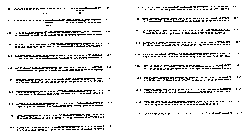
The DNA sequence (Fig. 9) reveals an open reading
frame of 1092 by starting with an ATG codon at position
204 and finishing at position 1296 with a TAA stop codon.
The open reading frame corresponds to a protein of 364
amino acid residues. Ten nucleotides upstream of the
methionine codon is a sequence, AAGGAG, that is comple-
mentary to the 3' end of the 16S rRNA of E. coli (Shine,
J. and Dalgarno, L. Proc. Natl. Acad. Sci. USA, 71:1342,
1974). The spacing between the centre of this putative
ribosome-binding site (rbs) and the start codon is 13 by
in comparison to the average spacing of ZO by in E, coli.
The 5' flanking region, upstream of the proposed rbs,
3S shows the presence of possible promoters. The sequences or
the -10 region, TAAAAT (151-156), and the -35 region,
TTGCTT (127-132), show homology to the consensus of E.
WO 91/18926 ~ ~ ~ ~ ~ ~ PCT/SE91/00129
21
coli promoters (Rosenberg, M. and Court, D., Annu. Rev.
Genet, 13:319, 1979) and are identical with promoters
recognized by the E. coli RNA polymerase. The spacing
between the putative -10 and -35 sequences is 18 bp, which
is comparable with the favoured value of 17 bp.
Between position 1341 and 1359 there is an inverted
repeat with the potential to ~orm a stem and loop struc-
ture. This repeat does not, however, resemble a typical
rho-independent transcription terminator.
Protein D structure
The gene for protein D encodes for a protein of 364
amino acid residues deduced from the nucleotide sequence
(Fig. 9). The N-terminal amino acid sequence has typical
characteristics of a bacterial lipoprotein signal peptide
(Vlasuk et al., J. Biol. Chem. 258:7141, 1983) with its
stretch of hydrophilic and basic amino acids at the N-ter-
minus followed by a hydrophobic region of 13 residues, and
with a glycin in the hydrophobic core. The putative signal
peptide ends with a consensus sequence Leu-Ala-Gly-Cys,
recognized by the enzyme signal peptidase II (SpaseII).
The primary translation product has a deduced molecular
weight of 41,821 daltons. Cleavage by SpaseII would result
in aprotein of 346 amino acids with a calculated
molecular size of 40,068 daltons, in contrast to the
estimated size of the mature protein D of approximately 42
kilodaltons. Posttranslational modifications of the
preprotein may account for this discrepancy. Several
attempts to determine the amino-terminal amino acid
sequence of protein D were performed by applying about
1000 pmoles thereof in an automated amino acid sequences.
Since no amino acid phenylthiohydantoin derivatives were
obtained, the amino-terminal end of the single IgD-recep-
tor polypeptide chain is probably blocked.
Protein D expressed in E. coli JM83 carrying pHIC348
was analysed in immunoblotting experiments (Fig. 10).
Cytoplasmic, periplasmic and membrane fractions from cells
in late logarithmic phase were separated on a SDS-PAGE gel
- CA 02083172 2000-07-06
22
and electroblotted to an Immobilon*filter. A protein that
binds all three anti-protein D monoclonal antibodies
(16C10, 2066 and 19B4) and radiolabeled IgD could be
detected in all three fractions (lane 2-4) from E. coli
JM83/pHIC348 as a single band with an estimated molecular
weight of 42 kilodaltons, i.e. equal or similar to protein
D prepared from H. influenzae (lane 1, Fig. 10).
The nucleotide sequence and the deduced amino acid
sequence of H. influenzae 772 protein D were compared with
other proteins of known sequence to determine homology by
using a computer search in the EMBL and Genbank Data
Libraries. Apart from similarities in the signal sequence
no homology was found.
SUMMARY
A novel surface exposed protein of H. influenzae or
related Haemophilus species is described. The protein
named protein D is an Ig receptor for human IgD and has an
apparent molecular weight of 42,000. Protein D can be
detected in all of 116 encapsulated and non-encapsulated
isolates of H. influenzae studied. The protein from all
strains shows in addition to the same apparent molecular
weight immunogenic similarities since protein D from all
strains interacts with three different mouse monoclonal
antibodies and monoclonal human IgD. A method for
purification of protein D is described. Cloning of the
protein D gene from H. influenzae in E. coli is described
as well as the nucleotide sequence and the deduced amino
acid sequence corresponding to a molecular weight of
41,821 daltons including a putative signal sequence of 18
amino acids containing a consensus sequence,
Leu-Ala-Gly-Lys for bacterial lipoproteins.
*Trademark