Note: Descriptions are shown in the official language in which they were submitted.
:, ~1, ~~~ t.s, -r'f ~ ~ n~~
S~w.e i_:11.5 ~ ?~7
ATRCO 1.0-033
MEDICAL VENTILATOR
EACKGROUND OF THE INVENTION
This invention pertains to apparatus for
medical ventilation and, more garticularly, to apparatus
for controlling the inspiratory and expiratory flow and
pressure of gases within a medical ventilator.
Medical ventilators, particularly anesthesia
ventilators, generally offer only limited ventilatory
modes of operation to the tree3ting physician or
anesthesiologist. These ventilators often comprise a
flow-control valve for controlling inspiratory flow and
adjustable mechanical means, if any, for controlling
expiratory flow. Such mechanical means, however, are
35 difficult to monitor and provide little flexibility.
Medical ventilators offering more comprehensive
ventilatory options generally are not available in
anesthesia ventilators and, moreover, require
complicated pneumatic hardware. This requirement
increases the exgense of manufacturing and requires
independent control of several pneumatic valves and
circuits.
SUI~iI~IARY OF THE INVENTTON
The present invention provides apparatus for
controlling both the flow and pressure of gases in a
medical ventilator, throughout the respiratory cycle,
using a single inspiratory flow-control valve. This
valve provides complete control of pressure or flow at
any paint within the pneumatic circuit during both
inspiration and expiration. Appropriate feedback
signals preferably are provided to the valve's
controller to provide inspiratory closed-loop flow
control and continuous inspiratory and expiratory
closed-loop pressure control. The present invention,
therefore, provides for an anesthesia ventilator modes
of ventilation normally available only in an advanced
intensive care unit (ICU) ventilator, such as, e.g.,
constant positive airway pressure (CFAP), pressure
z~~~~
-2- tb:o ~3asd3o~
support ventilator (PSV) and adjustable PEEP. Also, by
removing the ventilator's bellows assembly and providing
inspiratory flow directly to the patient°s mouth, the
present invention provides an advanced, multi--functional
TCU ventilator.
~n one aspect, the present invention comprises
a first inspiratory conduit for directing a flow of gas
from a terminal space, an inspiratory flow-control means
for controlling the inspiratory flow of the gas into the
terminal space, and an expiratory conduit for directing
an expiratory flow of the gas from the terminal space.
Means are provided for isolating the expiratory flow
from the first inspiratory conduit. Additionally,
pressure-control means are provided for causing the
35 gas's pressure within the expiratory flow to track the
pressure within the first inspiratory conduit.
The terminal space may comprise, e.g., a
bellows assembly or the patient's mouth. Tn the latter
case, the medical ventilator functions as an advanced
rCU ventilator, and the gas comprises respiratory gas
for the patient's breathing.
The inspiratory flow--control means preferably
comprises a proportional solenoid valve, and the means
for isolating preferably comprises a second inspiratory
conduit and means for prohibiting the gas from flowing
from the second inspiratory conduit to the first
inspiratory conduit.
The pressure-control means preferably includes
a diaphragm or balloon-type valve and means for
connecting the first inspiratory conduit to tire back
chamber of the diaphragm valve. In one embodiment, the
connecting means connects the first inspiratory conduit
directly to the back chamber of the diaphragm valve and
comprises means for releasing the gas to the surrounding
atmosphere. ~n a second embodiment, the connecting
means comprises a two-position solenoid valve. In one
of the solenoid valve's two positions, the valve
provides means for releasing the gas to the surrounding
-~- ~ ~~ <.yr1 $.~ ~T
~~ ~5a,~~t~f~
atmosphere and for connecting the first inspiratory
conduit to the back chamber of the diaphragm valve. In
the ether of solenoid valve's two positions, it provides
means for preventing the release of gas to the
surrounding atmosphere and for connecting a supply of
gas at a predetermined pressure to the back chamber of
the diaphragm valve. The connecting means also may
comprise means for releasing the gas to an exhaust space
having a pressure below that of 'the surrounding
atmosphere.
In a further aspect of the present invention,
the medical ventilator comprises processing means and
pressure feedback means for detecting the pressure of
the gas within the first inspira~tory conduit. The
pressure feedback means provides a signal indicative of
this pressure to the processing means, and the
processing means in turn controls the inspiratory
flow-control means to cause the pressure within the
first inspiratory conduit to track a desired pressure.
The feedback means alternatively may be placed within
the terminal space or at any other location within the
pneumatic circuit.
~7Cn another aspect of the present invention,
the feedback means may be a flow sensor to detect the
inspiratory flow, and the processing means provides
means for controlling the inspiratory flow-control means
to cause this flow to track a desired flow.
Other objects, features and advantages of the
present invention will be more readily apparent Exam the
detailed description of the preferred embodiments set
forth below, taken in conjunction with the accompanying
drawings.
DESCRTPTION Og' THE DRAWINGS
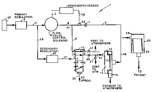
FIGURE 1 is a functional black diagram of a
medical ventilator in accordance with the present
invention;
y~~, e~,~»~..-y.~~
~.~ .. ~'~ c.9
FIGURE 2 is a schematic diagram of an
alternative embodiment for the connecting valve shown in
FIGURE 1;
FIGURE 3 is a schematic diagram of a second
alternative embodiment for the connecting valve shown in
FIGURE It
FIGURE 4 is a functional block diagram of a
second embodiment of a medical ventilator in accordance
with the present invention.
l0 ,~~~AII~ED DESCRIPTION OF THE PREFERRED EP~BODIME1~1~S
A ~aedical ventilator 1 in accordance with the
present invention is shown in FIGURE 1. A source of gas
30 enters anedical ventilator 1 through primary
regulator 2. Primary regulator 2 maintains this gas at
a pressure suitable for use by the ventilator. For
example, a source having a pressure of 50 psi is
regulated to exit from primary regulator 2 at a pressure
of 26 psi. The gas exiting primary regulator 2 enters
source conduits 3 and 29.
2o The gas in source conduit 3 enters
flow-control valve 4. This valve controls the magnitude
of the flow of gas passing through it, and is itself
controlled by microprocessor 32 via signals transmitted
on Sine 5. Microprocessor 32 controls the flow
according to a predetermined pressure or flow waveform
selected by the operator or technician and provided to
the microprocessor by a waveform generator =not shown).
The flow-control valve 4 is preferably a high-grade
proportional solenoid valve, but single or multiple
pulse-width modulated (PWri) two-position valves also may
be used.
Gas from flow-control valve ~i enters two
conduits, inspiratory conduit 6 and pressure-control
conduit 8. Check valve 7, at the end of conduit 6,
allows gas to flow into conduit 9 only if the pressure
in conduits 6 and 8 is greater than the pressure in
conduit 9. If the pressure in conduits 6 and 8 is less
a~''~ f' '~' ~~ A~z;~
~, ,.~r~a~,~*ar~
_5_
than the pressure in conduit 9, on the other hand, check
valve 7 closes to prevent the flow of gas from conduit 9
back into conduit 6.
Pressure-control conduit 8 terminates at input
parts A and C of connecting valve 10. This valve may be
a five-port, two-position solenoid valve, as shown in
FIGURE 1. In its deactivated position, ,shown in
FIGURE 1, pressure-control conduit 8 :is connected to
input parts A and C, which are, respectively, connected
to output ports E and D. Output port E vents to the
atmosphere through conduit 11 and pneumatic resistor 12.
This resistor has a relatively high pneumatic resistance
for reduced gas consumption. Rather than being a fixed
resistor, pneumatic resistor 12 may be variable and
controlled by microprocessor 32 to optimize performance
and gas consumption.
Output port D vents to conduit 13, °through
pneumatic resistor 26, and into expiratory valve 14,
preferably a diaphragm or balloon-type valve. Gas from
conduit 13 enters back chamber 15 of exgira~tory valve
14. If the pressure in back chamber 15 is greater than
the pressure in expiratory conduit 19, diaphragm 17
effectively seals the gas present in conduit 19 from
flowing through exhaust 16 and into the atmosphere. On
the other hand, if the pressure in conduit 19 is greater
than the pressure in back chamber 15, diaphragm 1~ opens
and the gas in conduit 19 flows through expiratory
valve ~4 and out through exhaust 16.
A safety valve 27 may be disposed on
conduit 13. This valve opens when the pressure within
this conduit exceeds a predetermined safe level. Safety
valve 27 may be located at other places within the
pneumatic circuit, such as, e.g., within conduits 6 or
9. Preferably, safety valve 27 opens at a pressure of
approximately 1.4 psi.
Connecting valve 10 is moved to its activated
position in response to a signal from microprocessor 32
on control line 22. In its activated position,
'~ ~.~~ q ~ K T! A-~ T2
~.J v ~~~Z.57. ii~d
-6-
pressure-control conduit 8 is disconnected from ports A
and C of connecting valve 10 and is connected to ports F
and I3, both of which are blocked. Also, in this
position, port G, which also is blocked, is connected to
conduit 11, and conduit 13 receives gas from sealing
conduit 21, across ports J and I. Secondary regulator
20 receives gas from conduit 29, preferably at a
pressure of approximately 25 psi, and transmits this gas
at a lower pressure, preferably at approximately
1.4 psi, to seal diaphragm 17 during normal operating
conditions.
Gas flowing into conduit 9 enters the outer
chamber 24 of bellows assembly 23, integrates to
pressure, and compresses bellows 25. Gases within these
bellows, generally containing anesthetic agents, are
transmitted to the patient. ~y omitting bellows
assembly 23 and terminating conduits 9 and 19 directly
at the patient's mouth, however, medical ventilator 1
can function as an ICiJ respiratory ventilator.
FIGURES 2 and 3 show alternative embodiments
for connecting valve 10. As shown in FIGURE 2, the
five-port, two-position solenoid valve shown in FIGURE 1
can be replaced with two solenoid valves in series,
namely, one two-part two-position solenoid valve and one
four-port two-position solenoid valve. These valves are
shown in FIGURE 2 in their deactivated positions, with
pressure-control conduit 8 connected across ports A and
13 to conduit 11, and across parts C and D to conduit 13.
In their activated position, conduit 21 is connected
across ports J and I to conduit 13, and pressure-control
conduit 8 is sealed by ports F and H.
FIGURE 3 shaves another embodiment for
connecting valve 10. In this embodiment, three two-
port, two-position solenoid valves are connected in
series. These valves are shown in their deactivated
positions, with pressure-control conduit 8 connected
across ports A and B to conduit 11, and across ports C
and D to conduit 13. In the activated position,
.-~ .~' fl Tv ,t-. ~r-B A
4 ,~ a ~Y ~9 rl 1 r~4i't~
-~-
conduit 21 is connected across ports J~ and I to
conduit 13, and pressure-control conduit ~ is sealed by
ports F and H.
Microprocessor 32 can control the pressure
within outer. chamber 24 of bellows assembly 23 during
both inspiratory and expiratory flow solely by
controlling flow-control valve 4. In this
pressure-control mode, connecting valve 10 remains in
the deactivated position shown in FIGURS 1 during both
inspiratory and expiratory flow. To increase the
pressure of gas within outer chamber 24,
microprocessor 32 commands flow-control valve 4 to
increase the flow of gas from source conduit 3 into
conduits 6 and 8. The introduction of additional gas
into conduits 6 and ~ eventually results in the pressure
within these conduits exceeding the pressure within
conduits 9 and 19 and outer chamber 24. Upc>n this
occurrence, check valve 7 opens and gas flows from
conduit 6, through conduit 9 and into cuter chamber 24.
This gas compresses bellows 25 and causes gas within
these bellows to enter the patient.
Since connecting valves ZO is deactivated,
pressure-control conduit g is in pneumatic communication
with back chamber 15 through parts C and D of connecting
valve 10 and conduit 13. The pressures of gas within
back chamber 15 of expiratory valve 14, therefore,
continuously tracks the pressure of gas within
conduits s and 8. As a result, when check valve 7
opens, diaphragm 17 closes because the pressure within
back chamber 15 (equal to that within conduit 6 and 8)
exceeds the pressure within expiratory conduit 19 (equal
to that within conduits 9 and outer chamber 24).
6~hhen shack valve 7 opens, most of the gas
flowing from flow-control valve 4 enters outer
chamber 24. A portion of this gas., however, is vented
to the atmosphere through parts A and S, and conduit il,
but this loss is small because the pneumatic resistance
of pneumatic resistor 12 is high. In order to further
a ,~~~ y ~r i lr~ .rp ,
y.~i.~~1'~~L:J~~
diminish this loss, however, connecting valve 30 may be
activated during all, or during a portion of, the period
of flow into outer chaBCber 24.
To decrease the pressure within outer
chamber 24, microprocessor 32 commands flow-control
valve 4 to reduce the flow of gas from source conduit 3
into conduits ~b and 9. Eventually, this reduced flow
causes the pressure within conduits 6 and 8 to drop
below that within conduits 9 and 19, and check valve ?
30 closes. Again, since the preasure within back
chamber Z5 tracks that within conduits 6 and 8,
diaphragm 15 opens when this valve closes. The opening
of diaphragm 1? enables an expiratory flow of gas from
outer chamber 24 to the atmosphere through expiratory
Z5 conduit 1.9 and exhaust ~.6.
Medical ventilator 1 can control the pressure
of this expiratory flow through flow-control valve 4.
During expiration, gas within conduits 6 and 8 vents to
the atmosphere through ports A and B of connecting
20 valve 3.0, pneumatic resistor 12 and conduit 31. If the
flow through flow-control valve 4 is increased to
increase this venting, the pressure within conduits 6
and 8 also increases. Since the pressure within back
chamber 15 tracks that within conduits 6 and 8, this
25 increased pressure is transmitted to the expiratory flow
of gas from expiratory conduit 19 and outer chamber 24.
This transmission occurs because the increased pressure
within back chamber 15 reduces the opening of
diaphragm ~.? and, therefore, increases the pressure
30 within the gas flowing from expiratory conduit 39.
The expiration of gas from outer chamber 24
eventually causes the pressure within this chamber, and
within conduits 9 and 19, to fall below that within
conduits 6, 8, 13 and back chamber 15. Upon this
35 occurrence, expiratory valve 14 again closes and check
valve ? again opens to cause a renewed inspiratory flow,
controlled by flow-control valve 4, into outer
chamber 24.
W :a.5~ s
Through control of only flaw--control valve 4,
therefore, medical ventilator 1 controls the pressure
within the inspiratory flow into, and the expiratory
flow from, outer chamber 2~4 of bellows assembly 23.
This control provides the anesthesia ventilator with
wide range of ventilatory-mode options such as, e.g.,
adjustable positive expiratory end pressure (PEEP).
Also, as indicated above, by deleting bellows
assembly 23 .and terminating inspiratory conduit 9 and
expiratory conduit 19 directly at l:he patient's mouth,
medical ventilator 1 provides a mufti-functional ICU
ventilator capable of providing all known (and unknown)
ventilatory modes through c~ntrol of only flow-control
valve 4. A hospital, therefore, can use medical
ventilator 2 as both an anesthesia ventilator and an ICU
ventilator.
Ey providing pressure-feedback signals to
microprocessor 32 from any point within the pneumatic
circuit, such as, e.g., fram inspiratory condu~.~t 6 via
sensor 33, flow-control valve ~d can be controlled by
microprocessor 32 to control pressure within the
pneumatic circuit in a closed-loop fashion. In
accordance with such control, microprocessor 32 xesponds
to the actual pressure measured by sensor 33 (Pc) with
commands to flow-control valve 4 to reduce the magnitude
of any deviation between this actual pressure and a
desired pressure. A waveform generator (not shown)
provides a signal to microprocessor 32 indicative of
this desired pressure.
3o For closed-loop pressure-control, connecting
valve to again remains in its deactivated position (as
shown in FIGURE 1). If at~any given instant of time
(clock cycle), the actual pressure (Pc) sensed by
sensor 33 is less than the desired pressure indicated by
the desired pressure waveform, microprocessor 32
responds by commanding an increased flow of gas from
flow-control valve 4. This increased flow raises the
pressure within conduits ~ and 8 and, therefore,
a
~.W .~Jt.311t''aJ~
-10-
decreases the magnitude of this differential. Also, if
during this particular instant of time check valve '7 is
open and expiratory valve 1~4 is closed (i.e., Pc is
greater than the pressure within conduits 9 and 19 and
outer chamber 24), this increased pressure is
transmitted to outer chamber 24 through an inspiratory
flow into this chamber from conduits 6 and 9. On the
other hand, if during this particular instant of time,
check valve 7 is closed and exgiratory valve 14 is open
~i.e., Pc is less than the pressure within conduits 9
and 19 and outer chamber 2~), then this increased
pressure is transmitted through the expiratory flow from
this chamber through conduit 19 and expiratory valve 14.
This transmission occurs via pressure-control conduit 8,
ports C and D of connecting valve 10 and conduit 13 to
back chamber 15 of expiratory valve 14. The increased
pressure in conduit 8, of course, also results in an
increased flow to the atmosphere through ports A and B
of connecting valve 10, pneumatic resistor 12 and
conduit 11. As explained above, the increased pressure
within back chamber 15 decreases the extent to which
expiratory valve 14 is open and, therefore, increases
the pressure within the expiratory flow from outer
chamber 24.
On the other hand, if the actual pressure (Pc)
sensed by sensor 33 is greater than the desired pressure
indicated by the desired pressure waveform,
microprocessor 32 responds by commanding a reduced flow
from flow-control valve 4. In a manner analogous to
increasing the flow through this valve, the resultant
decreased pressure within conduits 6 and 8 is
transmitted to outer chamber 24 via conduit 9, if an
inspiratory flaw is occurring into this chamber, or via
back chamber i5 and expiratory conduit 19, if an
expiratory flow is occurring from this chamber. By
controlling only flow-control valve ~4, therefore, the
pneumatic circuit of medical ventilator 1 enables
continuous closed-loop pressure-control throughout the
~' ~~ A,'"~
i~~ m: ~'J1.J'1:1i'-ys1
_~i_
respiratory cycle, .~.e., during both inspiratory flow
into, and expiratory flow from, bellows assembly 23 (or
the patient's mouth).
.Also, continuous closed-loop pressure control
can be provided regardless of the location of sensor 33.
.Sensor 33 can be located, e.gr., within conduits 9 or 19,
outer chamber 24 or within the patient's mouth or other
respiratory organs. Since a treating physician or
anesthesiologist often is most conce:rneG'I with the actual
pressures existing within the patient's respiratory
organs, such a location is particularly advantageous for
sensor 33 regardless of the presence or absence of
bellows assembly 23).
The provision of continuous closed-Loop
control also is advantageous if a spontaneous breath or
cough occurs at any point during the pressure cycles
commanded by the desired pressure wavefarm. For
example, if a spontaneous breath occurs during
expiratory flaw from outer chamber 24, a pressure
differential immediately occurs between conduits 6 and 9
causing check valve 7 to open. The opening of this
valve causes a flow of gas into outer chamber 24 to
support this breath. This action also causes a drop in
the magnitude of Pc which causes microprocessor 32 to
command an increased flow from flow-control valve 4.
This increased flow further supports the spowtaneous
breath and, moreover, raises the pressure within back
chamber 15 back to the target pressure indicated by the
desired pressure waveform. ~;ventually, the pressure
within outer chamber 24 is raised back to the desired
pressure, check valve 7 closes, and expiratory flow
resumes at the desired pressure, again controlled by
expiratory valve 14.
~i patients cough during inspiratory flow is
facilitated in an analogous manner. The resultant
increased pressure within outer chamber 24 closes check
valve 7 and opens expiratory valve 14 to release this
pressure. Concurrently with this action, the raised
r~~.'~f~' 'l'r1 ~_' "Z
FL,I v~~J'%.52?llvJ~~
-12-
pressure within conduits 6 and 8 causes
microprocessor 32 to reduce 'the flow from flow-control
valve 4. This reduced flow causes a further pressure
drop within back chamber 15 to further support the
release of pressure from outer chamber 24. Eventually,
Pc drops to the desired pressure, expiratory valve 14
closes, check valve "7 reopens and inspiratory flow
resumes at the desired pressure:. ~Cn this manner,
therefore, spontaneous inhalations and exhalations arc
supported and facilitated, and continuous closed-loop
pressure-control is maintained. Also, since the
pressure within bellows assembly 23, or equivalently,
patient mouth pressure, can be made 'to follow any target
pressure waveform, medical ventilator 1 can function as
a high performance 3~CU ventilator capable of per:~orming
constant positive airway pressure (CPAP), pressure
support ventilation (PSV), positive end expiratory
pressure (PE~Pj and other known and unknown vent:ilatory
modes of control.
Medical ventilator 1 also is capable of
inspiratory flaw-control. Tn this made of operation,
the volume of gas flowing through conduits c and 9 is
regulated during inspiration, and connecting valve 10 is
switched to its activated position during inspiration.
When connecting valve 10, is activated, gas from
secondary regulator 20 at approximately 1.4 psi flows
through conduit 21, connecting valve 10 (across ports J
and 1), conduit 13, pneumatic resistor 26, and into back
chamber 15 of expiratory valve 14. Back chamber 15
reaches a pressure of approximately 1.4 psi, therefore,
which seals diaphragm 1? and expiratory conduit 19 to
prevent gas from flowing through this conduit to
exhaust 16. Also, pressure-control conduit 8 is closed
by input ports F and H of connecting valve 10. All gas
from flow-control valve 4, therefore, flows through
conduits 6 and 9 and into outer chamber 24, and this
flow is controlled by flow-control valve 4 in response
to control signals from microprocessor 32 on line 5.
d~,'ns~W,,~.
-13-
Closed-loop control of this flow may be
achieved by transmitting flow-magnitude feedback
signals, from sensor 33, to microprocessor 32 on
line 28. The microprocessor then controls flow-control
valve 4 such that the actual volume of gas flowing
through the inspiratory conduits tracks a target
waveform, provided to the microprocessor from a target
waveform generator (not shown).
In response to a signal from
20 microprocessor 32, connecting valve 10 is switched to
the deactivated position, shown in FIGURE 2, and the
flow from flow-control valve 4 is terminated (or set to
provide some positive end back pressure (PEEP) to the
back of diaphragm 1?). Gas in book chamber 25 vents to
15 the atmosphere through expiratory conduit 13, ports D
and C of connecting valve 10, ports A and ~ of this
valve, and finally through conduit 11. The pressure
within outer chamber 24 and conduits ~ and 19 eventually
exceeds the pressure within conduits 6 and ~3 and back
20 chamber 15, and, as a result, check valve 7 closes,
diaphragm 17 opens, and gas leaves outer chamber 24 and
passes through conduit 39, expiratory valve 14, and out
exhaust 16.
Medical ventilator 1 comprises several safety
25 features. For example, in the event of a Bower
shut-down, flow-control valve 4 fails in a zero flow
position, and connecting valve 10 fails in the
deactivated position shown in FIGURE 3. The entire
pneumatic circuit, therefore, is vented to the
30 atmosphere.
~. more hazardous type of failure occurs if
flow-control valve 4 becomes stuck in a wide-open or
full-flow position which would drive the pressure in
conduit 6 and bellows assembly 25 to a high level. In
35 this event, however, safety valve 27 opens to prevent
the pressures within the pneumatic circuit from
exceeding a safe level. Preferably, the safety valve is
mechanical, and is triggered by pressures greater than
°~~' ~' il ~n ,n ~'-T ~~
-14 - bid '1r X91.9 "i ? ~4~:
1.4 psi, once safety valve 27 is activated, flow from
flow-control valve 4 vents through pressure-control
conduit 8, across ports C and D of connecting valve 10,
and out safety valve 27 to the atmosphere. This flow
also produces a negative pressure difference between the
pressure in back chamber 15 of expiratory valve 14 and
conduit 19. This differential allows diaphragm 17 to
ogen, and further exhausts the flow :in conduit 1~ to the
atmosphere. Safety valve 27 may be located at other
points within the pneumatic circuit, such as, e.g.,
within conduits 6 or 9.
In another embodiment of the present
invention, conduit 11 is vented to a sub-atmospheric
pressure to provide a negative pressure bias for
expiratory valve 14. This venting can be effected by,
e.g., connecting conduit l1 to a venturi (not shownj.
since a negative pressure in conduit 11 is tran:aferred
to back chamber 15 of expiratory valve 14, the
expiratary valve°s response time would be improved by
such venting and its resistance lowered, especially at
low PE7EP levels. Also, the existence of a negative
pressure would further tend to open expiratory valve 14
in the event of a failure by flow-control valve 4. This
improved performance, however, would be traded off
against the cost of increased components and additional
gas consumgtion.
F1GTIRE 4 shows another embodiment of the
present invention with similar elements similarly
numbered. In this embodiment, connecting valve 10,
pneumatic resistor 26, and safety valve 27 have been
deleted, and pxessure-control conduit 8 is connected
directl~.y to conduit 11 and back chamber 15 of expiratory
valve 14. Also, a diaphragm or balloon-type valve,
safety valve 127, has been added which has a continuous
pressure, preferably 1.4 psi, applied to its back
chamber 135 from secondary regulator 20.
~'~y!1 fi ~i'w~!
W sw.il_l~ljr'~c!~~
-15-
The embodiment shown in FIGURE 4 regulates the
pressure of gas flowing into, and out of, bellows
assembly 23, in either an ogee or closed-loop fashion,
in the same manner as the embodiment shown in FIGURE 1.
~1n aver-pressure condition is prevented, however, by
secondary regulator 20 and safety valve 127. If the
pressure in pressure-control conduit 8 is driven above
1.4 psi, safety valve 127 spans ~t:a vent gas to the
atmosphere. This venting alas produces a negative
pressure difference between the pressure in back
chamber 15 and that in conduit 19, allowing expirato~y
valve 14 to span and exhausting gas in conduit 19 to the
atmosphere.
The embodiment shown in FIGURE 4 also can
35 operate in a flaw-control mode by calculating, and
providing to microprocessor 32, the pressure and flow
characteristics of pneumatic resistor 12. Since the
instantaneous pressure within conduit 8 is known to the
microprocessor (via signals from sensor 33),
microprocessor 32 can calculate for any given instant of
time the volume of gas flowing into the atmosphere
through conduit 11. Ey deducting this flaw from the
total flow through flow-control valve 4 and conduit 6,
the total inspiratory flaw through conduit 9 and into
bellows assembly 23 can be controlled.
As with the previous embodiments, the bellows
assembly may be omitted from the embodiment shown in
FIGURE 4 and inspiratory gas transmitted directly to the
patient's mouth.
Although particular embodiments of the present
invention have been shown and described, many varied
embodiments incorporating the teachings of the present
invention may be easily constructed by those skilled in
the art. The foregoing description of the preferred
embodiments, therefore, should be taken as illustrating,
a~ather than limiting, the invention as defined in the
following claims.