Note: Descriptions are shown in the official language in which they were submitted.
WO91/1~ rCT/US90/0~2
- 1. 2~33699
PROCESS FOR ~SE IN RADIOSURGERY
FIELD OF INVENTION
This invention relates to a process for aiding in the
planning of radiosurgery, it includes visualizing, in real
; time three dimensional viewing, with the aid of a computer
,; , , .
usins stereoscopic views, the treatment volume and
surrounding structures.
The treatment areas can be represented from information
derived from CT scan slices or a MRI scanner both of which
produce two dimensional cross sectional views of the area.
These structural areas are then rotated for viewing at any
angle utilizing a real time computer display. This computer
enhanced display will form the stereoscopic three
dimensional views utilized in the radiosurgical procedure.
. - .
The treatment doses, which are units of treatment which
can be produced by a Gamma knife as well as other -
radiosurgical equipment, are represented by a volume display
.~.i:
;~ outlining approximately the fifty percent isodose curve.
These volumes, essentially spherical for the Gamma knife,
are referred to as "shots", which are then through the
present process moved in three dimensional space and shown
, . ~:
in real time on three dimensional displays.
The process of the present invention is adaptab~e or
use in stereoscopic angiography which is the showinc O r the
blood vessels in the brain. By the employment of two
simultaneously displayed stereoscopic orthogonal views,
",.,~ .
:.~
. ~.~' ' . . ' . '
' '.'~,.. ' ' ' ' , . ,' . ` ' .,' , ., , . ' "
''~.', ', ' ' - ~ ' " ', " ' ' ' '- ", . ,' " '
~ WO91/1&~W PCT/US90/0~n2
2CS3~9~
2-
which may be rotated ninety degrees to each other, the
precise locating of the X,Y, and Z coordinates can be
achieved. A cursor may be moved in three dimensional space
on the stereoscopic angiography display with simultaneous
viewing of the cursor in both the orthogonal views. By
being able to view it simultaneously in real time, projected
onto both orthogonal views, precise localization of the
.3 .
blood vessels can be obtained.
The scan slices determine from any of the studies, CT,
:. ~
MRI, and angiography can be simultaneously displayed on one
or more computer monitors as cross sections. Real time
display of the shot approximations are shown in cross
section in all of the planes simultaneously. The shots can
.,.~
then be moved in real time on all of these cross sections to
determine how they interact within the slices and each
~' other.
PRIOR ART
The mechanical and electronic equipment and
' apparatuses employed in stereotactic surgery is presently
, available and their function for use in the specific field
is well known in the profession.
.
A condensed introduction to the required physical
apparatuses necessary to practice the present inventive
procecs can be found in an article entitled ~Stereotactic
Neurosurgery Planning on a PC Based Work Station~, Journal
.
~ .
:. .
:.,
.::~
~ ' .
WO 91/18644 rcr/usso/03072
2C~3G99
3.
of Digital Imaging 1989. For 3-D reconstructed CT images :-
see Journal Computer Assist To~ography - Vol. 12 No. 1,
1988, Technical Note Holography of ~ D Surface Reconstructed
CT Images. See also the article Stereoscopic Co~puted
Three-Dimensional Surface Displays; Radiographics ~ol. 5 No.
, 6, November 1985.
,, ~
~, ~
,.
BRIEF DESCRIPTION OF THE DRAWINGS ~ -
The inventive process described in this application -
- will be best understood by reference to the accompanying
drawings in which:
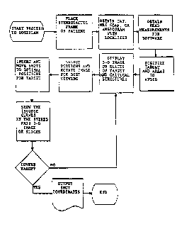
. Fig. 1 is a flow chart of the step by step application
1' .
of the process, and `-`
: " :. -
Fig. 2 is a flow chart of the display program of the
process as it is implemented. ~-
. . . .
DESCRIPTION OF THE INVENTION
Stereotactic surgery performed throush the employment
of image sources which def ine the cerebral structure in ~-
cooperation with a computerized work station iDcluding the
applicable software, is an acceptable medical procedure.
The method of developing the necessary images as well as the
trajectories of the shots of high energy ionizing radiation,
is achieved through the use of the stereotactic frame which
develops the coordinate geometry. The treatment areas are
~ ,
':::! `
~ ~.
'
., " ~
9 ~ I Q3~. .Z- - _
2CS~99 4 . lPEA/U~ 1 3 JU~l99~
represented from information derived from modalities such as
CT ~RI and DSA.
As disclosed hy Fi3. 1 the process ~egins with the
?lacement of a stereotactic frame on the ~atient and through
it resulting yraphic points are determined. In step t:lo
these refPrenced graphics are overlaid with scan slices
o5tained through CT or MRI methods which disclose the areas
to be treated in its relation to surrounding safe structure.
In step three the head m~asurements are then
coordinated into the pre-exlsting software so that target
areas and safe structure may be digitizet. In the third
step the 3-D images of target and safe structural areas are
displayed. In the following step selective portions of
these images may be colorized and rotated to afford all
possible line of sight viehing so that prospecive shots may
be optimally positioned onto the target areas~, correspondin2
to its configuration including length, width and depth.
In the final step the perspective isodose curves are ~^~
then imposed upon the images. At this stage it may be
determ;ned whether or not the shots correspond to the
target's imagery. This continuous unobstructed viewing from
different rotated angles allows for real time display of the
target area safe structure as well as shot positions and
volumes. ~
The process permits the adjustment of each individual ~- .f~i
shot's volume and position enabling the operator to assure
that there are no shot overlays which would produce an ~-
un.qanted degree of radiation. The stereoscopic display of
the shots and area to be tr~ated affords the optimum
placem~nt of the isodose curve.
SUBSmUTESllER ~:
.. .., ., . . . . . . ; ~
~ . - ': . , .: ,
2c~,699 Pt~TI~S & / O 3 0 7 2
5 IP~A/US 1 3JUll932
If only one raphic screen is available for display the
slicos aroun~ the active shot ~n~ as many additional slices
~s possible are displayed. If multiple graphics screens are
availa~l~ all slices aro displayed. Shot manipulation in
real time, is displayed on tho slices using the same control
functions as for the third dimensional display-
The foregoing descriSed process allows the operator toenter the outline of the treatmPnt volume (shots) and other
regions of interest (tumors or effected structure) omittins
or permitting exclusion by definition critical structures ~ ,~
-that are to be spared. The display is constructed from a ^ '
series of transverse axial images. These outlines are then ~ i
displayed as a wire frame stereo pair image set. By -
utilizing a display system ~qhich allows each of the images
to be viewed with the appropriate eye, the structures are
; displayed as a three dimensional model. :~
Approximations'of the shots are then superimposed on
: -. :
~ ~ this display and can be moved in stereotactic space. The ~ ~
.~ ..
entire image may then be rotated in any direction for --
optimal viewing of the targets and shots. Portions of the
entire volume may be selected for viewing to allow better
visualization of the shot positions rela'tive to the target
edges.
This novel function allows the operator to obtain a
very effective approximation for the posit10ning of the
shots to cover the target, and th~ ability to avoid the
critical structures. After pl~cement of the shots, their '`
calculated isodose contours can be'displayed as a
stereoscopic palr superimposed on the digitized image and it
to may be rotated for close examination.
, .
r~ sussnTuTE SH~
, ~,., . , .. .~ . . .
. . i~ . . . ' ~
. , .. , . . ~ .
pG~1U~ 307
2c~3~9~ 6. lPE~ JU~ 9
The following data, repr2sented in the flow chart of
Fig. 2 is ad~ed to the existing software and includes, as
st2p ona digitizin; CT or ,~21 ima3es as well as di~itizing
slices from the computed Axial Tomogr~ph~ scan/MRI digitiz d
imagos, including la?lace and other od3e d~tection
al~orithms. Step two utilizes a mouse dijitizer for ~ -
solection of display curves adjusting display
characteristics as well as shot positions. This step
~ermits the editing of curves and/or deletin~ curves, and
will illustrate the reference distance as required.
The final s.ep requires calculating the dose metr;c
c nter from orthogonal images using prior programming
including utilizing the digitizer input from the established
ax2s as well as the input from the dose metric center To
the following may be added a manual input of scal2 readin~s.
It is required that one must generate minimum/maximum ~ ~
X's Y's for the given Z from Angio outline to correlat~ to ~- -
the transverse axial CT or MRI images. Also to yenerate
cross sectional plots from digitized angiographic plots to
correlate to cross sections on other studies.
The dose metric center calculations may be made from A~
and LAT views. Angiographic outlines are digitiz~d from
othogonal views for display. Angiographic volumes may also ~ :
be digitized from otho~onal views for display. Digitizin~
the angiographic volumes from orthosonal storeoscopic vie.~s
for display throush the use of coplanar slices. ?lanning
the angiographic volume from the orthogonal stereoscopic
views using shot approximations. Generatin; an ansio
predicted outlines from CT digital slices and emplpying a
3-D mouse input for imagery information.
SU8STIl'UTE SHEE~ ~ ~
rF.r~ ' : ' "` ~ "'' ' '
~ . ' . - .' ', ' ~ ' ~ .
,'''~''' ','. ,'~ `' ' ' " ' ' `'' .' ' '"
' ' ' ' ' '' " ~ I ,'' '. '
P~T~US 90/03~7~
2~s~99 7 IPEA/US 1 3 JUL 1992
Tho program may include a 'irst menu which will ?ermit
the dls?lay an~ adjustment of color images including color
t~ hide imagery as ~ell as color to dis~lay imagery; to;.le
~is~13y vf axes; toggle hidden line r~moval on/off delay;
color c~ange, color C0~2i screen labels an~ finally a ~-
printout instructional manual.
Ano.her menu may inclu~e tata directed to the shots
such that thcy can be modifiec', ~dited and selncted for
displ~y. Each shot can be moved or all re~uired shots may
be blocked to~,ether for movemont to new coordinates a
printout of shots can be obtaine~ indicating saved shots and
loadet sbots with all shot information transferred to Kula.
The first menu then p_rmits the rotation and adjustmert
of the images and shot volumes. It permits the display of ~;~
all curves except those of the shots as well as showin~ the
shots without the other curves so as to-illustrate how they ~ ,~
all interact. The menu will display ali points outside a
specific distance from the Z coordinate of the center or all
shots outside spscific distance from the center. ~ -,
The second menu p?rmits the actlvation of a cursor ~z
drone which has a variable siz- in th~ Z direction and which ~ -
when activated will cause the display of the curves whicl~ ~,
fall~within the 7 range pointet to by the tron~. The s~con~
menu will activat~ the graphic display between the
stereoscopic 3-D display and the slices as well as
simu1taneously displayin3 the slices with shot
re,pres ntations imposed thereon.
.
, '.
SV8STITUTE SHEET
if.. "' .(, " ' . . ' . .'`' `' ' " ... '`, ~ ;
WO91/1&~ PCT/US90/03072
2~S3~1~39
This novel function allows the operator to obtain a
very effective approY~imation for the positioning of the
shots to cover the target, and the ability to avoid the
critical structures. After placement of the shots, their
calculated isodose contours can be displayed as a
stereoscopic pair superimposed on the digitized ima~e and it
to may be rotated for close e~amination.
While I have illustrated and described the preferred
process for carrying my invention into effect, this is
capable of variation and modification without departing from
the spirit of the invention. I therefore, do not wish to be
limited to the precise details of progression set forth in
the described process, but desire to avail myself of such
variations and modifications as come within the scope of the
appended claims.
.
. : . , :
:'.- ' - . ': : ' '- : '
. ' - . -:
' ~ ,' ' ':' i
, ' , ' '
': , :, .