Note: Descriptions are shown in the official language in which they were submitted.
2 ~83~
PG 1-
8267
RWA
FEMORAL CUTTING GUIDE
BACKGROUND OF THE INVENTiON
Field of the Invention
This invention relates to an orthopedic instrument for guiding a saw biade for
shaping the distal end of a human femur to receive an endoprosthetic femoral
component.
Description of the Prior Art
Instruments used to prepare bone surfaces and to place femoral endopros-
thetic components in the knees perform two basic functions.
First is the control of power and other tools to provide accurate fixation
surfaces of bone that match implant geometry. Second is to posltion fixation
surfaces relative to bone and soft tissue architecture to approprlately orient the
prosthetic component.
In addition, instruments must provide the necessary flexibility to
accommodate the variations in geometry and surgical complications that are
encountered within the patient population. They must also meet the disparate needs
of surgeons who wish to follow different methodoiogies when performing the
surgical replacement of the knee.
Conventional femoral components usually have five planar fixation surfaces
which match the bone to the implant. Thus the femur must be prepared to have a
distal cut surface, a posterior cut surface, an anterior cut surface, an anterior
chamfer cut surface and a posterior chamfer cut surface.
Some existing femoral components have marginally different forms of fixation
surface, ie. no posterior chamfer, or the anterior chamfar surface formed as a curved
surface rather than a flat one. In general it has been found that flat surfaces are
nevertheless advantageous since they are easier to prepare using oscillating saws.
A number of different surfaces can be used to control the positioning of the
essentially planar blades of front or side cutting oscillating saws for shaping the
planar surfaces.
~Q~8~
Flat metallic bloGks on which the saw blade is rested, obviously rely to some
e~tent on the skill of the surgeon to avoid tilting of the saw blade, as may happen
when the saw enco~lnters a localized harder section of bone, or when the saw blade
has a long travel beyond the guide surface.
Slots having small clearance relative to the thickness of the saw blade may
also be used. In general these offer better control of the saw blade than blocks, but
they can impede visibility at the operative site. Simple slots do not provide
clearance for the tooth set on the saw blade, but a number of solutions have been
proposed to this problem. These include variable thickness slots formed by
assemblies of elements.
The slot is temporarily made deeper to allow passage of the saw blade teeth
and subsequentiy reduced to more closely hold the main body of the biade in
position. Alternatively, the slot is made open ended on one side so that the blade
may be introduced into it from the side without having to pass the teeth through the
slot. Variations in the design of the saw blade itself have also been used. These
may have zero net set on the teeth or provide a local clearance behind the teeth so
that the total blade and tooth form can be passed through a close clearance slot.
Block type cutting guides are shown in U.S. Patents 4,474,177,4,487,203,
4,502,483,4,524,766 and 4,567,885.
Fulcrum type cutting guides are described in U.S. Patent 4,718,413 and also
in U.S. Patent 4,892,093. These consis~ of an upper and a lower guide surface
which are linearly separated along the plane of intended cut by the saw blade. By
providing a separation between the two surfaces the saw blade, including its tooth
set, may be introduced between the two surfaces and then biased against them to
control the cutting plane. The separation of the guide surfaces normal to the plane
of operation of the saw biade is matched to the thickness of the saw blade. The
choice of orientation of the guide surfaces is chosen so that any deviation by the
surgeon in maintenance of the contact between the saw blades and either of the
guide surfaces results in conservative removal of bone, which may be corrected
subsequently. The guide of U.S. Patent 4,892,093 sits on the already prepared
distal femur and provides for the cutting of four additional cuts.
2 ~3 ~
The femoral components may be located with six degrees of freedom relative
to the patients fernoral geometry. These can be expressed in a cartesian manner
relative to orthogonal anatomical reference planes.
Angulation: Varus Valgus,
Flexion-Extension,
Internal-External Rotation.
Linear Position: Inferior-Superior,
Anterior-Posterior
Medial-Lateral.
To position the component on the bone, a number of datum features of the
patients anatomy and their relative location as controlled by soft tissue structures at
the knee may be utilized.
Two major schools of though exist as to the optimum method to provide
15 consistent functional placement. The first is independent femoral anatomical
placement. In this method the femoral component is positioned on the femur by
referencing datum features on the femur itself.
The second is referenced to the tibial position. In this method the position of
the femoral component is controlled relative to the proximal cut of the tibia. The
20 ligaments and other soft tissue structures at the knee joint will in this case affect the
femoral components position. The positional referencing, according to different
methodologies, is performed surgically prior to placing the femoral component.
A third method is varus-valgus and flexion-extension. Angulation of the
component in planes is usually performed simultaneously. The reference datum is
25 either the femoral shaft or the line joining the center of the knee and the hip joints.
Two major methods for accomplishing this are currently used.
First is intramedullary alignment. A rod is introduced through the center of
the knee into the intramedullary space and passed up the inside of the femur to the
internal isthmus, picking the axis of the femoral shaR. This technique has been
30 found to be very reliable, but is thought by some surgeons to be overly invasive and
in patients where there is excessive bowing of the femoral shaR, or where the
intramedullary space is blocked, for example by a long stemmed hip implant, it may
not be available.
2~88~6 l~
The second is extramedullary alignment. An external guide rod is aligned
with the anterior corlex of the femur, or from the center of the knee to the femoral
head.
The posterior condyles of the femur are used in the anatomical approach. In
5 the referenced technique the internal-external rotation is controlled by balancing of
the flexion gap so that the medial and lateral compartments of the joint are equally
spaced or tensed.
Inferior-Superior positioning is controlled in the anatomical approach by a
fixed amount of bone being resected from the distal femur. The amount of resection
10 is normally the same as the thickness of the distal portion of the implant component
where bone stock has not been eroded away. In the referenced technique the
amount of bone to be removed is adjusted relative to the proximal tibial cut to allow
for the total thickness of both the femoral and tibial components.
In the anatomical approach the anterior-posterior position of the femoral
15 component may be referenced to a number of alternative features at the distalfemur. These include the posterior condyles, where an amount of bone is resectedfrom the posterior condyles which corresponds either to the posterior thickness of
the femoral component or to some proportion or fixed amount in excess of this.
Alternatively, anterior features or the distal femur may act as references, usually
20 either the anterior cortex or the deepest point of the patella groove. In cases where
a large intramedullary stem is to be used, the position of the femoral componentmay need to be chosen to match the position of the implant stem within the intra-
medullary canal in which it must fit. In the referenced approach a posterior resection
of the femur is performed so that the flexion gap of the joint matches the thickness
2~ of the femoral and tibial components. In general all these approaches result in
either an anterior or poslerior cut being performed. Subsequently the opposite cut
is performed so that the implant will fit between these resected surfaces.
The medial-lateral placement of the component is usually performed by eye
to match the rim geometry of the resected bone surface performed by all the
30 previous cuts. In cases where a large intramedullary stem is used, the position may
be dictated by the fit of the stem into the intramedullary cavity.
CA 02088867 1997-12-10
Current techniques generally require the sequential use of alignment
and cutting guides. In all current systems multiple cutting guides are needed to
fully prepare the distal femur for the implant. Because these sequential operations
require the assembly and disassembly of instrument configurations and the use of
intermediate datums cut onto the bone, there are penalties in terms of time of
operation and accuracy. The current invention is intended to address these
inadequacies while incorporating the flexibility to allow for alternative operative
approaches to be used in placement of the femoral component.
SUMMARY OF THE INVENTION
The present invention provides a femoral cutting guide for guiding
system a cutting device for preparing the distal end of a human femur to receive
an endoprosthetic femoral component comprising: a base component including a
pair of side walls; a plurality of spaced apart guide rods extending between said
side walls for guiding the cutting device for forming a distal femoral surface, an
anterior femoral surface, a posterior femoral surface, and anterior and posterior
chamfered femoral surfaces for receiving a femoral component; an accessory
removably attachable to one of said rods; means for aligning said base component
on the bone, including an intramedullary rod and an element having a generally
circular hole for allowing the insertion of the intramedullary rod therethrough, said
20 element removably attachable to said accessory, said hole adapted to be
positioned adjacent the intercondylar notch of the distal femur when said element
is attached to said accessory and said accessory is attached to one of said rods;
and pin elements for attaching said base component to medial and lateral bone
surfaces after alignment, said pin elements extending through holes in said side
64680-680
CA 02088867 1997-12-10
walls, said holes located proximally of said guide rods when said cutting guide is
mounted on said femur.
These and other advantages of the present invention will become
apparent from the following description of the accompanying drawings, which
disclose several embodiments of the invention. It is to be understood that the
drawings are to be used for the purposes of illustration only and not as a definition
of the invention.
BRIEF DESCRIPTION OF THE DRAWINGS
In the drawings, wherein similar reference characters denote similar
10 elements throughout the several views;
FIG. 1 is a side elevation of a conventional femoral component;
FIG. 2 is a diagram showing the various reference directions for a
knee;
FIG. 3 is an isometric view of a base component of the orthopedic
instrument according to the present invention;
FIG. 4 is a front elevation of the base component shown in FIG. 3;
FIG. 5 is a plan view of the component as shown in FIG. 3;
- 5a -
64680-680
~ ~8 i3 8 6 ;4j
-6-
FIG. 6 is a partial cross-sectional rear elevation of the component as shown
in FIG. 3 with the partial cross-sectional portion taken on the line 6-6 of FIG.7;
FIG. 7 is an end view of the component as shown in FIG. 3;
FIG. 8 is a cross-sectional view taken on the line 8-8 of FIG. 5;
FIG. 9 is an isometric view of a posterior condylar alignment accessory for
use with the base component;
FIG. 10 is a side elevation of the accessories shown in FIG. 9;
FIG. 11 is an end view of the accessories as shown in FIG. 9;
FIG. 12 is a plan view of the accessories as shown in FIG. 9;
FIG. 13 shows the posterior condylar alignment accessory in position on the
base component and carrying an extramedullary alignment and drill guide;
FIG. 14 is a front view of the drill guide as shown in FIG. 13;
FIG. 15 is an end elevation of the drill guide as shown in FIG. 14 partly in
section;
FIG. 16 shows the posterior condylar alignment accessory carrying a sizing
stylus;
FIG. 17 is an isometric view of the base component provided with condylar
defect screws;
FIG. 18 shows the base component and posterior condylar alignment
accassory provided with an intramedullary boss and an intramedullary rod;
FIG. 19 is a side view ot various accessories in position on the base
component;
FIG. 20 is a plan view of the accessories in position as shown in FIG. 19;
FIG. 21 shows a!ternative accessories in position on the base component;
FIG. 22 is a plan view of the accessories shown in FIG. 21;
FIG. 23 shows an exploded assembly of the apparatus and including spacer
blocks for application to the bone;
FIG. 24 is a diagrammatic view showing the use of spacer blocks; and
FIG. 25 is a plan view of three spacer blocks for use in the assembly as
30 shown in FIG.23.
2 ~
DESCRIPTION OF THE PREFERRED EMBODIMENT
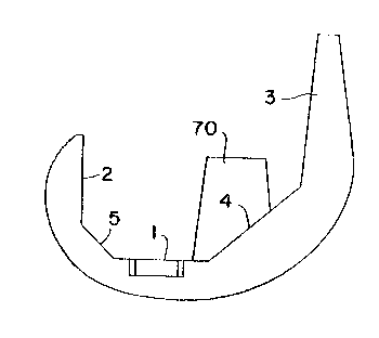
FIG. 1 is a side elevation of a conventional femoral component having five
planar fixation surfaces which match the bone to the implant. Reference numeral 1
indicates tha distal cut surface, reference numeral 2 the posterior cut surFace,5 reference numeral ~ the anterior cut surface, reference number 4 the anterior
chamfer cut surface and reference numeral 5 the posterior chamfer cut surface.
The femoral component can be located with six degrees of freedom, relative
to the patients femoral geometry and FIG. 2 shows the various reference d!rections
for a patients knee. The various degrees of freedom can be expressed in a
10 cartesian manner relative to orthogonal anatomical reference planes as follows:
Angulation: Varus-Valgus, Flexion-Extension,
Internal-Exterl1al Rotation.
The Linear Position: Inferior-Superior,
Anterior-Posterior and Medial-Lateral.
The preferred embodiment of the orthopedic instrument of the present
invention comprises a base component which is used with various accessories. Thebase component, as shown in FIGS. 3-8, comprises two side plates 6, 7 joined by a
number of parallel guide members 8. These structures are used to control the
20 positioning of an oscillating saw blade so that it matches the shape of the femoral
component to be implanted. The geometry of guide members 8 allows for the
cutting of all five cuts to place a femoral component without any repositioning of the
base component relative to the bone.
Each side plate 6, 7 has a pair of angled through holes 9 such that four
25 elongated pins (not shown) can be used to position the instrument in place on the
distal femur. The positioning of these is such that the pins pass into bone thatnot be removed from the femur during preparation for the implant. The parallel
guide member 8 joining the side plates 6, 7 are either manufactured integrally with
them, for example by casting or machining, fabricated by welding or assembled, for
30 example by screwing and dowelling.
Seven of the parallel guide members 8 are shown in this embodiment as part
threaded headed rods 10 which are screwed across from on plate 6 to the other 7.This allows for the use of alternative diameter rods to accommodate differing
thicknesses of saw blade as sold by various manufacturers. Differing thicknesses of
-8 -
saw blade may also be used for each of the cuts. A thin short blade may be most
appropriate to the access and cutting of the posterior and posterior chamfer surface,
where the required travel of the saw blade is short and longer blades may result in
less accurate cuts due to excessive movement at the cutting teeth. When cuning
5 the anterior and distal cuts, a longer and thicker blade may be needed to give the
required cut length and stiffness of blade to avoid deviation when harder sections of
bone are encountered. In addition, the use of separable rod structures allows them
to be manufactured from harder materials, or to be coated in some way to minimize
wear and the generation of metallic debris due to the rubbing action of the saw
1 0 blade.
The size of the guida members is kept as small as possible to maximize the
visibility of the bone through the cutting frame, consistent with providing enough
control of the saw blade. The other three guide members 8 are provided by cross
bars 1 1, 12 and 13, 19 which have guide surfaces 1~, 15, 16, 17, 18 and 25
1 5 respectively.
The directions of cut between the guide surfaces form five different cuts
which are indicated by arrows 20, 21, 22, 23 and 24 in FIG. 8. This figure showshow the base component is located on a femur to be prepared. The shape of the
original femur is indicated by broken line 26 and the shaped surfaces to align with
20 the surfaces 1, 2, 3, 4 and 5 shown in FIG. 1 carrying the same reference numerals.
Between the two guide surfaces 16, 17 used for the anterior and posterior
chamfer there are portions of two halves of a female screw threaded hole 30. These
aliow threaded bolts 31 to be inserted to stabilize the base component against the
distal femur when bone loss is present due to degenerative changes as shown in
25 FIG. 17.
A male boss feature 37 is provided on the cross bar 12. This and a ledge
portion 38 on the cross bar 13 form means of a~taching a number of accessories. A
counterbore 39 is provlded on cross bar 11. This allows accessories which are used
across a range of guide sizes to be appropriately positioned relative to the intra-
30 medullary stem on the femoral component for any of the base component sizes.
~8~6'~t'
The cross bar 19 carries an extension which provides an anterior anchor 26
and is provided with a drill guide 27. This anterior anchor 26 allows a hole to be
drilled into the anterior cortex of the femur to allow a fixation pin 28, shown in F!G. 8,
to be inserted, which provides a significantly enhanced stability for the instrument on
the bone.
A posterior condylar alignrnent accessory 40 is shown in FIGS. 9-12, which
attaches to the base component using the boss 37 and ledge 38 arrangement via a
finger screw 41 as shown in FIG. 13, which screws into a threaded hole 48 in theboss 37 after passing through a hole 49 in a main portion 42, provided with two thin
arms 43 which can be located against the posterior condyles. These are made as
thin as possible while maintaining enough stiffness and toughness to withstand
repeated operative use and act as a condylar sled.
The main portion 42 has a slide 44 which is used as a means of attaching
further accessories. This is illustrated in FIG. 13 which shows accessory 40 with its
condylar sled and extramedullary alignment guide 60 attached.
Anteriorly, accessory 40 has an inclined parallel slot 45 and hole 46 which
are used in conjunction with a sizing stylus 50, as shown in FIG. 16. A nnarker line
crosses the slot 45. The angulation of the holes 45 and slot 45 allows a single
stylus 50 to be used to check sizing of the base component prior to performing any
cuts. Stylus 50 is arranged to move its indicating tip 51 along the locus of the front
of the anterior flanges of a range of sizes of femoral component relative t~ posterior
arms 43. When the sizing stylus 50 is pushed up against the anterior cortex of the
fesnur, the appropriate femoral component size is indicated by the position of marker
line 47 relative to a scale 52 on the stylus 50.
The extramedullary alignment and drill guide 60 depicted in FIG. 13 is shown
in FIGS. 14 and 15 and comprises a guide block 61 and extension 62. Guide block
61 is shaped to fit into slide ~4 in posterior condylar alignment accessory 40 and a
peg 63 engages into counterbore 39 on cross bar 11 as shown in FIG. 15. Guide
block 61 is provided with a clamp member 67, one end of which is threaded at 68 to
receive a locking hand nut 69. ~hus the guide may be securely fastened to
posterior accessory 40. A retaining screw 67a engages the clamp member 67 to
prevent complete removal thereof when the hand nut 69 is released. Guide 60 may
g ~ ll
-1 o-
be similarly positioned into the other positioning accessories that fit onto the base
component, which have slideways equivalent to slideway 44 on posterior accessory40.
A number ot parallel through holes 64 in drill guide 60 are used with long
5 alignment rods 65 as shown in FIG. 21 to orient the instrument assembly relative to
the femoral geometry. These are positioned to overlie the anterior surface of the
femur. Alternative holes are available to allow placement of the guide rods as close
to the patient as possible. The preferred technlque is to align to the femoral head
from the center of the knee for varus-valgus alignment. Flexion-extension alignment
10 is performed by moving the base component until alignment rod 65is parallel with a
line joining the center of the knee and the greater trochanter. Alternative extra-
medullary alignment methods may use the line of the anterior fernoral shaft for
varus-valgus orientation. In this case an alternative drill guide (not shown) is used in
which holes 64 for the alignment rod are angled to compensate for the valgus angle
15 of the femoral shaft relative to the mechanical axis of the femur, ie. the line between
the hip and knee centers.
In the preferred embodiment drill guide 60 also incorporates a drill bushing
66 which extends through clamp member 67. This allows an entry hole to be
formed into the intramedullary cavity. The position of this entry hole is controlled so
20 that it corresponds with the position of the intramedullary stem 70 on the femoral
implant. In this way a further degree of repeatability of technique is gained, current
methods relying on ~'eyeballing" of this entry hole, and the removal of bone is such
that it will not compromise the fixation of the intramedullary feature of the implant in
this area. In existing techniques the intramedullary entry hole may siamese with the
25 fixation surface prepared for the implant's intramedullary feature.
As described with regard to FIG. 17, threaded bolts 31 can be provided
which act as condylar defect screws. These can be placed in the screw holes 30 of
the base component when it is positioned against the distal femur and allow the
guide to be repositioned to make allowance for bone loss caused by degenerative
30 changes, or to adjust tha varus-valgus orientation of the bone guide.
An intramedullary boss 75 is provided as shown in FIG. 18. The boss 75 fits
into the slide 44 on the posterior condylar accessory 40. It also similarly fits in the
alternative positioning accessories to be described. The boss 75 is available in a
number of different valgus angles as indicated by reference numeral 76, and can be
2~h'g~
fitted in two opposite senses to suit either right or left limbs. The boss forms a
guide for the placement of an intramedullary rod 77 which is passed up into the
bone to align the assembly with the femoral canal.
As discussed previously, surgeons may have different preferences in their
choice of alignment method and datums. The current system aims to include as
much versatility as possible without comprising the ease of performing any one of
the approaches when chosen.
There are various alternative femoral alignment methods: Anterior-Posterior
Position. The instrument described so far used the posterior condyles to position
the femoral component. Alternative instruments for other methods are shown in
FIGS. 19, 20, 21 and 22 in which the same reference numerals are used to indicate
similar parts to those used in other figures.
An intramedullary alignment accessory is shown in FIG. 19 which allows the
intramedullary rod 77 to be used as the datum for positioning of the femoral
component. This is necessary when long intramedullary stems are used. This
accessory makes use of an alternative means for locating the extramedullary and
pilot drill guide and the intramedullary boss, and consists of a posterior condylar
alignment accessory with adjustable condylar sleds. In this construction a device 80
similar to accessory 40 is provided but arms 43 are replaced by a pair of adjustable
gauges 81 which can move in relation to main portion 42, and which can be
clamped by a screw clamp 82. An alignment gauge 83 similar to sizing stylus 50
can be moved in hole 46 and slot 45 and locked by clamp 84. A further differenceis that the boss 75 is replaced by a support member 85, which is located in slide 44
but also has a peg a6 which acts in a similar manner to peg 63 on the drill guide 60.
An anterior cortex/patella groove alignment accessory is shown in FIGS. 21
and 22 which allows anterior structures of the femur to be used to place the femoral
component, lhe various parts described in the other figures being assembled
together as shown.
The instruments described so far intend neutral alignment in internal-external
rotation. According to the way that the proximal tibia is resected, it may be
advantageous to externally rotate the femoral component relative to the natural
anatomy. This is achieved by the construction shown in FIG. 23 in which spacer
blocks 90 can be used which fit on to the arms 43 of accessory 40. FIG. 25 shows
g ~
-12-
three blocks 90 which are marked as shown at 91 to show the degree of offset. FIG.
24 shows how the rotation is achieved. The bone in this figure and in FIG. 25 being
indicated by reference numeral 100.
To use the instrument, the base component is initially assembled with the
5 chosen femoral alignment accessory. In lhis case the technique will be described
with respect to use of posterior condylar alignment accessory 40. The extra-
medullary alignment and drill guide 60 is then introduced into slide 44 on the
alignment accessory so that pin 63 on the guide engages against the bottom face of
counterbore 39 in cross bar 11 and a long alignment rod 65 is placed in a hole 64
10 so that the rod lies just above the skin on the patient's thigh and guide 60 is
clamped by hand nut 69. The assembled instrument is then positioned so that
alignment rod 65 passes over the center of the hip and is parallel to the femorai
shaft in the sagittal plane. In cases of condylar bone loss, the condylar defectscrews 31 are introduced and adjusted to stabilize the base component relative to
15 the damaged distal femur.
With the instrument correctly aligned, sizing stylus 50 is introduced into
posterior condylar alignment accessory 40 and pressed up against the anterior
cortex. The size indication is then read from the scale marking 52. If the size
reading does not correspond to the femoral guide currently in position, then the20 base component is replaced with one of the appropriate size. The procedure
performed so far is then repeated. Sizing stylus 50 is removed. With the appro-
priately sized assembly s~orrectly positioned extramedullary alignment and drili guide
60 is used to drill a pilot hole into the intercondylar area.
The extramedullary alignment and drill guide 60 is then removed and
25 replaced with an intramedullary boss 75, appropriately oriented for the left or right
limb and of a valgus angle setting determined from preoperative x-rays or surgeon
preference. An intramedullary rod 77 is then introduced through the boss 75 until it
engages into the isthmus of the femoral canal. The assembly consequentially
repositioned relative to this new datum in its flexion extension and varus-valgus
30 alignment. Anterior-posterior position is now reset by pressing the posteriorcondylar alignment accessory's skids 43 up against the posterior condyles. If intra-
medullary alignment is not possible or required, the preceding steps are omitted.
~ i~ 3 r~ 7
-13-
The positioned assembly is now pinned in place onto the distal femur using
four pins passed through the holes 9 in the side plates 6, 7. These are introduced
either by hammering, drilling or screwing, and may have heads to allow tensile
capture of the jig. A hole is now drilled in the anterior cortex of the femur using the
5 drill guide 27 on the anterior anchor 26 and the fixation pin 28 is inserted to stabilize
the assembly.
All accessories are now removed from the base component. The cuts for the
placement of the femoral component are now made using an oscillating saw.
Preferably, these will be made in the following order: 1) anterior cut, 2) posterior cut,
10 3) posterior chamfer cut, 4) anterior chamfer cut, 5) distal cut.
The distal femur is resected last to allow the piece of bone which will be
removed to support the guide while the other cuts are being made.
Where there is any doubt about the choice of component size, based on the
stylus measurement and~or preoperative templating the largest of the possible sizes
15 is chosen first. The anterior cut is made first to assure that the correct size is being
used and this ensures that the initial anterior cut will not have removed bone needed
for the fixation of a smaller sized component.
While several examples of the present invention have been described, it is
obvious that many changes and modifications may be made thereunto, without
20 departing from the spirit and scope of the invention.