Note: Descriptions are shown in the official language in which they were submitted.
WO92/10tS2 '~ 2 2 ~ ~ PCT/US91/0669
BACKGROUND OF THE INVENTION
This invention relates to a method and apparatus for
adjusting the shape of components of the eye and more
particularly to making fixed changes in the corneal curvature
to correct refractive error.
Deviations from the normal shape of the corneal surface
produce errors of refraction in the visual process. The eye
in a state of rest, without accommodation, focuses the image
of distant objects exactly on the retina. Such an eye enjoys
distinct vision for distant objects without effort. Any
variation from this standard constitutes ametropia, a
con*ition in which the eye at rest is unable to focus the
image of a distant object on the retina. Hyperopia is an
error of refraction in which, with the eye at rest, parallel
rays from distant objects are brought to focus behind the
retina. Divergent rays from near objects are focused still
further back. In one aspect of hyperopia, the corneal surface
is flattened which decreases the angle of refraction of rays
as they pass through the refractive surfaces of the cornea,
causing a convergence or focus of the rays at a point behind
the retina. The retina is comprised partially of nerve fibers
which are an expansion of the optic nerve. Waves of light
falling on the retina are converted into nerve impulses and
carried by the optic nerve to the brain to produce the
sensation of light. To focus parallel rays on the retina, the
hyperopic eye must either accommodate, i.e., increase the
convexity of its lens, or a convex lens of sufficient strength
to focus rays on the retina must be placed before the eye.
Myopia is that refractive condition in which, with
accommodation completely relaxed, parallel rays are brought to
focus in front of the retina. One condition which commonly
causes myopia is when the corneal curvature is steepened, thus
the refraction of rays is greater as the rays pass through the
',
.. :
~ ,,
0 92/10152 2 ~ ~ ~ 2 ~ 5 PC~r/US91/06691
refractive surfaces of the cornea, and the over-refracted rays
converge or focus in front of the retina in the vitreous of
the eye. When the rays reach the retina they become
divergent, forming a circle of diffusion and consequently a
blurred image. A concave lens is used to correct the focus of
the eye for myopia.
The normal treatment of these classic forms of refractive
error of the eye is with the use of eyeglasses or contact
lenses, both of which have well-known disadvantages to the
user. It has been estimated that 60 million pairs of
eyeglasses and 3 million pairs of contact lens are sold
annually.
Recent research has been directed to operative techniques
to change the refractive condition of the eye. Such
techniques are generally referred to as "keratorefractive
techniques". Two such techniques are more particularly called
keratophakia and keratomileusis. Keralomileusis involves the
regrinding of a corneal lamella into a meniscus or hyperopic
lens to correct myopia or hyperopia. A corneal optical lathe
has been especially developed for this procedure and is also
used in the keratophakia procedure, when a homograft ground
into a convex lens is placed interlamellarly to correct
aphakic hypermetropia. The homograft tissue (corneal lamella)
is frozen with carbon dioxide. The homograft is cut as a
contact lens would be, i.e., to the optical power required to
effect the desired optical correction of the cornea. In
keratomileusis, the anterior corneal lamella is shaped by the
lathe and in keratophobia, it is the corneal stroma of a donor
eye that is shaped by the lathe. These techniques have a
broad application in the correction of high hyperopic and
myopic errors. These procedures require radial cutting of the
cornea about the periphery of the graft which weakens the
cornea so that pressure from fluids below the incisions pushes
. ~, ~ ,:'
:'' . : .: - ,
: - ~
~ , , ,: . :
2 ~ 9 2 ,~ ~ PCT/US91/066~
up under the cuts and flattens the curvature of the cornea.
This flattening of the cornea results in refractive errors to
the eye not compensated for by the graft. Suturing in these
operations also causes radial asymmetry of the cornea which
consequently promotes astigmatic error in this regard.
Sutures also cause scarring of the corneal tissue, which scar
tissue loses its transparency. Surgical correction of
astigmatism is accomplished by asymmetrically altering the
corneal curvatures. The effect of a peripheral distorting
force may be easily visualized by imagining an inflated
balloon with a spherical surface being compressed between the
palms of the hands. Because the volume of air in the balloon
is constant, the surface area remains constant. The
previously spherical anterior surface is distorted meridional
as a result of compressing the diameter between the hands so
that the curvature changes without changing the circumference
of the surface. The meridian passing over the balloon between
the extended fingers steepens, while the uncompressed meridian
at right angles thereto flattens as its diameter lengthens in
proportion to the shortening of the compressed diameter. This
demonstrates the effect that may result from slight variations
in the symmetrical patterns or intentional asymmetrical
patterns attempted to be accomplished during surgical
procedures and attendance suturing. It is thus seen that
present procedures in keratorefractive techniques are best
limited to situations where other more standard corrective
practices are found ineffective. It is readily seen that the
limiting factors in such surgical techniques is the gross
complexity involved not only with multiple incisions in
corneal tissue for affecting the procedures but also complex
suturing patterns, resulting in gross restructuring of the
eye. The eye is thus faced with a difficult job of adjusting
to this trauma.
- ,
': ' . '
'
~092/10152 2 ~ Q r~ ~ 8 5 PCT/US91/06691
Over the past few years developments have been made in
the use of lasers as a means to reshape the cornea in an
attempt to get rid of refractive errors. In these processes,
pulsed lasers remove tissue from the cornea by shaving off or
vaporizing portions of the corneal surface to cause it to
flatten. The most common type is an Exemer laser. The
fundamental effect of such a laser on tissue is a
photochemical one, the breaking of molecular bonds with so
much energy that the tissue fragments fly from the surface at
supersonic speeds, leaving behind a discreet space. The
process has been designated as ablative photodecomposition or
photoablation.
The use of Exemer lasers require delivery of the beam to
the eye in a controlled manner requiring that the homogenous
beam be appropriately managed and focused because the optical
elements must withstand the high energy photons and because
the beam must be shaped to a non-uniform configuration to
create the new non-uniform optical surface of the cornea.
Such delivery system contains multiple components including
lenses to expand or focus the beam, mirrors to direct the
beam, modulators to homogenize the beam, masks to shape the
beam, and detectors to measure the intensity and configuration
of the beam. Current models range from a simple collection of
lenses and masks to complex robots with components that
control not only the laser parameters but also the optical and
mechanical components. Because the process is dealing with
submicron (less than .0000l of a meter) accuracy, great
demands are placed upon such systems for stability, even
though the interaction of the laser and tissue lasts only
microseconds.
Using the system requires exquisite technical and
biological control to modulate corneal shaping.
Another laser treatment process focuses light, like a
. .. . . . . . .
.. . , - . .:~ .
: . . .
. ~ :
WO92/10152 PCT/US9t/066' ~-
2~ 6
magnifying glass, to boil away tissue one cell at a time, ~
instead of carving away the surface. One problem is adequate -
control to prevent the process from cutting through a layer of ~ . corneal tissue known as Bowman's membrane--a section of the
eye that does not regenerate.
.,.. ~....... :. -, . . .
:
. . -
- .
` '' , ~ ' '. . ' ~ ' '
. .
O 92/10152 ~ ~ 3 ~ ~ ~ 5 PC~r/US91/06691
SUnIMUiRY OF THE INrVENTION
It is therefore an object of the present invention to
provide a new and improved keratorefractive technique
involving method and apparatus for changing the shape of the
optical zone of the cornea to correct refractive errors of
hyperopia (far-sightedness), myopia (near-sightedness), and
astigmatism, whereby a non-spherical surface exists on the eye
system and the simplicity of the technique virtually
eliminates the chance of error or further complications
resulting from gross disturbances of the eye system.
With this and other objects in view, the present
invention contemplates a method and apparatus that can only be
described as scraping, sculpting, or removing portions of the
cornea for the purposes of correcting refractive error in
human cornea. For the purposes of this invention and that of
said co-pending application, S.N. 450,672, the action will be
called "scraping".
Another object of the invention is to provide mechanical
apparatus capable of easily being used by a surgeon for
scraping the cornea in order to correct refractive errors of
hyperopia, myopia, and astigmatism which includes means to
provide consistency in depth and configuration of the surface.
Another object of this invention is to provide method and
apparatus for scraping the cornea wherein the cornea is
maintained in a more rigid posture during the procedure to
eliminate flexure of the cornea and thus provide greater
accuracy in determining predicable amounts of corneal material
to be removed. This is accomplished by creating a vacuum in
the operative space above the cornea during the process.
Specifically, the method objects of this invention
involve the surgical reprofiling of the corneal portion of an
eye of humans, to change the corneal radius and thus correct
refractive errors. The steps include creating a placido ring
.. . .
. ~ . .
: ~
: ' - , ` '
, ~ . . ~ :
WO92/10152 ~ PCT/US91/066~
?.~3
keratograph of a simulated cornea having the correct
refractive qualities. Next, an actual keratograph of the
cornea is created. The two kerotographs are compared to
determine the amount of refractive error, i.e. whether it
would be hyperopia, myopia, or astigmatism.
A reprofiling tool is constructed to include a plurality
of scraper blades of shape sufficient to change a corneal
radius to that of the simulated cornea. The reprofiling tool
is then positioned within a holding sleeve that is
contiguously positioned upon said eye such that the scraper
blades will contact the cornea. A vacuum is created in the
chamber above the cornea wherein the scraping tool is
positioned. The scraping tool is then rotated or oscillated
with the axial movement of the scraping tool being changed and
indexed until the corneal radius has been corrected to that of
the simulated or ideal cornea.
The apparatus used to achieve the objects of this
invention specifically includes a cylindrical positioning ring
having a resilient vacuum ring means on its bottom side for
temporary attachment to the sclera portion of an eye which
surrounds the cornea that is to be reprofiled. A plurality of
positioning pins exist on the top side of the positioning ring
and a vacuum means is provided for communication with the
vacuum ring. A cylindrical holding sleeve includes means at
the bottom of the holding sleeve to interconnect with the
positioning pins of the cylindrical positioning ring. A
flexible and preferably clear tubing member extends from the
bottom of the holding sleeve to seal against the cornea. Fine
Screw threads of a given pitch, preferably about 40 threads
per inch, are formed on the interior or exterior portion of
the holding sleeve. Threadably connected thereto is a guide
sleeve having screw threads of the same pitch as the threads
of the holding sleeve for rotatable attachment with the
.. : . .; - .. - :
, - . . ~ - :
~-, - : .
. - ~ -, , ~ :
.: ' -
,; - . . . . .
,O 92/10152 P ~ /US91/06691
holding sleeve. A scraping tool is adapted to be rotatably
and axially received within the positioning ring, the holding
sleeve, and the guide sleeve. A seal means is provided
between the scraping tool, the guide sleeve and/or holding
sleeve. A collar means existing on the scraping tool allows
it to be rotatably supported upon the guide sleeve. A
plurality of blades at the bottom of the scraper tool are
designed to be of a shape sufficient to scrape away portions
of the cornea to achieve the desired corrective curvature.
' ` :- .-':, ' . ' : '
:-
.. .. .
W092/10152 ~ 'j PCT/US91/066
BRIEF DESCRIPTION OF THE DRAWINGS
Figure l is a schematic illustration of a horizontalsection of the eye.
Figure 2 is a schematic illustration of a hyperopic eye
showing adjustment of the cornea to shorten the radius of
curvature.
Figure 3 is a schematic illustration of a myopic eye
system showing adjustment of the cornea to increase its radius
and thus flatten the corneal slope.
Figure 4 is a detailed schematic illustration of a
horizontal section of the frontal portion of an eye showing
the various layers of the cornea.
Figure 5 is an exploded view showing the basic components
of the apparatus of this invention.
Figure 6 is an end view of the reprofiling tool taken
along the line 6-6 of Figure 5.
Figure 7 is a top view of the tool holder taken along the
line 7-7 of Figure 5.
Figure 8 is a top view of the positioning ring taken
along the line 8-8 of Figure 5.
Figure 9 is an assembly view, partly cut-away to show the
apparatus of the invention.
".. .
.
- .,
~ - .
~092/101~2 2 ~ ~ 2 2 ~ ~ PCT/US91/06691 ~ ~-
DETAILED DESCRIPTION OF T~E PREFERRED EMBODIMENT
Before explaining the present invention in detail, it is
to be understood that the invention is not limited in its
application to the details of the construction and arrangement
of parts illustrated in the accompanying drawings. The
invention is capable of other embodiments and of being
practiced or carried out in a variety of ways. It is to be
understood that the phraseology and terminology employed
herein is for the purpose of description and not of
limitation.
Referring first to Figure 1 of the drawings, a horizontal
section of the eye shows the globe of the eye resembling a
sphere with an anterior bulged spherical portion 12
representing the cornea. Thus the eye is actually comprised
of two somewhat modified spheres placed one in front of the
other. The anterior of these two segments is the smaller more
curved cornea.
The globe of the eye consists of three concentric
coverings enclosing the various transparent media through
which the light must pass before reaching the sensitive
retina. The outermost covering is a fibrous protective
portion, the posterior five-sixths of which is white and
opaque and called the sclera 13, and sometimes referred to as
tne white of the eye where visible to the front. The anterior
one-sixth of this outer layer is the transparent cornea 12.
A middle covering is mainly vascular and nutritive in
function and is comprised of the choroid 14, ciliary body 15
and iris 17. The choroid generally functions to maintain the
retina. The ciliary muscle is involved in suspending the lens
and accommodation of the lens. The iris is the most anterior
portion of the middle covering of the eye and is arranged in
a frontal plane. It is a thin circular disc corresponding to
the diaphragm of a camera, and is perforated near its center
:: .: . - . :: . : . . .
: : : : :
,.. ~ ~ -
:- . - : - . .
.
:. - , ,
. . .
' ~ ' - . : .
WO92/10152 PCT/US91/066g
2~ 85 12
by a circular aperture called the pupil 19. The size of the
pupil varies to regulate the amount of light which reaches the
retina. It contracts also to accommodation, which serves to
sharpen the focus by diminishing spherical aberration. The
iris divides the space between the cornea 12 and the lens 21
into an anterior chamber 22 and posterior chamber 23. The
innermost portion of covering is the retina 18, consisting of
nerve elements which form the true receptive portion for
visual impressions.
The retina is a part of the brain arising as an outgrowth
from the fore-brain, with the optic nerve 24 serving as a
fibre tract connecting the retina part of the brain with the
fore-brain. A layer of rods and cones, lying just beneath a
pigmented epithelium on the anterior wall of the retina, serve
as visual cells or photoreceptors which transform physical
energy (light) into nerve impulses.
The vitreous 26 is a transparent gelatinous mass which
fills the posterior four-fifths of the globe. At its sides it
supports the ciliary body 16 and the retina 18. A frontal
saucer-shaped depression houses the lens 21.
The lens 21 of the eye is a transparent bi-convex body of
crystalline appearance placed between the iris 17 and vitreous
26. Its axial diameter varies markedly with accommodation.
A ciliary zonule 27, consisting of transparent fibers passing
between the ciliary body 16 and lens 21 serves to hold the
lens in position and enable the ciliary muscle to act on it.
Referring again to the cornea 12, this outermost fibrous
transparent coating resembles a watch glass. Its curvature is
somewhat greater than the rest of the globe and is ideally
spherical in nature. However, often it is more curved in one
meridian than another, giving rise to astigmatism. A central
third of the cornea is called the optical zone with a slight
flattening taking place outwardly thereof as the cornea
.......
. .: . ,, . ~ , ' :
: . . - .
- : :
: - .
:
VO 92/10152 ~ !3 c~ 3 5 PC~r/US91/06691
13
thickens towards it periphery. Most of the refraction of the
eye takes place on the surface of the cornea.
Referring next to Figure 2 of the drawings, the globe of
an eye is shown having a cornea 12 with a normal curvature
represented by the solid line 39. If parallel rays of light
41 pass through the corneal surface 39 of Figure 2, they are
refracted by the corneal surfaces to converse eventually near
the retina 18 of the eye. The diagram of Figure 2 discounts,
for the purposes of this discussion, the refractive effect of
the lens or other portions of the eye. The eye depicted in
Figure 2 is hyperopic and thus the rays of light 41 are
refracted to converge at point 42 behind the retina. If a
peripheral band of pressure is applied inwardly at the chord
43 of the cornea, the walls of the cornea are caused to
steepen. This is because the volume of fluids within the
anterior chamber 22 remains constant, thus the anterior
portion of the cornea, including the optical zone (inner third
of the cornea) steepens in slope to form a curvature (shown in
exaggeration) following the dotted line 44. The rays of light
41 are then refracted from the steeper surface 44 at a greater
angle to direct the refracted rays into focus at shorter
distance, such as directly on the retina 18.
Figure 3 shows a similar eye system to that of Figure 2
except that the so-called normal corneal curvature of Figure
3 causes the light rays 41 to refract into focus at a point 46
in the vitreous which is short of the retinal surface 18.
This is typical of a myopic eye. If chord 43 of the cornea is
expanded uniformly outwardly as shown by the arrows, the walls
of the cornea are flattened. Light rays 41 refracted by the
now-flattened corneal surface will be refracted at a smaller
angle and thus converge at a more distant point such as
directly on the retina 18.
Referring now to Figure 4, a more detailed drawing of the
" -
.
: . :: - , : , .
~' ' ', , ' '., ' ' :
-, . , - ' ,. .
. , ' ' : . : . .
,:;- . ~ ' ~. ;
`-,
WO92~1015~ PCT/US91/0669
~ ?~2a~ 14
anterior portion of the globe shows the various layers of the
cornea comprising an epithelium 31. Epithelial cells on the
surface thereof function to maintain transparency of the
cornea. These epithelial cells are rich in glycogen, enzymes
and acetylcholine and their activity regulates the corneal
corpuscles and controls the transport of water and
electrolytes through the lamellae of the stroma 32 of the
cornea.
An anterior limiting lamina 33, referred to as Bowman's
membrane, i5 positioned between the epithelium 31 and the
substantia propria or stroma 32 of the cornea. The stroma is
comprised of lamella having bands of fibrils parallel to each
other and crossing the whole of the cornea. While most of the
fibrous bands are parallel to the surface, some are oblique,
especially anteriorly. The fibrous bands within alternate
lamella are at a near right angle to bands in the adjacent
lamella. A posterior limiting lamina 34 is referred to as
Descemet's membrane. It is a strong membrane sharply defined
from the stroma and resistant to pathological processes of the
cornea.
The endothelium 3~ is the most posterior layer of the
cornea and consists of a single layer of cells. The limbus 37
is the transition zone between the conjunctiva 38 and sclera
13 on the one hand and the cornea 12 on the other.
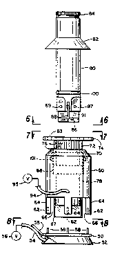
Referring now to Figures 5-9, the assembly of the basic
parts of the apparatus are shown in exploded and assembled
views. These parts comprises a cylindrical positioning ring
50 having a resilient vacuum ring 52 extending from the bottom
side of the positioning ring for contact with the eye of the
patient being treated. A vacuum hose 54 provides
communication from the inside of the resilient ring 52 and a
vacuum pump source means 56 as a means to retain the assembled
parts upon the eye for surgical procedures herein described.
; . .: : : . . : :
:- .. ~ . ~ ' :
~, , .: ~
: ,: : .
:.......................... : . .~
VO92/10152 ~v ~ 2 ~ ~ PCT/US91/06691
A plurality of positioning pins 58 are provided on the top
side of the positioning ring to receive the cylindrical
holding sleeve 60, the pins being adapted to be received
through openings 62 in legs 64 of holding sleeve 60. Visual
inspection openings 66 are provided between the legs for use
by the surgeon or technician performing the process. A
translucent, flexible, e.g. vinyl, cylinder 67 is centrally
positioned at the bottom of the holding sleeve, which, when in
use, will contact the outer portion of the cornea. The
interior of the cylindrical holding sleeve 60 includes a
plurality of screw threads 68 along a portion of its length,
the threads being a very fine thread, e.g. of a pitch equal to
40 threads per inch. An indicia or marker 70 is provided in
the body of the cylindrical holding sleeve so as to provide a
visual measuring point for the surgeon relative to the
rotatable position of a micrometer-like guide sleeve 72 which
includes exterior threads to match threads 68 of the
cylindrical holding sleeve. The guide sleeve includes an
outer knob portion 74 and indicia generally designated by the
numeral 76, e.g. millimeter markings on the lower portion of
the guide sleeve which function to provide the amount of axial
movement of the guide sleeve. The interior surface 78 of the
cylindrical holding sleeve is adapted to rotatably receive a
scraping tool 80. A conduit 94 communicates the interior of
the holding sleeve with a vacuum source 95. The scraping tool
includes a collar 82 which is adapted to rest upon the top
surface 83 of the guide sleeve 72 for movement upwardly or
downwardly therewith. The top end of the scraping tool can
include a knurled grip portion 84 for rotation and/or
oscillation by the surgeon. Positioned along the body of the
scraping tool is O-ring 100 to seal against interior surface
78. An upper 0-ring 101 frictionally engages the threads of
a guide sleeve 72 to prevent inadvertent turning. This, along
- -
: ', . ~ -
:
- - - . : . . :
,. : , '
WO92/10152 PCT/US91/0669
2~ 285
16
with the cylinder 67, forms a vacuum chamber above the cornea
98. At the bottom of the scraper tool are a plurality of
blades 86 and 88 which are retained within the body of the
scraping tool 80 by pins 87, 89, and 91. The blades 86 and 88
are retained transverse to the longitudinal axis of the
scraping tool 80. The blades 86 and 88 as used in the
invention are of surgical steel. The scraping tool 80 of
Figure 5 is adapted to provide a scraping operation upon the
cornea over the top center thereof for myopia refractive
error, i.e. near-sightedness, which will effectively lengthen
the corneal radius of curvature as described relative to
Figure 3. To correct for hyperopia (far-sightedness), the
scraping tool blades as show~ and described in Figure 8 of
Serial No. 450,672 are utilized. The blades are adapted to
contact the outer anterior portion of the cornea in order to
shorten the effective radius thereof, that is, the blades will
be adapted to contact the area A as shown in Figure 2 whereas
the scraping tool 80 of Figure 5 will be adapted to sculpt the
area B of Figure 3.
The operation of the apparatus and methods of surgery are
accomplished by first taking optical measurements of the eye
as to the shape of the cornea and to determine the refractive
error, for example, the shape the cornea should have in order
for that eye to operate in an optically correct manner--i.e.
correct refractive errors. Typically, a kerotograph
photographic image using a placido-ring target such as
described in U.S. Patent 3,797,921 is used. The photograph is
of reflected light from the placido rings upon a standard
spherical surface of the same size as the cornea in question,
creating an image in the same manner as a topographic contour
map. Subsequently, the topographic survey of the eye to be
corrected is made for comparison purposes and to provide the
surgeon with the necessary information for correcting the
,: , .: :
:: ' -' '
-- .
~092/10152 PCT/US9t/06691
17
refractive errors. Once this occurs, the operation will
proceed by placing the positioning ring 50 over the eye. The
size of this ring may vary for different operations but is
preferably of size wherein the resilient vacuum ring 52 will
rest upon the sclera of the eye concentric about the cornea.
Once the cylindrical positioning ring 50 is in place, the
cylindrical holding sleeve 60 is then positioned thereupon by
the- engagement of openings 62 with positioning pins 58.
Resilient vinyl seal tubing or cylinder 67 contacts the outer
circumference of the cornea to seal therewith. Thereafter,
the scraping tool 80 is inserted within the cylindrical
holding sleeve 60 to a position where the bottom of the knife-
edge blades 86 and 88 will initially contact the cornea. As
shown in U.S. Application S.N. 450,672, and incorporated
herein by reference, the determination of contact of the tool
blades with the cornea can be achieved electrically. See
Figure ll of the aforesaid application. By rotating the guide
sleeves 72 in incremental amounts as dictated by the caliper
or measuring scales 70 and 76, the surgeon can continue to
increase the depth of the scraping operation. Scraping of the
cornea occurs by hand rotation of the scraping tool 80
although other mechanical or motor operated means are within
the scope of this invention.
In myopic conditions, the scraping tool 80 of Figure 5 is
utilized. During the operation, the knife-edge blades press
upon the corneal surface which becomes depressed and thus
gives a larger contact surface with the blades resulting in a
larger diameter of sculptured surface. The scraping action is
accentuated in proportion to the pressure between the cornea
and the blade. With a partial vacuum formed in the chamber
above the cornea, the cornea becomes less yieldable and semi-
rigid. The extent of rigidity is a function of the amount of
vacuum. Pressures between five-eighths (5/8) to three-fourths
,: ' . - ~ ,: ~
WO92/101~2 ~ PCT/US91/0669.
?J8 ~
18
(3/4) atmosphere or six (6) to ten (lO) inches of Mercury (Hg)
appear to be preferable, although not limiting to the purposes
of this invention. It has been found that this allows the
tool to have a greater positive 'feel' during the procedure
with more predictable results in removal of corneal material
to achieve the correct contour. The resulting effect is a
lengthening of the refractive radius in that portion of the
cornea under the blade. When the tool is removed, the cornea
returns to its normal contour except that the radius over the
top center thereof is now longer than it was initially. As a
result, refractive light through the cornea now focuses upon
the retina. The scraping action occurs by the surgeon in
incremental movement by rotating the guide sleeve 72 relative
to the cylindrical holding sleeve 60 utilizing the incremental
measuring indicia 76 relative to pointer or other indicia 70.
As one example, the guide sleeve is graduated into 25
micrometer divisions to provide .OOl" adjustments for each
marked division of rotation. Through use, the surgeon or
technician begins to decide the amount of downward movement
needed to achieve the required changes in the cornea by the
rotation and/or oscillation of the knives. The rotation for
a period of a few seconds will result in removal of small
amounts of corneal material from the cornea. The tool can be
removed and/or kerotographic photographs taken to determine if
the refractive error has been corrected. Since the apparatus
and the surgical methods deal with very small increments of
movement in the corneal reprofiling process, it is essential
that the first contact setting be precise and accurate. Many
times this can be done by visual means by the surgeon and in
other instances electrical detecting means as previously
described can be provided between the cornea and the tool
blade to provide an exact setting of the tool which permits
repeatable amounts of corneal removal.
~. ~ . . .
: ~ . , ~ .
.
: ~: . , ., : .- . . . .
- .,
,., ~ . . ; . ., : :
.: . .- . : , .