Note: Descriptions are shown in the official language in which they were submitted.
20g~
DENTAL APPLIANCE FOR TREATMENT OF
SNORING AND OBSTRUCTIVE SLEEP APNEA
Field of the Invention
This application relates to a dentally retained
intra-oral appliance worn at night for treatment of snoring
and obstructive sleep apnea. The appliance maintains the
patient's mandible in an anterior, protruded position to
prevent obstruction of the pharyngeal airway. The appli-
ance allows a limited degree of lateral movement of the
mandible relative to the upper jaw in the protruded posi-
tion to prevent aggravation of the tempromandibular jointand associated muscles and ligaments.
Background of the Invention
Snoring and obstructive sleep apnea are typically
caused by complete or partial obstruction of an individ-
ual's pharyngeal airway during sleep. Usually airway
obstruction results from the apposition of the rear portion
of the tongue or soft palate with the posterior pharyngeal
wall. Obstructive sleep apnea is a potentially lethal
disorder in which breathing stops during sleep for 10
seconds or more, sometimes up to 300 times per night.
Snoring occurs when the pharyngeal airway is partially
obstructed, resulting in vibration of the oral tissues
during respiration. These sleep disorders tend to become
more severe as patients grow older, likely due to a pro-
gressive loss of muscle tone in the patient's throat and
oral tissues.
Habitual snoring and sleep apnea have been
associated with other potentially serious medical condi-
tions, such as hypertension, ischemic heart disease and
strokes. Accordingly, early diagnosis and treatment is
recommended. One surgical approach, known as uvulopalato-
'~
094~1
pharyngoplasty, involves removal of a portion of the soft
palate to prevent closure of the pharyngeal airway during
sleep. However, this operation is not always effective and
may result in undesirable complications, such as nasal
regurgitation.
A wide variety of non-surgical approaches for
treating sleep disorders have also been proposed including
the use of oral cavity appliances. It has been previously
recognized that movement of the mandible (lower jaw)
forward relative to the maxilla (upper jaw) can eliminate
or reduce sleep apnea and snoring symptoms by causing the
pharyngeal air passage to remain open. Several intra-oral
dental appliances have been developed which the user wears
at night to fix the mandible in an anterior, protruded
(i.e. forward) position. Such dental appliances essential-
ly consist of acrylic or elastomeric bite blocks, similar
to orthodontic retainers or athletic mouthguards, which are
custom-fitted to the user's upper and lower teeth and which
may be adjusted to vary the degree of anterior protrusion.
United States patent No. 4,901,737, which issued
to Toone on 20 February, 1990, exemplifies the prior art.
Toone discloses an intra-oral appliance for reducing
snoring which repositions the mandible in an inferior
(open) and anterior (protrusive) position as compared to
the normally closed position of the jaw. Once the dentist
or physician determines the operative "snore reduction
position" for a particular patient, an appropriate mold is
taken of the maxillary dentition and of the mandibular
dentition for formation of the appliance template. The
Toone appliance includes a pair of V-shaped spacer members
formed from dental acrylic which extend between the maxill-
ary and mandibular dentition to form a unitary mouthpiece.
In an alternative embodiment of the Toone invention, the
spacer members are formed in two pieces and a threaded rod
~9~
-3
is provided to enable adjustment of the degree of mandibu-
lar protrusion or retrusion after the mouthpiece is formed.
European patent application No. 0,312,368 pub-
lished April 19, 1989 also discloses an intra-oral device
for preventing snoring. This device consists of a U-
shaped mouthpiece which conforms to the upper dental arch
of the user and includes a sloped, lower ramp for engaging
the mandibular dentition. Normal mouth motions, such as
the clenching of the jaw, will cause some of the mandibular
dentition to engage the underside of the ramp, thereby
camming the lower jaw forward to increase the spacing
between the base of the tongue and the posterior wall of
the pharynx.
While prior art dental appliances have proven
effective in maintaining the mandible in a protruded
position to improve airway patency, they often result in
undesirable side effects. One of the most common side
effects is aggravation of the tempromandibular joint and
related jaw muscles and ligaments, especially in individ-
uals who have a tendency to grind their teeth during sleep.
Aggravation of the tempromandibular joint has been associ-
ated with a wide variety of physical aliments, including
migraine headaches. Accordingly, many individuals suffer-
ing from sleep apnea and snoring disorders are not able to
tolerate existing anti-snoring dental appliances for long
periods of time.
The need has therefore arisen for a dental appli-
ance for treatment of snoring and sleep apnea which will
maintain the mandible in a preferred anterior position, but
which will also allow a limited degree of lateral excursion
of the mandible relative to the upper jaw to avoid discom-
fort to the tempromandibular joint and related muscles and
ligaments.
4 2~94411
Summary of the Invention
In accordance with the invention there is pro-
vided an intra-oral dental appliance to be worn by a
patient during sleep for treatment of obstructive sleep
apnea and snoring. The dental appliance includes an upper
member conforming to the patient's maxillary dentition, a
lower member conforming to the patient's mandibular den-
tition, and connecting means for releasably coupling theupper and lower members together. The connecting means
adjustably maintains the lower member in an anterior,
protruded position relative to the upper member while
permitting a limited degree of lateral movement of the
lower member relative to the upper member in the protruded
position.
The upper and lower members are preferably
flexible bite blocks formed from elastomeric material.
Preferably the connecting means is secured to an anterior
region of the upper and lower bite blocks and includes a
first element bonded to an undersurface of the upper bite
block; a second element bonded to an upper surface of the
lower bite block; and an elongated connector for releasably
coupling the first and second elements together.
Preferably the connector has an upper end which
is fixedly connectable to the first element and a lower end
which is loosely connectable to the second element to
permit a limited degree of movement of the lower bite block
relative to the connector. Advantageously, the second
element has an internal cavity for capturing the lower end
of the connector, the cavity having an opening formed on an
upper surface of the second element through which the
connector extends. The connector preferably consists of a
stylus having a threaded upper portion and an enlarged head
2~94411
~_ 5
formed on its lower end having a diameter exceeding the
size of the cavity opening.
The upper element preferably consists of a reten-
tion plate having a plurality of internally threadedapertures formed therein for releasably receiving the
threaded portion of the stylus. The threaded apertures are
spaced apart at regular intervals to enable incremental
adjustment of the degree of anterior protrusion of the
lower bite block relative to the upper bite block.
The second element preferably consists of a guide
box having an upper surface, vertical sidewalls, and an
open bottom end, the guide box having a hollow area between
the sidewalls comprising the internal cavity and an aper-
ture formed on the upper surface comprising the cavity
opening. The dimensions of the cavity preferably exceed
the size of the stylus head and the cavity opening is lat-
erally elongated to permit a limited degree of lateral
movement of the lower bite block relative to the stylus.
The guide box further includes a base plate for releasably
covering the guide box bottom end, the base plate having an
aperture formed therein to permit access to the stylus head
captured within the internal cavity.
The retention plate and the base plate each
further include a plurality of retention apertures spaced
around the periphery thereof to enable acrylic to flow
through the plates when the plates are initially bonded to
the upper and lower bite blocks.
Preferably, the upper and lower bite blocks
further include a first pair of bite pads formed on the
undersurface of the upper bite block and projecting down-
wardly therefrom and a second pair of bite pads formed onan upper surface of the lower bite block and projecting
upwardly therefrom for slidably engaging the first pair of
2094411
_ -6
bite pads. The bite pads are located in a posterior region
of the bite blocks to limit closure of the patient's jaw.
A method of treating snoring and obstructive
sleep apnea by adjustably maintaining a patient's mandible
in a protruded position is also disclosed comprising the
steps of (a) casting an upper bite block by taking a mold
of the patient's maxillary dentition; (b) casting a lower
bite block by taking a mold of the patient's mandibular
dentition; (c) securing a first retention element to the
undersurface of the upper bite block in an anterior region
thereof; (d) securing a second retention element to an
upper surface of the lower bite block in an anterior region
thereof, the second element comprising an upwardly pro-
jecting connector having a lower end loosely capturedwithin a cavity formed in the second element; (e) deter-
mining the preferred degree of mandibular protrusion
required to alleviate the patient's sleep apnea and snoring
symptoms; and (f) releasably securing an upper end of the
connector to the first retention element at a fixed posi-
tion corresponding to the patient's preferred degree of
mandibular protrusion as determined in step (e).
A connecting assembly for use in an intra-oral
dental appliance to be worn by a patient during sleep for
treatment of obstructive sleep apnea and snoring is also
disclosed, the dental appliance comprising an upper bite
block conforming to the patient's maxillary dentition and
a lower bite block conforming to the patient's mandibular
dentition. The connecting assembly includes a first
element securable to an undersurface of said upper bite
block in an anterior region thereof; a second element
securable to an upper surface of said lower bite block in
an anterior region thereof; and an elongated connector for
releasably coupling said first and second elements to-
gether, said connector having an upper end which is fixedly
connectable to said first element and a lower end which is
2094411
_ -7
loosely connectable to said second element to permit a
limited degree of movement of said lower bite block rela-
tive to said connector.
Brief Description of the Drawings
In drawings which illustrate the preferred
embodiment of the invention,
Figure 1 is a schematic, side elevational view of
the upper pharyngeal region of a typical patient suffering
from obstructive sleep apnea or snoring.
Figure 2 is a schematic, side elevational view of
the patient of Figure 1 fitted with the applicant's dental
appliance to maintain the patient's mandible in a protruded
position, thereby averting obstruction of the pharyngeal
airway.
Figure 3 is an enlarged, exploded view of the
connecting assembly of the applicant's invention for
connecting the upper and lower bite blocks shown in dotted
outline.
Figure 4 is an isometric view of the applicant's
dental appliance.
Figure 5 is a top, plan view of the dental
appliance of Figure 4.
Figure 6 is a front, elevational view of the
dental appliance of Figure 4.
Figure 7 is an enlarged, cross-sectional view
taken along section lines 7-7 of Figure 6 and looking in
the direction of the arrows.
2094411
-8-
Figure 8 is an enlarged, cross-sectional view
taken along section lines 8-8 of Figure 6 and looking in
the direction of the arrows.
Figure 9 is an enlarged, cross-sectional view
taken along section lines 9-9 of Figure 5 and looking in
the direction of the arrows.
Detailed Description of the Preferred Embodiment
Figure 1 is a schematic, side elevational view of
the upper pharyngeal region of a typical patient suffering
from obstructive sleep apnea or snoring. When the patient
is asleep the oral cavity tissues relax and the distal
(rear) portion of the tongue 10 tends to slide rearwardly
toward the posterior pharyngeal wall 12. This may result
in partial or complete occlusion of the pharyngeal airway
14 in the circled region 26. As the volume of airway 14
diminishes, the velocity of the air passing the oral cavity
tissues tends to increase. This may result in vibration of
the oral cavity tissues, such as the uvula 16 which is a
fleshy projection suspended from the soft palate 18 over
the root of tongue 10. The vibration of these oral tissues
causes the snoring sound.
Figure 2 is a schematic, side elevational view of
the individual of Figure 1 fitted with the applicant's
dental appliance 20. Dental appliance 20 causes the
individual's mandible 22 to be moved to an anterior, pro-
truded position relative to the upper jaw 24. This causes
the dorsal surface of the tongue 10 to move forwardly away
from the posterior pharyngeal wall 12. This in turn
results in an increase in volume of the pharyngeal airway
14 as shown in the circled region 26. It has been shown
that maintaining airway 14 substantially open during sleep
9 209~411
alleviates the undesirable symptoms associated with ob-
structive sleep apnea and snoring.
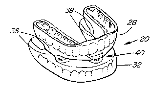
Figure 4-6 depict the preferred structure of
dental appli~nce 20 in further detail. Appliance 20
consists of an upper bite block 28 shaped to conform to the
maxillary dentition 30 and a lower bite block 32 shaped to
conform to the mandibular dentition 34. Bite blocks 28, 32
are preferably constructed from an elastomeric material.
Each bite block 28, 32 includes stainless steel dental
wires 36 (Figure 5) to stabilize appliance 20 and ensure
that it securely engages the wearer's teeth.
As best shown in Figures 4 and 6, the posterior
region of each bite block 28, 32 includes hard acrylic bite
pads 38 which provide a stop to the closure of the jaw and
which support the wearer's tempromandibular joint as dis-
cussed further below.
As shown best in Figures 3 and 9, bite blocks 28,
32 are united in an anterior region of appliance 20 by
means of a connecting assembly generally designated 40.
Assembly 40 includes a maxillary retention plate 42 which
is bonded to upper bite block 28 in the anterior palatal
region behind the central incisors and a mandibular guide
box assembly 44 which is bonded to an anterior region of
lower bite block 32. Retention plate 42 and guide box
assembly 44 are preferably bonded to respective bite blocks
28,32 with heat-cureable dental acrylic. Retention plate
42 and guide box assembly 44 are connected by means of a
stylus 46 as described further below. Stylus 46 preferably
comprises a threaded portion 47 and an enlarged head 48.
Guide box assembly 44 consists of a guide box 50
having an elongated aperture 52 formed on its upper surface
and a milled-out cavity 54 formed beneath aperture 50.
Assembly 44 also includes a base plate 56 which may be
2094411
--10--
releasably fastened to guide box 50 with screws 58. Screws
58 extend through apertures 60 formed in plate 56 and are
received in internally threaded apertures 62 formed on
either side of guide box 50 which are alignable with
apertures 60 (Figures 3 and 9).
As shown best in Figure 8, the elongated guide
box aperture 52 is preferably kidney-shaped to correspond
to the natural range of motion of the patient's jaw. Guide
box 50 is positioned so that the convex curvature of
aperture 52 faces forwardly.
As best shown in Figure 3, the threaded portion
47 of stylus 46 is inserted through guide box aperture 52
before guide box 50 and base plate 56 are fastened together
as aforesaid. Since the diameter of the stylus head 48 is
larger than aperture 52, the stylus head 48 is effectively
captured within the cavity 54 formed in guide box 50.
Cavity 54 is sufficiently large to permit stylus head 48 to
move vertically and laterally to a limited extent.
Base plate 56 has a central aperture 64 to allow
access to stylus head 48 (Figure 9). Accordingly, stylus
46 can be manually turned using a screwdriver or some other
- 25 suitable tool after guide box 50 and base plate 56 have
been fastened together.
Base plate 56 also includes a plurality of
countersunk, spaced-apart retention apertures 66. Reten-
tion apertures 66 are provided to allow dental acrylic toflow upwardly through base plate 56 to facilitate bonding
of guide box assembly 44 to the lower bite block 32. Aper-
tures 66 are countersunk to increase the surface area
available for bonding. Preferably, the base plate 56 is
larger than guide box 50 so that the dental acrylic flowing
through retention apertures 66 will surround the perimeter
of guide box 50, as best shown in Figure 7.
2094411
As best shown in Figures 3, 4 and 7 , maxillary
retention plate 42 includes a series of internally threaded
apertures 68 which are spaced apart at regular intervals.
Apertures 68 are provided for receiving the threaded
portion 47 of stylus 46. Maxillary retention plate also
includes a plurality of retention apertures 66 to allow the
acrylic to flow through plate 42 to facilitate its bonding
to upper bite block 28.
Preferably maxillary retention plate 40 and guide
box assembly 44 are constructed from commercially pure
titanium or some other metal which is non-reactive with
oral fluids. Stylus 46 and screws 58 are preferably
fabricated from stainless steel.
Dental appliance 20 is custom-fitted to suit the
requirements of each individual patient. Usually the first
step in the fitting procedure is for the dentist or phys-
ician to assess the natural range of motion of the pa-
tient's jaw and the likely degree of pharyngeal occlusion.
This may be determined by physical examinations, sleep
studies, x-rays and the like.
Molds of the patient's existing maxillary and
mandibular dentition 30, 34 are then taken to enable
casting of U-shaped bite blocks 28, 32. As indicated
above, bite blocks 28, 32 are preferably formed of an
elastomeric material. Dental wires 36 are embedded in bite
blocks 28, 32 to provide structural stability. Opposing
pairs of bite pads 38 are formed on the undersurface of
upper bite block 28 and the upper surface of lower bite
block 32 in a posterior region of appliance 20 (Figures 3
and 5). Bite pads 38 are constructed from hard dental
acrylic and are provided to limit closure of the patient's
jaw and prevent overeruption of the posterior teeth.
209~411
-12-
After bite blocks 28, 32 have been fabricated as
aforesaid, they are united by means of connector assembly
40 (Figure 3). Guide box 44 and stylus 46 of assembly 40
are loosely coupled together as described above so that the
threaded portion 47 of stylus 46 protrudes upwardly through
the elongated aperture 52 formed in guide box 50 (Figure
3).
Retention plate 42 is bonded to the undersurface
of upper bite block 28 and guide box assembly 44 is bonded
to the upper surface of bite block 32 by means of heat-
cureable dental acrylic. The soft acrylic flows through
retention apertures 66 formed on retention plate 42 and
base plate 56 to ensure that connecting assembly 40 is
securely set in place as the acrylic hardens. As explained
above, retention apertures 66 are countersunk to increase
the surface area available for bonding to the dental
acrylic.
After dental appliance 20 has been fabricated as
described above, bite blocks 28, 32 are releasably coupled
together by inserting the threaded portion 47 of stylus 46,
which extends upwardly from guide box 50, into one of the
mating apertures 68 formed in maxillary retention plate 42
(Figures 3, 7 and 9). Apertures 68 are spaced approximate-
ly 0.5 mm apart to allow the dentist or physician to make
small adjustments in the relative position of bite blocks
28, 32 and hence the degree of anterior protrusion of the
patient's mandible 22. Preferably apertures 68 should be
spaced to allow for a total adjustment range of approxi-
mately 7 mm. The inferior position of mandible 22 (i.e.
the degree of opening of the jaw) may also be incrementally
adjusted by varying the extent to which stylus 46 is
screwed within a selected aperture 68. Stylus 46 may be
turned with a screwdriver or other suitable tool insertable
through base plate aperture 64 to engage stylus head 48.
X~94~11
-
-13-
In practice, appliance 20 is easily insertable
within the mouth of a patient for wear during sleep.
Appliance 20 is initially adjusted to advance mandible 22
between 25% and 75% of the patient's maximum protrusive
capability. Typically approximately 5-8 millimetres of
mandibular protrusion and approximately 4-6 millimetres of
inferior opening are initially provided (Figure 2). This
is in contrast to some prior dental appliances where an in-
ferior opening in the range of 10-20 millimetres is recom-
mended. The inventor's studies suggest that in manypatients the tongue 10 has a greater tendency to slide
posteriorly as the degree of jaw opening increases.
Displacement of the tempromandibular joint is also more
likely if the jaw is fixed in a wide open position for long
periods of time. Accordingly, dental appliance 20 is set
to open the jaw the minimum amount possible while still
allowing the patient to breathe comfortably through the
mouth.
Opposed bite pads 38 formed on bite blocks 28, 32
provide a stop to complete closure of the jaw as best shown
in Figures 4 and 6. This prevents overeruption of the
posterior teeth during the wearing of appliance 20 and
provides support to the tempromandibular joint and associ-
ated ligaments and muscles.
After the patient has been fitted with dental
appliance 20 he or she is carefully monitored to determine
if further adjustments are required. For example, if the
patient's snoring or apnea episodes have not been complete-
ly eliminated, then the degree of mandibular protrusion may
be incrementally increased by unscrewing stylus 46 from its
initial setting, advancing lower bite block 32 forwardly,
and inserting stylus 46 into an adjacent aperture 68 formed
on maxillary retention plate 42 (Figure 7). The degree of
inferior opening of the mandible 22 can also be readily
adjusted to suit the needs of a particular patient by
209~
-14-
altering the extent to which the threaded portion 47 of
stylus 46 is screwed within a selected aperture 68. If the
patient experiences discomfort from wearing appliance 20,
then the dentist or physician can readily adjust the lower
bite block 28 to a more retruded and/or a less inferior
position.
A key feature of the applicant's invention is
that connecting assembly 40 allows a limited degree of
lateral movement of the patient's mandible 20 relative to
the upper jaw 24 while still maintaining mandible 20 in the
preferred protruded position. While the upper threaded
portion 47 of stylus 46 is fixed in a selected aperture 68
formed in maxillary retention plate 42, the stylus head 48
is not fixed relative to lower bite block 32. Rather,
stylus head 48 is loosely captured within cavity 54 formed
in guide box 50, as best shown in Figures 7-9. This allows
lower bite block 32 and hence mandible 22 to travel in a
lateral excursion relative to stylus 46 (i.e. in the
direction of the arrows shown in Figures 6, 8 and 9). The
extent of lateral travel of mandible 22 is restricted by
the size of aperture 52 formed on the upper surface of
guide box 50 and also the size of guide box cavity 54
(Figure 8).
As best shown in Figures 3 and 8 and as discussed
above, guide box aperture 52 is preferably kidney-shaped to
correspond to the natural range of motion of the patient's
jaw joint. In one embodiment of the invention, the size
and shape of aperture 52 could be customized to suit the
anatomy of each particular patient, such as by performing
gothic arch tracings to determine the natural range of
motion of the patient's jaw anatomy.
Prior art intra-oral devices which maintain
mandible 22 in a fixed, protruded position can lead to
serious side effects, particularly in patients prone to
2094~11
-15-
nocturnal bruxism (teeth grinding). Such prior art devices
may result in displacement or aggravation of the patient's
tempromandibular joint and associated muscles and liga-
ments. The applicant's dental appliance 20 effectively
overcomes this problem by allowing a predetermined degree
of lateral movement of mandible 22 in the protruded posi-
tion, while still maintaining acceptable airway patency.
As will be apparent to those skilled in the art
in the light of the foregoing disclosure, many alterations
and modifications are possible in the practice of this
invention without departing from the spirit or scope
thereof. For example, more than one connecting assembly 40
could be provided. Further, the connecting assembly or
assemblies 40 could be mounted in a posterior rather than
an anterior region of dental appliance 20. In another
alternative embodiment, guide box assembly 44 could be
inverted so that elongate aperture 52 is formed on a top
plate covering the open upper end of a guide box defining
internal cavity 54. Other equivalent means for loosely
coupling stylus 46 to lower bite block 32 may also be
envisaged. Accordingly, the scope of the invention is to
be construed in accordance with the substance defined by
the following claims.