Note: Descriptions are shown in the official language in which they were submitted.
2~~~~~~
ENDOSCOPIC CUTTING APPARATUS
mechnical Field
The field of art to which this invention relates is
surgical instruments, in particular endoscopic surgical
instruments.
to
Eackaround of the Invention
Endoscopic surgical techniques and procedures have become
widely accepted both among the medical surgical community
and the patient population. There are numerous benefits
associated with the use of endoscopic surgical techniques
rather than conventional open surgical techniques. These
benefits include reduced avenues for infection, shortened
post-operative recuperation period, decreased hospital
stay and decreased scarring. It is not unusual for the
post operative period to be shortened from weeks with
conventional open surgery to several days with endoscopic
surgical procedures, and outpatient endoscopic surgery is
becoming more and more typical. The term endoscopic as
used herein is defined to include endoscopic,
laparoscopic, arthroscopic, and thorascopic.
In a typical endoscopic surgical procedure, the abdominal
cavity of a mammal is typically insufflated with a sterile
gas, such as carbon dioxide, in order to provide increased
maneuvering room within the body cavity for endoscopic
instruments. This procedure is typically referred to as
inducement of pneumoperitoneum. Then, conventional
trocars are inserted into the patients body cavity through
SEN-123
2C~~J~~~
- 2 -
the surrounding skin, tissue and musculature of the body
cavity wall. A conventional trocar typically consists of
a trocar cannula which houses an elongated trocar
obturator. Trocar obturators typically have a piercing
point, although other types of obturators are also
available having blunt tips. Once the trocar has been
positioned within the body cavity, proximal to the target
surgical site, the trocar obturator is typically removed
leaving the trocar cannula in place as a pathway to and
from the target surgical site. The surgeon will place
various types of endoscopic surgical instruments thorough
the trocar cannulas in order to access the target surgical
site where the surgical procedure will be performed.
Examples of endoscopic instruments which have been
developed for use with endoscopic surgical techniques
include ligating clip appliers, electrosurgical
instruments, endoscopes, tissue graspers, needle graspers,
cannulas, tissue manipulators, endosurgical scissors, and
the like.
Although endosurgical procedures and techniques offer many
advantages, there are some deficiencies associated with
these procedures and techniques. In particular, when the
surgeon is operating using endoscopic surgical procedures,
he is typically using an endoscope which is positioned
within the body cavity through a trocar. The endoscope is
typically connected to a video camera and the output from
the video camera is displayed on a video monitor. The
surgeon typically views the display on the video monitor
as he manipulates instruments within the body cavity to
access the target surgical site and perform the actual
surgical procedures. The video display provides the
surgeon with only two- dimensional input and there is a
consequent loss of depth perception. This lack of depth
SEN-123
- 3 -
perception may result in the surgeon over-shooting or
under-shooting the target surgical site as he attempts to
position various endoscopic instruments within the body
cavity.
As can be appreciated, the internal organs of a mammal are
very tightly packed within the body cavities. Therefore,
the surgeon must exercise extreme care when maneuvering
instruments through a body cavity to a target surgical
site. This can be particularly difficult since, as was
mentioned previously, the surgeon is working in a three
dimensional space while viewing a two dimensional output.
The degree of care which must be exercised by the surgeon
is increased further when the surgeon is attempting to
maneuver cutting instruments to the target surgical site.
The cutting instruments which have been developed for use
in endosurgical procedures consist of conventional
endosurgical scissors and the like. The surgeon must be
careful when maneuvering endoscopic cutting instruments ,
for example, endosurgical scissors, through a body cavity
so that no internal organs or blood vessels are
accidentally nicked or cut. In addition, it has been
observed that endoscopic surgical scissors do not cut with
the same efficiency as a conventional scalpel.
What is needed in this art is an endoscopic surgical
cutting apparatus which will not accidentally nick or cut
internal organs or blood vessels but which has improved
cutting properties.
Summary of the Invention
It is an object of the present invention to provide an
endoscopic cutting apparatus which can be maneuvered
SEN-123
~0~~6~~
-4_
through a body cavity without the cutting blade being
exposed.
It is a further object of the present invention to provide
an endoscopic cutting apparatus which has a means far
engaging tissue or a blood vessel prior to and during
cutting.
It is another object of the present invention to provide
an endoscopic cutting apparatus which has improved tissue
and blood vessel cutting characteristics.
Accordingly, an endoscopic cutting device is disclosed.
The cutting device comprises a tubular frame having a
proximal end and a distal end. A handle is mounted to the
proximal end of the tubular frame. Hook means are mounted
to the distal end of the tubular frame for engaging tissue
or blood vessels prior to and during cutting. The hook
means has track means contained therein for receiving a
cutting blade means. The cutting blade means is mounted
to the frame and is moveable within said track means to
cut tissue or blood vessels engaged within the hook means.
Actuating means are mounted to the frame for moving the
cutting blade means.
Yet another aspect of the present invention is a method of
cutting tissue or blood vessels in an endoscopic procedure
using the above-described endoscopic cutting apparatus.
Still yet another aspect of the present invention is the
combination of a trocar cannula and the above-described
endoscopic cutting apparatus.
SEN-123
2fl9j~~~
- 5 -
Other features and advantages of the invention will become
more apparent from the following description and
accompanying drawings.
Brief Description of the Drawinos
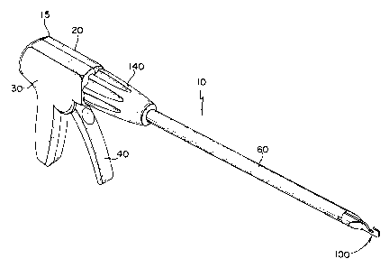
FIG. 1 is a perspective view of the endoscopic cutting
apparatus of the present invention.
FIG. 2 is a perspective view of the cutting apparatus
inserted through a trocar cannula into a body cavity.
FIG. 3 is an enlarged fragmentary view of the distal end
of the cutting apparatus showing the distal end of the J
hook member.
FIG. 4 is a partial perspective view of the distal end of
the J-hook member after the blade has cut a blood vessel
contained within the isolation hook.
FIG. 5 is an exploded perspective view of the cutting
apparatus of FIG. 1.
FIG. 6 is a partial perspective view of the distal end of
the J-hook member prior to folding to form the isolation
hook.
FIG. 7 is a partial perspective view of the distal end of
the J-hook member and the distal end of the blade carrier
and blade.
FIG. 8 is a partial top plan view of the cutting assembly
of the cutting apparatus.
SEN-123
- s -
FIG 9, is a side view partially in cross-section
of the endoscopic cutting apparatus in a first, at-rest
position.
' FIG 9A. is an enlarged, partial detailed view of the
rotatable connection between the plunger plate and the
blade carrier of the cutting apparatus of FIG 9 showing
the distal end of the plunger plate and the proximal end
of the blade carrier.
FIG. 10 is a detailed view of the cutting apparatus
similar to FIG. 9 in a second, actuated position.
FIG. 10A is an enlarged, partial detailed view of the
apparatus of FIG. 10 similar to FIG 9A showing the distal
end of the plunger plate and the proximal end of the blade
carrier after the cutting blade is actuated.
Best Mode For Carrvinq Out the Invention
The endoscopic cutting apparatus 10 of the present
invention is illustrated in FIG. 1, FIG. 2 FIG. 5, FIG. 9
and FIG. l0. The endoscopic cutting apparatus 10 is seen
to have a hollow frame 15 consisting of left handle 20 and
right handle 30. The left handle 20 is seen to have
downwardly extending handle grip 22 and elongate section
24. The distal end 26 of left handle 20 is seen to have
opening 27 for receiving the proximal end 62 of support
tube 60. Extending from the inner wall of elongate
section 24 is the journal bearing 28. Also extending from
the inner wall of handle 22 is the spring mounting pin 21.
The right handle 30 is similarly seen to have downwardly
extending handle grip 32 and elongate section 34. The
distal end 36 of the right handle 30 is seen to have
SEN-123
_ 7 _
opening 37 for receiving the proximal end 62 of support
tube 60. Extending from the inner wall of elongate
section 34 of right handle 30 is journal bearing 38.
Referring to FIGS 5, 9, and 10, the trigger 40 is seen to
be an elongate member. The yoke members 42 extend
upwardly from either side of the tap of the trigger 40.
Yoke members 42 contain slots 43. Cylindrical shaft
members 44 extend from either side of the trigger 40 below
the yoke members 42. Trigger 40 is pivotally mounted in
journal bearings 28 and 38 by inserting cylindrical shaft
members 44 therein. Trigger arm 45 containing hole 46 is
seen to extend proximally from the trigger 40.
The support tube 60 is an elongate tubular member having
proximal end 62 and distal end 64. The flange member 63
is seen to extend radially outward from proximal end 62 of
support tube 60.
The plunger plate 70 is seen to be an elongate plate
member having proximal end 72 and distal end 74.
Extending from distal end 74 are upper and lower hook
members 76 which form C-shaped slot 77. Located in the
proximal end 72 of the plunger plate 70 is the drive hole
73. The plunger plate 70 is mounted to trigger 4o by
placing the proximal end 72 of the plunger plate 70
between the yoke members 42 such that the drive hole 73 of
the plunger plate 70 is in alignment with the slots 43 of
the yoke members 42. Then the drive pin 50 is inserted
through the slots 43 and the drive hole 73.
Referring now to FIGS. 5,6,7 and 8, the blade assembly 100
is seen to consist of the blade carrier 105 and the blade
115. The blade carrier 105 has distal end 107 and
SEN°123
2~~~6
_8_
proximal end 109. Extending proximally from proximal end
109 are a pair of opposed coupling prongs 111. Mounted to
the distal end 107 of blade carrier 105 is the blade 115.
Blade 115 is seen to have distal cutting edge 117. The
blade 115 is mounted to the distal end 107 of the carrier
105 using conventional mounting methods including riveting
and welding.
The J-hook member 90 is an elongate plate-like member
having proximal end 91 and distal end 93. Located in the
distal end 93 of the J-hook member 90 is the isolation
hook 95 containing vessel hook aperture 96 and blade track
97. As can be seen in FIGS. 6, 7 and 8, the isolation
hook 95 is formed by folding a section 98 of the distal
end of the J-hook member 90 over and onto itself and then
cutting out the vessel hook aperture 96 using conventional
methods including gas burning, laser cutting, and the
like. The isolation hook 95 may also be formed by cutting
out apertures prior to folding in two places on the distal
end 93 of the J-hook member 90 so that when the section 98
of the distal end of J-hook member 90 is folded over to
form the isolation hook 95, the apertures are in
substantial alignment to form the vessel hook aperture 96.
J-hook member 90 is seen to have proximal mounting hole
92.
Referring to FIGS. 5, 9, and 10, the shroud 80 is seen to
be an elongate cylindrical member having distal end 84 and
proximal end 82. The shroud 80 has elongate axial slot 87
for receiving J-hook member 90 and blade assembly 100.
Located at the proximal end 82 of the shroud 80 are the
grooves 81 and the mounting hole 86. The J-hook member 90
and blade assembly 100 are mounted within slot 87 of
shroud 80. The J-hook member 90 is attached to the shroud
SEN-123
N
~~3~~
_ g
80 by the retaining pin 132 which is inserted through the
hole 86 located in the proximal end 82 of shroud 80 and
through the hole 92 located in the proximal end 91 of J-
hook member 90. The blade assembly 100 is slideably
mounted within the slot 87 of shroud 80. The distal end
107 of the blade carrier 105 and the blade 115 are
slideably contained within the track 97 of isolation hook
95. The 0-ring 134 is mounted into the groove 81 distal
to the mounting hole 86. The shroud 80 containing blade
l0 assembly 100 and the J-hook member 90 is mounted within
support tube 60. The shroud 80 containing the blade
assembly 100 and the J-hook member 90 is secured to the
support tube 60 by the retaining ring 130 which is snapped
into the groove 81 proximal to the mounting hole 86.
The plunger coupling 120 is seen to be a bushing-like
member having outwardly extending proximal flange section
122 and axial cylindrical section 128. Axial cavity 125,
having a rectangular cross-section, extends through the
flange 122 and the cylindrical section 128 ( see FIG. 9A).
The flange section 122 of the plunger coupling 120 is
rotatably engaged within the C-shaped slot 77 of the
plunger plate 70. The proximal coupling prongs 111 of
blade carrier 105 are engaged within the axial cavity 125
of the plunger coupling 120 thereby allowing rotation of
the blade assembly 100 with respect to plunger plate 70.
As can be seen in FIG.5, proximal end 62 of the support
tube 60 is rotatably mounted within the distal ends 26 and
36 of the left handle 20 and the right handle 30,
respectively, thereby allowing rotation of support tube 60
with respect to the frame 15. The frame 15 is formed by
mounting the right handle 30 to the left handle 20 using
conventional mounting methods such as ultrasonic welding,
SEN-123
- to -
bonding, fastening and the like. The support tube 60 is
prevented from displacing longitudinally by the shoulders
29 and 39 contained in the left handle 20 and right handle
30, respectively. The return spring 160 is seen to be
connected on one end to the pin 21 in the left handle 20
and on the other end to the hole 46 contained in the
trigger arm 45. The spring 160 provides a
counterclockwise biasing force on the trigger 40. The
knob ring 150 is mounted to the knob 140 in a conventional
manner to form a knob assembly which is rotatably mounted
to the distal end of frame 15 and keyed to the proximal
end of support tube 60 to assist in rotating the support
tube with respect to the frame 15.
The cutting apparatus l0 is actuated by squeezing the
trigger 4o causing it to rotate in a clockwise manner
about journal bearings 28 and 38 as shown in FIG. 10. As
the trigger 40 is rotated, the yoke members 42 also rotate
causing the drive pin 50 to displace longitudinally.
Since the drive pin 50 is engaged by the plunger plate 70
at drive hole 73, the rotation of trigger 40 also causes
plunger plate 70 to displace distally in a longitudinally
axial manner. The plunger coupling 120, which is
rotatably engaged in the C-shaped slot 77 of the plunger
plate 70, also engages the proximal members 111 of the
blade assembly 100. Displacement of the plunger plate 70
also causes the blade assembly 100 to be displaced within
the slot 87 of the shroud 80 and the track 97 of the J-
hook member 90. This causes the blade 115 to displace
distally through the vessel hook aperture 96 contained in
the isolation hook 95. This allows the blade 115 to sever
tissue or blood vessels contained within the vessel hook
aperture 96 (also shown in FIG.4). The blade assembly
100, the plunger coupling 120, the plunger plate 70, the
SEN-123
~~~j~~~
_ 1l, _
drive pin 50 and the trigger 40 are returned to an at-rest
position ( see FIG. 3) when the trigger is released
because of the counterclockwise bias farce of the return
spring 160 acting an the trigger arm 45 of triggex 40.
The support tube r0, shroud s0, J-hook member 90 and blade
assembly x,00 can rotate with respect to the left handle 2a
and right handle 30 and the plunger plate 70. The flange
63 of the support tube 60 is rotatably mounted in the
frame 15. And, plunger coupling 120 is rotatably engaged
within the C-shaped slat 77 of the plunger plate 70
thereby allowing the blade assembly 100 to rotate with
respect to the plunger plate 70. Rotating the knob 144
causes the support tube 60 and the assembly contained
~,5 therein to xotate with respect to the frame 15 as seen ~.n
fTG.2.
In another embodiment of the cutting apparatus of the
present irwention (not shown in the drawings) extension
means are mounted to the frame 15 to extend and retract
isolation hook 95. The extension means will typically
consist of an extension plate slideably mounted, axially,
in tie frame 15 and rotatably connected at its distal end
to the proximal end of J~hoak member 90 which will also be
slideably mounted. Typically, a button slideably mounted
to the extexior of the frame ~.5 and connected to the
extension plate will actuate the extension plate causing
the ,I-hook member 9o to slide axialJ.y within the slat 87
of shroud S0. This in.turn will cause the ~.solation hook
95 to slide into and out of the distal end of the
apparatus 10. Zn tha.s emmadiment of apparatus 10, the
isolation hook 95 is typically in a retxdcted position
prior to insertion into a trocar cannula. mhe distal end
of the apparatus ~.0 is maneuvered proximal to the target
S~,N-123
CA 02095655 2003-11-27
-12-
blood vessel or tissue at which time the isolation hook 95 is
extended by actuating the extension mechanism and the apparatus
10 is then used in a manner as previously described.
The endoscopic cutting apparatus 10 of the present invention
will be constructed from materials conventional in this art.
The materials include plastics such as polycarbonate, nylon,
ZO polyetherimide and nitrile as well as the 300 and 400 series
stainless steels and the like and equivalents thereof. The edge
117 of the blade 115 will be manufactured using conventional
blade edge forming methods including grinding with conventional
grinding apparatus and the like. The apparatus 10 is typically
packaged in conventional packaging materials. The endoscopic
cutting apparatus 10 is typically sterilized after packaging and
prior to use using conventional sterilization techniques. It is
particularly preferred to sterilize the apparatus 10 using
cobalt-60 generated radiation, although other types of
sterilization including autoclaving and ethylene oxide
sterilization may be used.
The endoscopic cutting apparatus 10 may be used in conventional
endoscopic techniques including cholecystectomy, appendectomy,
anastomosis, heria repair and the like. Endoscopic surgical
techniques and procedures are widely known, e.g., endoscopic
surgical techniques are disclosed in the following publications:
Textbook of Laparoscopy, Jaroslav Hulka, M.D., Grune & Stratton,
Inc., New York (1985) and Laparoscopy for Surgeons, Barry A.
Salky, M.D., Igaku-Shoin, New York (1990). When utilizing
endosurgical techniques, initially a patient is typically
anesthetized using a sufficient dose of anesthesia
- 13 -
effective to induce an anesthetized state. Conventional
anesthesiology techniques and procedures are utilized
including, where needed, the use of an endotracheal tube
and a ventilator. The next step after the application of
anesthesia is the insufflation of the body cavity
containing the target surgical site. This is done using
conventional techniques and equipment. The gases which
are typically used for insufflation include conventional
sterile gases such as carbon dioxide and the like. After
the body cavity has been insufflated sufficiently so that
the surgeon has room to effectively manipulate and
maneuver instrumentation within the body cavity, several
conventional trocars are inserted in a conventional manner
through the body cavity wall into the body cavity, for
example, the abdominal cavity. Conventional trocars
typically comprise a piercing obturator concentrically
housed in a trocar cannula. After the trocars are
inserted, the piercing obturators are then removed from
the trocar cannulas leaving the trocar cannulas as
pathways to the body cavity. Conventional endoscopic
instrumentation is inserted through the cannulas including
endoscopes, staplers, sutures, cannulas, electrosurgical
instruments, ligating clip appliers, and the like. The
instruments are maneuvered to the target surgical site
where a surgical procedure is performed. The surgeon
views the interior of the body cavity and the target
surgical site by observing the output from the endoscope.
Conventional endoscopes typically are connected to video
cameras and the output displayed on a video monitor.
One of the crucial endoscopic techniques which must be
mastered by the surgeon when utilizing endoscopic
procedures is the ability to maneuver instruments in a
three-dimensional body cavity while observing a two-
SEN-123
~~j~~~
- 14 -
dimensional visual output on the video display of the
endoscope. This requires both skill and judgment based
upon the experience of having performed previous
operations. When using conventional endoscopic scissors,
the surgeon must locate the vessel or tissue to be cut and
engage the vessel or tissue between the blades of the
scissors. The surgeon maneuvers the endoscopic scissors
in position about the intended cut and then makes the cut
in the tissue. ors previously mentioned, the physician is
maneuvering in a three-dimensional body cavity, however,
he only has two-dimensional input from the video display
of the endoscope. Therefore, the surgeon must use extreme
care in maneuvering the scissors to the tissue or blood
vessel where the cut is to be made. The surgeon must use
judgment in deciding whether the tissue or blood vessel is
properly contained within the jaws of the conventional
scissors when cutting since the scissors do not provide
for positive retention. There is also an element of
hazard present requiring extra care as the surgeon
attempts to maneuver the cutting instrument in the
relatively cramped space in the body cavity. The hazards
include accidental nicking or cutting of organs or blood
vessels or tissue. In addition, endoscopic scissors tend
not to cut well and become dull very rapidly.
Using the endoscopic cutting apparatus 10 of the present
invention, these deficiencies are eliminated. The cutting
mechanism 10 of the present invention is inserted through
a trocar cannula into a body cavity and maneuvered by the
surgeon to the target surgical site where a blood vessel
or tissue is to be cut, as shown in fIG. 2. The
endoscopic cutting apparatus 10 is a cutting instrument
which can engage and cut tissue and which may also be used
to maneuver tissue or blood vessels. The surgeon
SEN-123
- 15 -
positions the blood vessel or tissue within the vessel
hook aperture 96 of the J-hook member 90. The surgeon can
see on the endoscope video monitor that the tissue or
blood vessel is positively contained within the vessel
hook aperture 96. The surgeon then actuates the blade
assembly 100 by squeezing the trigger 40 which causes the
blade assembly 100 and the blade 115 to travel distally
such that blade 115 displaces distally through track 97 in
isolation hook 95 thereby cutting the blood vessel or
l0 tissue engaged within the vessel hook aperture 96 in the
isolation hook 95 of J-hook member 90.
It will be appreciated by those skilled in the art that
the cutting apparatus 10 of the present invention can be
used not only in endoscopic surgical procedures but also
in conventional open procedures. It will also be
appreciated that the apparatus 10 may, if one were willing
to accept whatever disadvantages may be present, if any,
be inserted through a small slit directly into a body
cavity without a conventional trocar.
The fo~.lowing example is illustrative of the principles
and practice of the present invention although not limited
thereto.
ExamQle
A mammal is prepared for surgery using conventional
surgical techniques. A sufficient dose of a conventional
anesthesia is administered using conventional
anesthesiology techniques effective to induce an
anesthetized state. The abdominal cavity of the patient
is then sufficiently insufflated using conventional
insufflation equipment and techniques with carbon dioxide
gas to produce an effective pneumoperitoneum. Three
SEN-123
~~J~~3~aJ
- 16 -
trocars are then inserted through the abdominal wall of
the mammal into the abdominal cavity. The trocars are
conventional trocars having elongated obturators with
piercing tips concentrically housed in trocar cannulas.
The trocar obturators are then removed leaving the trocar
cannulas as pathways to the abdominal cavity. An
endoscope is inserted through one of the trocar cannulas.
The output from the endoscope is displayed on a video
monitor. The surgeon observes the interior of the
abdominal cavity on the video monitor and maneuvers
instruments into position using the video monitor display.
The endoscopic cutting apparatus 10 of the present
invention is inserted through one of the trocar cannulas.
The surgeon maneuvers the distal end of the apparatus 10
to a position proximate to a target blood vessel which is
to be ligated. The surgeon then positions the blood
vessel within the vessel hook aperture 96 in the isolation
hook 95 of ,T-hook member 90. The blood vessel is
positively engaged within the aperture 96 in isolation
hook 95 and the surgeon is easily able to observe this
positive engagement on the endoscope video monitor. The
surgeon then manipulates the position of the blood vessel
with the apparatus 10. Typically, prior to cutting, the
blood vessel or tissue is ligated with a conventional
ligating clip appl.ier which is used to apply ligating
clips to the blood vessel or tissue on either side of the
intended cut. The surgeon then actuates the cutting
apparatus 10 by squeezing the trigger 40 such that the
cutting blade 115 advances distally through the blood
vessel contained within the vessel hook aperture 96 in
the isolation hook 95. As the surgeon releases the
trigger 40, the blade 115 is automatically retracted from
the vessel hook aperture 96 in isolation hook 95. The
surgeon then withdraws the distal end of the apparatus 10
SEN-123
_ 17 _
from the body cavity and out through the trocar cannula.
The surgeon then removes the trocar cannulas and closes up
the wounds using conventional techniques including
stapling, suturing, and/or taping.
The endoscopic cutting apparatus 10 of the present
invention provides a means for cutting tissue or blood
vessels. The possibility of inadvertently cutting or
nicking blood vessels, tissue or organs is minimized.
Tissue or blood vessels are easily positively engaged
within the vessel hook aperture of the isolation hook 95.
The surgeon is readily able to see on the endoscopic video
display that tissue or blood vessels are positively
retained and engaged within the vessel hook aperture 96
of isolation hook 95 prior to cutting. When using
endoscopic scissors, the surgeon must use skill and
judgment to determine when the tissue is within the open
scissor blades. There is no positive tissue or blood
vessel retention provided by conventional endoscopic
scissors, the surgeon is not able to perceive from the
endoscopic video display whether or not the tissue or
blood vessel is absolutely positioned within the scissor
jaws. In addition, the cutting blade 115 of the apparatus
l0 cuts quickly and repeatedly without the blade edge 117
becoming dull. In contrast, it is known that endoscopic
scissors become dull very quickly. Another advantage of
the apparatus 10 of the present invention is that the
isolation hook 95 can be easily manufactured in one
preferred embodiment by folding the section 98 of the
distal end 93 of the J-hook member 90 over and onto itself
to form the track 97 and the isolation hook 95.
8EN-123
- 18 -
Although this invention has been shown and described with
respect to detailed embodiments thereof, it will be
understood by those skilled in the art that various
changes in form and detail thereof may be made without
departing from the spirit and scope of the claimed
invention.
to
20
30
SEN-123