Note: Descriptions are shown in the official language in which they were submitted.
12'~ ;g~2
FLUID CONDUCTING TEST STRIP WITH TRANSPORT MEDIUM
d Of The Invention
The present invention relates to a reagent
S strip which allows a user to quantitatively determine
the concentration of an analyte in a liquid test sample.
More specifically, the present invention relates to a
reagent strip which combines a porous transport medium
with a testing pad capable of quantitatively indicating
the concentration of an analyte in a liquid test sample,
such as glucose or cholesterol in whole blood.
Backaround Of The Invention
Numerous test devices have been developed for
the analysis of body fluids in order to determine the
concentration of specific analytes in test samples.
These include devices for detecting, f or example,
glucose, cholesterol, proteins, ketones, uric acid,
phenylalanine, or enzymes in either blood or urine.
Often tests are used to diagnose or treat a particular
disease, such as diabetes or high blood pressure.
Two general types of test strips are in common
use. Older devices require the application of a drop of
blood to the top surface of a reagent pad, allowing the
drop to react for a timed interval, removing the drop by
wiping or blotting, and then determining the analyte
concentration either visually or through the use of a
reflectance photometer. Newer devices simplify the
procedure by allowing the user to apply a drop of blood
to a test strip while it is inserted in a meter.
Optical and electronic elements within the meter detect
the presence of blood, automatically start a timing and
measuring process, and complete the analysis without
removal of blood from the strip.
VIA EXPRESS MAIL ~0. HE~46~5991X
M'~ILED MAY 12, 1992
2~9~9~2
- 2 -
While the commonly used test strips are widely
acclaimed and entirely satisfactory for many
applications, the user of such test strips must bring
the finger from which the blood drop has been obtained
to the meter, rather than simply bringing the strip to
the finger. Also, the deep red coloration of blood in
the strip can interfere with a visual confirmation of
the amount of color formed. Visual confirmation is a
desirable indication of proper operation of the meter
which reinforces user confidence in the accuracy of the
concentration measurement. These disadvantages can be
especially troublesome when the user is a patient
suffering from disabilities related to a disease, such
as diabetes, who must determine his own analyte
concentrations. Such patients may have difficulty
performing mechanical procedures required to operate
conventional reagent strips.
Accordingly, there exists a need for a reagent
strip which provides a test that requires only a single
step in which the user can apply an unmeasured sample of
whole blood and determine the concentration level of an
analyte in a whole blood sample, either visually or
through electronic viewing means.
Summarv Of The Invention
The invention provides a reagent strip for
measuring the concentration of an analyte in a test
sa~ple. The reagent strip comprises a testing pad and a
porous sample transport medium. The testing pad is
formed from an anisotropic membrane which contains
spati~lly separated regions having differently sized
pores. One side of the testing pad has pores with
relatively small effective diameters. The side with
relatively small pores defines a testing surface. An
opposite side with relatively larger pores defines a
2a~9~2
~ 3 --
sample receiving ~urface. The testing pad contains a
color-forming reagent system which reacts selectively
with the analyte.
The transport medium is attached to the sample
receiving surface of the testing pad. The transport
medium is adapted to accept a whole blood sample and
transport a detectable portion of the blood sample to
the sample receiving surface. It is preferred that the
transport medium be capable of holding from about 10 to
about 50 microliters of blood, preferably about 35
microliters of blood and of passing from about 3 to
about 10 microliters of blood to the testing pad.
The transport medium is connected to the
testing pad by an adhesive layer, which may be a
continuous layer formed along an outer edge of the
testing pad, leaving a central portion of the testing
pad substantially unobstructed. Alternatively, the
adhesive layer may be discontinuous.
The reagent strip may optionally further
comprise a rigid support member that defines an aperture
which extends completely through the support member.
The support member also defines a free surface adapted
to contact and engage mechanical viewing means. The
viewing means may be utilized in conjunction with or as
an alternative to direct visual inspection for
evaluating a change in coloration at the testing surface
produced by the color-forming reagent system.
In another embodiment, adapted to facilitate
evaluation primarily by mechanical viewing means after a
color-change reaction, the reagent strip comprises a
rigid support member, a testing pad, and a porous
transport medium. The support member defines an
aperture which extends through the support member. The
support member has a free surface which is adapted to
contact and engage viewing means. A portion of the
2~9~982
- 4 -
suE~port member disposed about the aperture has a
predetermined and carefully controlled thickness which
can be used to locate the testing surface at a
reproducible distance from the viewing means.
The testing pad contains a color-forming
reagent system which is specific to the analyte of
interest. The testing pad is formed from an anisotropic
membrane and has a side with relatively small pores that
defines a testing surface. The testing pad also has an
opposite side with relatively large pores which defines
a sample-receiving surface. The testing surface is
attached to the support member so that the testing
surface faces and overlaps the aperture.
The transport medium is attached to the
sample-receiving surface and is capable of accepting a
whole blood sample. The transport medium transports a
detectable portion of the sample to the sample-receiving
surface where the color-forming reagent system causes a
change in coloration which can be evaluated at the
testing surface visually or by the use of viewing means,
such as a reflectance meter. When the free surface of
the support member is engaged by the viewing means, the
testing surface is conveniently and reliably maintained
at an optimum viewing distance relative to said viewing
means. Concentration determinations performed while the
free surface is engaged with the viewing means tend to
be relatively more accurate and more reproducible.
It is preferred that the thickness of the
portion disposed about the aperture be in the range of
about 0.002 to about 0.040 of an inch. The testing pad
preferably has pores which vary in size from about 25
micrometers on the side with relatively large pores to
about 0.3 micrometer on the side with relatively small
pores. The testing pad may be attached to the transport
medium by a continuous adhesive positioned along an
20~9~2
-- 5 --
outer edge of the testing pad. Alternatively, the
adhesive layer may be discontinuous and extend fully
across the receiving surface.
The invention also provides a method for
measuring the concentration of an analyte in a test
sample. The method is especially suitable for test `
samples which contain solid color bodies. A test strip
of the present invention is contacted with a transport
medium. The transport medium absorbs the test sample,
transports the test sample to the sample-receiving
surface, and distributes the test sample across the
sample-receiving surface.
At the receiving surface, the sample is
absorbed into the testing pad. The sample moves through
the testing pad, by capillary action, for example, and
encounters progressively smaller pores as it approaches
the testing surface. The smaller pores filter the test
sample and remove at least some of the solid color
bodies.
The testing reagent reacts chemically with the
analyte to vary coloration of the testing surface.
Coloration of the testing surface is compared to a
calibrated color standard in order to determine the
concentration of the analyte in the test sample.
The method may optionally further comprise the
step of locating the testing surface a reproducible
distance from a mechanical viewing means by contacting
the viewing means with a free surface of a rigid support
member attached to the testing surface.
These and other features of the invention will
be better understood in connection with the following
drawings and detailed description of the preferred
embodiments.
~ .,
2~93982
-- 6
Brief Description Of The Drawinqs
FIG. 1 is a perspective view of a preferred
embodiment of a reagent strip of the present invention;
FIG. 2 is a perspective view of another
preferred embodiment of a reagent strip of the present
invention;
FIG. 3 is a partially cutaway elevation view
of a viewing means with a reagent strip positioned to
enter and engage the viewing means;
FIG. 3A is an enlarged partial cross-sectional
view of the viewing means and the reagent strip of FIG.
3; and
FIG. 4 is a cross-sectional view, not to
scale, illustrating a printing process for applying a
discontinuous adhesive layer to a receiving surface of a
porous testing pad, suitable for use in fabricating a
reagent strip of the present invention.
petailed Descri~tion of the Preferred Embodiments
Whole blood samples can be applied to one side
of the reagent strip of the present invention and a
visual comparison of the analyte concentration level can
be made at the opposite side of the reagent strip.
Preferably, the color developed in the reagent strip is
stable for an extended period of time. In order to
facilitate either visual or electronic interpretation,
the reagent strip absorbs excess blood beyond that
needed for the test, thus preventing contamination of
the viewing means and adjacent areas. When
determination of the concentration is to be done through
electronic viewing means, the reagent strip is adapted
to be conveniently brought to the surface of a whole
blood sample and, thereafter, positively and
reproducibly engaged with the viewing means, without the
complications of timing or blood removal.
. ~ - ` !
' .
2i~39~
- 7 -
A reagent strip in accordance with the present
invention comprises a testing pad and a transport
me!dium. The testing pad is formed from an anisotropic
me!mbrane which has a small pore side and a large pore
side. For present purposes, an anisotropic membrane is
a membrane which has one or more differentiable spatial
regions, characterized by a different nominal effective
pore diameter. In the testing pad of the present
invention, the anisotropic membrane is oriented so that
a side which defines a sample receiving surface has
relatively larger pores than an opposite side which
defines a testing surface.
The thickness of the testing pad will be
sufficient to permit the formation of a colored reaction
lS product on a testing surface of the testing pad which is
opposite a side with relatively larger pores which
defines a sample receiving surface. A membrane having a
thickness in the range of about 50 to about 500 microns
is usually employed as the testing pad, with a thickness
in the range of about 100 microns to about 200 microns
being preferred. The testing pad has pores with
effective diameters in the range of about 0.1 to 1.0
micron, preferably about 0.3 to 0.6 micron, on the small
pore side. The large pore side has spores with
effective diameters in the range of about S to about 50
microns, with about 10 to about 20 microns being
preferred.
When blood separation is effected, most of the
colored components of whole blood reach a point in the
anisotropic membrane where relatively smaller pores
prevent the colored components from penetrating further
into the membrane. The balance of the sample is a
relatively clear fluid containing an analyte of interest
which can penetrate completely through the membrane. A
color change relating to the concentration of an analyte
'' ` ~ : I .
~ '
2~9~2
-- 8 --
in whole blood can be read, visually or by viewing
means, on the underside of the membrane substantially
free from interference caused by the highly colored
blood components which are separated in the anisotropic
membrane.
The testing pad does not deform substantially
upon wetting and, preferably, is relatively
incompressible. The testing pad may be composed of
porous polyamides, polysulfones, polyesters,
polyolefins, or cellulosics. Polysulfone is the
preferred material for the testing pad.
The transport medium is a porous medium
adapted to accept a whole blood sample and transport a
detectable portion of the sample to the sample receiving
lS surface. The sample is absorbed into the pores of the
transport medium and passed through the medium by, for
example, capillary action. The transport medium may be
composed of natural fiberc, such as cotton or paper, as
well as polyesters, polyamides, polyethylene, and other
synthetic polymers. Polyethylene is the preferred
transport medium material.
The transport medium has pores having an
effective diameter in the range of about 20 microns to
about 200 microns, preferably about 50 to about 100
microns. The transport medium is generally hydrophilic
or may be rendered hydrophilic by treatment with
surfactants compatible with red blood cells. One such
compatible surfactant is MAPHoS7~ 66 sold by Mazer
Chemical, a division of PPG Industries Inc. Chemicals of
Gurnee, Illinois. In a preferred embodiment, the
transport medium is capable of absorbing blood samples
of up to about 35 to about 40 microliters.
The transport medium may be, for example, a
filter paper or sintered plastic material, such as those
porous polyethylene materials commonly available from
3982
the Porex Corp. of Fairburn, Georgia. The transport
medium is generally fabricated to have a thickness of
about 0.025 inch, with about 0.25 inch width and about
1.0 inch length. The transport medium is treated with a
red blood cell compatible surfactant solution. Since
only about 3 to about 5 microliters of blood are
required to saturate the testing pad, the transport
medium will preferably possess a small void volume in
order not to require large volumes of blood. Excess
blood applied to the reagent strip is absorbed and held
in a portion of the transport medium which may extend
beyond the testing pad.
The testing pad is attached to the transport
medium by an adhesive layer. Emulsion-based pressure-
sensitive adhesives are preferred for this service,
including acrylic, rubber, and ethylene vinyl acetate
(EVA) based formulations. A suitable rubber adhesive is
sold under the trade designation Unita~ 13125 and a
suitable EVA adhesive is sold under the trade
designation Vetak G80525, both of such products being
commercially available from Imperial Adhesives of
Cincinnatti, Ohio. An acrylic adhesive commercially
available from Century Adhesives of Columbus, Ohio under
the tradename C-800 is especially preferred. However,
all of the adhesives tested were excessively hydrophobic
and tended to impede the passage of blood through the
adhesive layer. Mixing about 10 g of C-800 adhesive
with about 0.1 g of fumed silica and 0.15 milliliter of
a solution containing sodium dodecyl sulfate,
isopropanol, and water produced an adhesive with optimum
characteristics.
The adhesive may be placed in continuous
strips located only near the perimeter of the test pad,
leaving a central portion of the receiving surface of
the test pad substantially unobstructed.
2 ~ 8 2
Alternatively, the adhesive layer may be in the form of
a discontinuous array of adhesive in the form of dots,
patterns, or thin lines. If dots of adhesive are used,
it is preferred that they be arranged so that there are
about 60 to 70 dots to the inch. The dots may be placed
by use of a silk screening process. The discontinuous
adhesive layer may also be fabricated from commercially
available adhesive products deposited from a release
lining.
It is preferred that the adhesive be applied
in a discontinuous adhesive layer by a printing process
such as flexography. A continuous thin film of adhesive
is applied to the porous transport medium so that the
adhesive coats only the points of the surface which
lS contact a printing roller or printing plate surface.
Adhesive does not bridge the pores. Consequently, the
discontinuous pattern of adhesive precisely complements
the pattern of pores on the surface of the porous
transport medium. Suitable ~dhesives for this process
may include pressure-sensitive, wet-bond, or hot-melt
adhesives. The printing process is described in more
detail below.
Alternatively, when the transport layer is
composed of a material that fuses at industrially
practical temperatures, the transport layer may be
attached directly to the testing pad by an application
of heat and pressure. The transport layer is heated
until it begins to melt and then pressed against the
testing pad and cooled. Direct attach~ent of the
transport layer to the testing pad by fusion obviates
any need for a distinct adhesive layer.
The porous adhesive layer connects the
transport medium to the sample receiving surface of the
testing pad. The transport medium is adapted to accept
a whole blood sample and transport a detectable portion
2~9 .~9~2
-- 11 --
of the sample to the receiving surface. The sample may
be moved by capillary action. The transport medium
preferably extends past one or more ends of the testing
pad so as to form a reservoir for holding excess a~ounts
of blood sample which may be present during actual use.
It is usually more desirable to retain such excess
amounts of the blood sample in the transport medium,
rather than allowing the excess to drip upon the user or
upon the viewing means in an uncontrolled fashion.
Accordingly, it is preferred that the transport medium
be capable of holding from about 10 to about 50
microliters of blood, preferably about 35 microliters of
blood and of passing from about 3 to about lO
microliters of blood to the testing pad.
The testinq pad is impregnated with a color
forming reagent system specific to an analyte. Typical
analytes are glucose, cholesterol, urea, and many others
which will readily occur to those ~rdinarily skilled in
the art. Preferably, the color forming reagent system
includes an enzyme which selectively catalyzes a primary
reaction with the analyte of interest. A product of the
primary reaction may be a final dye which undergoes a
change in color that is detectable at the testing
surface. Alternatively, the product of the primary
reaction may be an intermediate which undergoes another
reaction, preferably also enzyme catalyzed, and
participates in a secondary reaction which, directly or
indirectly, causes a final dye to undergo a change in
color which is detectable at the testing surface.
An exemplary color-forming reagent system is
the system which is specific to glucose and contains
glucose oxidase, a peroxidase, and an oxidizable dye.
Glucose oxidase is an enzyme, usually obtained from
A~pe~ill~ Niger or Penicilli~m, that reacts with glucose and
oxygen to produce ~luconolactone and hydrogen peroxide.
-2~
The hydrogen peroxide so produced is catalyzed by a
peroxidase enzyme, such as horseradish peroxidase, in
the presence of final dye, such as alizarin cyanin green
or anazolene sodium. The final dye exhibits a color
change that may be observed at the testing surface.
Many other suitable color-forming reagent systems
specific to particular analytes are known in the art.
The testing pad is adapted to receive a whole
blood sample at the sample receiving surface and
transport a liquid portion of the whole blood sample
toward the testing surface. The liquid portion of the
sample is moved by capillary action. However, red blood
cells included in the whole blood sample are
substantially separated from the liquid portion of the
sample as it moves through the testing pad.
As the whole blood sample penetrates the
testing pad, it encounters progressively smaller pores
which serve to filter red blood cells and other color
forming particles from the sample. The liquid portion
of the sample which reaches the testing surface is
substantially clear and does not interfere with the
user's evaluation of any change in coloration caused by
the color-forming reagent system at the testing surface.
The color-forming reagent system is adapted to
produce a quantitative change in coloration at the
testing surface which is a function of the concentration
of the analyte in the whole blood sample. Although it
is preferred that the change in coloration is detectable
by the naked eye, the color change may be evaluated
visually or through the use of viewing means, or both.
A reflectance meter is a typical form of viewing means.
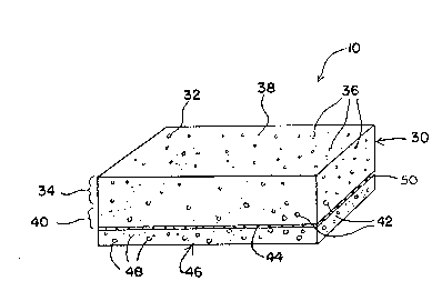
Referring now to FIG. 1, a preferred reagent
strip 10 includes a testing pad 30 which is formed from
an anisotropic membrane 32. The testing pad 30 has a
side 34 which contains relatively small pores 36. The
- l~B~3~2
side 34 with relatively small pores define~ a testing
surface 38.
The testing pad 30 also has an opposite side
40 having relatively large pores 42. The side 40 with
relatively large pores defines a sample receiving
surface 44.
A transport medium 46 is attached to the
sample receiving surface 44. The transport medium 46
contains relatively large pores 48. Connecting the
transport medium 46 to the sample receiving surface 44
is an adhesive layer 50. Preferably, the adhesive layer
50 is discontinuous.
In another embodiment of the invention, the
reagent strip includes a rigid support member which is
attached to a testing pad. The support member may be
fabricated of nylon-coated paper, MYLARI~, and other
materials which are chemically inert and relatively
rigid.
The rigid support member defines an aperture
which passes completely through the rigid support
member. A portion disposed about the aperture has a
carefully controlled and predetermined thickness which
is in the range of about 0.002 to about 0.050 of an inch
thick, preferably about 0.010 to about 0.020. At least
one surface defined by the support member is a surface
adapted to positively and reproducibly contact an
engaged viewing means.
A significantly higher degree of
reproducibility can be obtained when the reagent strip
of the present invention is evaluated using viewing
means, cUch as a reflectance meter, located a precisely
controlled distance from a testing surface of a testing
pad. Objective viewing means are generally more
reproducible than visual examination by subjective human
eyes. In addition, it has been found that small
2~ 9 .i98~
- 14 -
d:ifferences in the distance between the testing surface
and a lens of the viewing means can cause significant
changes in determination results.
Accordingly, the rigid support member is
fabricated with a portion disposed about the aperture
having a carefully controlled thickness. When the
surface of the support member is engaged with the
viewing means, the distance between the viewing means
and the testing pad is conveniently and reliably set.
Of course, other advantages accrue from the
incorporation of the support member. For example, the
support member can act as a shield which protects the
viewing means from contact with the liquid sample
carried by the testing pad.
The testing pad and an adhesive layer which
attaches the sample-receiving surface of the testing pad
to a transport medium are as described above.
The transport medium is substantially as
described above. However, the transport medium may
extend beyond the edges of the testing pad to form
regions which d~ not contact the testing pad and are
useful for storing excess sample. The regions which
extend beyond the testing pad are preferably attached to
the support member.
2S FIG. 2 illustrates a preferred reagent strip
100 having a rigid support member 120 defining an
aperture 122 and a free surface 124. The free surface
124 is adapted to engage viewing means (not shown). A
portion 126 of the support member which surrounds the
aperture 122 is fabricated with a known and carefully
controlled thickness.
A testing pad 130 is attached to the support
member 120 so that the testing pad 130 overlaps the
aperture 122. The testing pad 130 is formed from an
3S anisotropic membrane 132 having a side 134 with
20~9~2
relatively small pores 136 and a side 140 with
relatively large pores 142. At least a portion of the
testing surface 138 abuts the aperture 122. The side
134 with relatively small pores deflnes a testing
surface 138 which faces the support member 120. The
side 142 with relatively larger pores defines a sample-
receiving surface 144.
An adhesive layer 150, which is preferably
discontinuous, connects the sample receiving surface 144
to a transport medium 146. The transport medium 146
extends beyond the testing pad 130, having a region 147
which does not contact the testing pad 130. The region
147 is useful as a storage reservoir for excess amounts
of a blood sample. The transport medium 146 contains a
multitude of relatively large pores 148.
While the viewing means is not a part of the
present invention, an example of a suitable viewing
means i8 illustrated in FIG. 3 in which a reagent strip
300 of the present invention is inserted into a viewing
mean~ 350. The viewing means 350 is a hand-held
reflectance meter, battery operated and equipped with
display means 396. FIG. 3 depicts the orientation of
the reagent strip 300 with the viewing means 350 just
before insertion. An arrow in FIG. 3 indicates a
direction of movement to brin~ the reagent strip into
~ngagement with the viewing means 350.
A rotatable member 35S turns on an axis about
a pin 356 to present a calibration surface 357 to a
light source 360. Alternatively, the rotatable member
355 may be rotated to open a port 358 in which the
reagent strip 300 may be inserted. The calibration
surface 357 may be, for example, an unblemished white
surface, having a reflectance of approximately 100
percent. Alternatively, the calibration surface 357 may
2 ~
- 16 -
be a flat black surface representing zero percent
ref'lectance for calibration purposes.
When the reagent strip 300 is inserted into
the port 358, a free surface of the support member
contacts and engages a mating surface 359 which is
illustrated in FIG. 3A. When the reagent strip is
engaged, light rays 370 from the light source 360 pass
through a window striking the testing surface of the
reagent and reflecting to a light sensor 390. A signal
from the light sensor will be electronically compared to
similar signals received by the light sensor 390 when
the calibration surface 357 is presented to the light
source 360. The difference in signals is quantitatively
related to the concentration of an analyte in a blood
sample on the reagent strip 300, according to a
predetermined mathematical formula. The concentration
so determined is indicated digitally on the display
means 396.
When the reagent strip is intended to be
inserted into a meter, blood must be absorbed by the
reagent strip and retained so that it does not soil the
meter with repeated use. The major reservoir for blood
in the reagent strip is the transport medium. After
complete absorption of the sample, blood is held in the
transport medium as in a sponge. Unlike a sponge,
however, the presently favored material, surfactant-
treated porous polyethylene of the type commercially
obtainable from Porex Corporation, is essentially
incompressible, and blood is not squeezed out. However,
since the surfactant treatment renders the polyethylene
completely wettable, a film of blood remains in the
outer surface of the material. When an object comes in
contact with this film, surface tension can draw blood
back out of the transport medium, contaminating the
object.
- 17 -
It has been found that printing a film of
hydrophobic polymer onto the sample receiving surface of
the transport medium, preferably by flexographic
printing, improves the blood retention properties of the
reagent strip. When an appropriate polymer is applied,
blood is absorbed into the transport medium, but does
not transfer back to external contacting surfaces, even
when moderate pressure and sliding actions are applied.
A preferred hydrophobic polymer for this treatment is
GAF ES-22S, obtainable from GAF Chemicals Corp. of
Irvine, California, which is a monomethyl ether of
poly(methylvinyl ether/maleic acid). For example, a 30
w% ES-225 solution in ethanol can be printed onto the
transport medium using the flexographic printing method.
Another way to improve the blood retention of
the reagent strip is to modify the surface of the
transport medium by chemical reaction in such a way that
its surface energy is higher (more water-wettable) than
that of the native polyethylene, but not as high as the
surfactant-treated material. Blood is then drawn into
the transport medium, but will not be drawn out by
contacting surfaces.
The surface energy of a polyethylene transport
media can be suitably modified by corona-discharge,
which modifies polymer surfaces by introducing polar,
oxygen-containing groups into the polymer chains at the
surface. Alternatively, other means of suitably
chemically modifying the polymer surface may be
employed, such as other plasma treatments or solution-
based treatments. Polymers other than polyethylene canbe employed to form the transport medium, with a native
surface energy such that no treatment is necessary.
The invention also provides a method for
measuring the concentration of an analyte in a test
sample. The method is especially suitable for use with
2~ ~ 3
- 18 -
te!st samples that contain solid color bodies, such as
r~.d blood cells. A reagent strip is provided which
irlcludes a testing pad in which pores are
anisotropically arranged to produce a gradient in
effective pore size which extends from a sample-
receiving surface to a testing surface of the testing
pad. Pores closer to the testing surface are generally
smaller than those further from the testing surface. A
porous sample transport medium is attached to the sample
receiving surface.
A test sample, preferably of a biological
fluid having solid color bodies, which contains the
analyte is contacted with the transport medium. The
transport medium absorbs the test sample, transports the
test sample to the receiving surface, and distributes
the test sample transversely relative to a direction in
which the test sample is moved.
The test sample enter~ the testing pad at the
sample-receiving surface. Once inside the testing pad,
the test sample is further transported toward the
testing surface. As it travels along the gradient of
pore sizes, the test sample encounters progressively
smaller pores. The smaller pores filter the test sample
and at least some solid color bodies are separated from
the test sample. The portion of the test sample which
reaches the testing surface contains relatively few
solid color bodies.
The testing reagent c~emically reacts with the
analyte to produce a change in coloration which is
detectable at the test surface. The change in
coloration is evaluated by comparing the coloration of
the test surface to a calibrated color standard. The
color standards are prepared in advance using data from
testing pads and transport mediums exposed to samples
having ~nown concentrations.
2~9~9~
-- 19 --
~ he method may optionally further comprise the
step of locating the testing surface a reproducible
di~itance from a mechanical viewing means by contacting
the viewing means with a free surface of a rigid support
member attached to the testing surface. The rigid
support member has been described above. The testing
pad is attached to the support member so that at least a
portion of the testing surface overlaps the aperture and
is exposed to the viewing means.
FIG. 4 is a cross-sectional view of a printing
system 400 for applying a discontinuous adhesive layer
to a surface of a porous transport medium. Although not
essential to the invention, a flexographic printing
system, such as the printing system 400, is a convenient
means of applying a discontinuous adhesive layer which
does not bridge pores of the transport medium.
Referring now to FIG. 4, a doctor blade 410
forms a reservoir in which an adhesive supply 420 is
temporarily held. An anilox roller 430 turning counter-
clockwise as seen in FIG. 4, transfers a continuous film
440 of adhesive to a printing roller 450. The anilox
roller 430 is a steel cylinder with tiny engraved,
closely spaced pits which transfer a predetermined
amount of adhesive liquid from the adhesive supply 420
to the printing roller 450. The printing roller 450
turns clockwise as seen in FIG. 4.
A surface 460 of the transport medium is
brought into contact with the continuous thin film 440,
moving relative to the printing roller 450 from left to
right as depicted in FIG. 4. The surface 460 is made to
approach the printing roller 450 at a distance and with
a speed which causes the continuous thin film 440 to
adhere to the surfa~e 460 but not bridge pores 464.
Accordingly, a discontinuous adhesive layer 470 which
2~98~
- 20 -
does not bridge the pores 464 is applied to the surface
46~.
A continuous thin film 440 does not completely
leave the printing roller 450. Some remaining adhesive
480 does not adhere to the receiving surface 460 but is
instead carried along by the rotation of the printing
roller 450, and eventually returned to the continuous
thin film 440.
Descriptions of the invention and examples of
its use have been set forth to communicate the invention
fully, not to limit the scope of the invention in any
way. As should be apparent to those of ordinary skill
who study the specification, the invention may be
practiced in various embodiments. The scope of the
invention is intended to be as broad as the claims will
allow.