Note: Descriptions are shown in the official language in which they were submitted.
UTSH:165
2~0278~
N~HOD AND APPARATU~ FOR ~LAB~O~RAP~IC
MEA8UR~MENT A~D IHA~IN~
This applic~tion is a continuation in part of the
copending applications entitled "Method and Apparatus for
Measurement and Imaging of Tissue Compressibility or
Compliance," serial number 7/535,312,* filing date June 8,
1990, and "Transaxial Compression Technique for Sound
Velocity Estimation," serial number 7/438,695*, filing
date 11/17/89 (which is also the parent for application
serial number 7/535,312). Applicant incorporates said
applications serial nos. 7/438,695 and 7/535,312 by
reference herein and claims the benefit of said
applications for all purposes pursuant to
37 C.F.R. S 1.78.
.~
The U.S. Government may own rights in this
application and patents that may issue therefrom pursuant
to N.I.H. grants ROl-CA38515 and R01-CA44389.
This invention relates generaIly to a method and
apparatus for performing elastographic diagnosis of a
target body. Elastography is a system for measuring and
imaging elastic modulus and compressibility distributions
in an elastic tissue. It also has application to strain
35 profiling and improved sonographic measurement and ~ -~
imaging. This system is typically based on external
compression of a target body, and utilizes one or more
transducers, acting as or with a compressor, to generate pre~
and post-compression sonic pulses and receive the ~-
* see Canadian Patent File No. 2,068,740
210278~
resulting echo sequences (A-lines) from within the target
body. The pre- and post-compression echo sequence pairs
may then be cross-correlated or matched to determine the
strain along the path of the sonic pulses, and preferably
to yield a strain profile of the target body. This
strain profile may then be converted into a
compressibility profile or elastogram by measuring the
stre~s imposed by the compressing device and calculating
the e$astic moduli based on the stress and the strain
profile.
An elastogram may be considered to be a ~pecial form
of multi-trace sonogram, wherein each trace is a record
or display with depth within a target body of an elastic
modulus function of the body. A preferred elastic
modulus function for purposes of display is the inverse
of the bulk modulus, which provides a measure of
compressibility. As explained later in this description,
the inverse of the Young's moduli may usually be used
instead of the bulk moduli. Less preferred but also
helpful are the Young's moduli themselves. Methods for
making and usinq elastograms are described at length in
co-pending application no. 7/535,312.
While the methods described in application no.
7/535,312 produce greatly improved recordc and
understanding of structures of elastic tissues, it has
been observed that certain inaccuracies in the resulting
elastograms may arise. In particular, inaccuracies have
been observed if the transducer and compressor used to
compress and insonify a tissue are relatively small in
size relative to the depth (or thickness) of the target
body, giving rise to decreasing stress in the target body
as the distance increases from the compressor. Likewise,
inaccuracies have been observed in elastograms and
sonograms if the target body is not relatively
~ - : '' : `'!: :: :~: :: : -:. .~.; . .. .
`~
~ -3~
210278~
homogeneous with respect to sonic speed, giving rise to
strata through which sonic pulses travel at differing
velocities. The improved methods and apparatus for
elastography disclosed herein, while generally enhancing
the accuracy of elastograms, have particular application
in reducing the effect of such inaccuracies in both
elastograms and sonograms.
Traditional ultrasonic diagnosi~ is achieved by
transmitting ultrasonic energy into a target body and
generating an image from the resulting echo signals. A
transducer is used to both transmit the ultrasonic energy
and to receive the echo signals. During transmission,
the transducer converts electrical energy into mechanical
lS vibrations. Acquired echo signals produce mechanical
oscillations in the transducer which are reconverted into
electrical signals for amplification and recognition.
A plot or display (e.g., on an oscilloscope, etc.)
of the electrical signal amplitude versus echo arrival
time yields an amplitude line (A-line) or echo seguence
corresponding to a particular ultrasonic transmission.
When the A-line is displayed directly as a modulated
sinusoidal pattern at radio frequency ("RF"), it is
typically referred to as an RF or "undetected" siqnal.
For imaging, the A-line is often demodulated to a non-RF
or "detected" signal. -
~ .
Ultrasound techniques have been extensively used in
the field of diagnostic medicine as a non-invasive means
of analyzing the properties of tissue in vivo (i.e.,
living). A human or animal body represents a
nonhomogeneous medium for the propagation of ultrasound
energy. Acoustic impedance changes at boundaries of ~-
regions having varying densities and/or sound speeds
within such a target body. At such boundaries, a portion ~-
-4-
210278~
of the incident ultrasonic beam is reflected.
Inhomogeneities within the tissue form lower level
scatter ~ites that result in additional echo signals.
Images may be generated from this information by
modulating the intensities of pixelq on a video display
in proportion to the intensity of echo ~eguence segments
from corresponding points within the target body.
Conventional imaging techniques are widely used to
evaluate various diseases within organic tissue. Imaging
provides information concerning the size, shape, and
position of soft tissue structures using the assumption
that sound velocity within the target is constant.
Qualitative tissue characterization is carried out by
interpretation of the grey scale appearance of the
sonograms. Qualitative diagnosis largely depends on the
skill and experience of the examiner as well as
characteristics of the tissue. Images based only on
relative tissue reflectivity, however, have limited use
for quantitative assessment of disease state
Techniques for quantitative tissue characterization
u~ing ultrasound are needed for more accurate diagnosis
of disorders. In recent years many significant
developments have been achieved in the field of
ultrasonic tissue characterization. Some acoustic
parameters, e.g., speed of sound and attenuation, have
been successfully used for tissue characterization.
Tissue compressibility is an important parameter
which is used to detect the presence of diffuse or
localized disease. Measuring changes in compressibility
becomes important in the analysis of tissue for
pathological conditions. Many tumors are firmer than the
surrounding normal tissue, and many diffuse diseases
result in firmer or more tender pathology. Examples can
~ -5-
210278~
be found in diffuse liver disease, prostate cancer,
uterine fibroids, muscle conditioning or disease, breast
cancer disease, and many other conditions.
Traditionally, physicians routinely palpate various
regions of a patient's body to get an impression of
tissue firmness or tissue softness. This technique is a
foro of remotely trying to sense what i~ going on in
terms of tissue compliance. For example, in a liver, if
the compliance in an area is sensed to be different from
compliance in the surrounding area, the physician
concludes from the tactile sensations in his fingers that
something is wrong with the patient. The physician's ~
fingers are used to perform a qualitative measurement. ~-
In the last several years, a number of articles have
appeared in the literature that explore various
technigues for measurement and imaging of soft tissue
compliance and tissue motion using ultrasound. These
20 technigues rely on one of the following procedures: ;
Doppler ultrasound velocity measurements, cross-
correlation technigue~ to guantify motion in ti~sues, and
visual inspection of M-mode and B-mode images.
Additionally a Fourier feature extraction technique has
been proposed. Internal mechanical excitation (motion of
cardiac structures, arterial pulsation) or external
vibration sources of motion produce displacement of the
tissues under investigation. The di~placements of
different ti~sues are then analyzed by one of the e
technigues.
The amplitude and velocity of motion induced by
arterial pulsation is generally too low for evaluation
with Doppler velocity measurements. However, a number of
researchers have used pulsed Doppler and color flow
Doppler systems in conjunction with external mechanical
210278~
harmonic excitations to determine the elastic properties
of ti~sue. Using a low frequency external excitation
source, the velocity of propagation of mechanical waves
has been measured and relates to the modulus of
elasticity of the tissues. The velocity of vibration of
tissues under low freguency vibration excitation has been
used to determine their relative compressibility. This
technique has been termed "sonoelasticity" and produces
B-~cans which are "otained" with color coded relative
compressibility information. Sophisticated Young's
modulus measurements have been applied to determine
muscle elasticity as a function of contractility state by
measuring Doppler shifts due to very low frequency
excitations (lOHz). A similar approach using vibrations
in the 100-lOOOHz range has been proposed to study
dynamic muscle elasticity in vivo.
Cross-correlation techniques allow the use of either
internally or externally generated sources of mechanical
excit~tion due to their ability to quantify minute
motion~ of tissue. External harmonic excitation has been
used to assess motion of soft tissues with one
dimensional and two dimensional correlators. The
displacement and/or velocity of internally generated
motion also have been measured using one dimensional and
two dimensional correlators. Tissue strain caused by
arterial pulsation in the liver and by transmitted
cardiac motion in fetal lung have been proposed for
tissue characterization.
Visual inspection of ultrasound M-mode waveforms has
been used to study benign and malignant lesions in liver,
pancreas and breast and to observe the elasticity of
fetal lung. In magnified B-scans of the fetal thorax
paracardiac lung movements have been measured to classify
fetal lungs as stiff, intermediate or compliant. The
~ -7
210278~
examination of fetal lung sonograms has been used to
evaluate compre~sibility as an indicator of lung tissue
maturity.
But, one of the main difficulties in these methods ~ 5
is the lack of definition of the magnitude and direction
of the driving force. This difficulty applies to driving ~-
forces that are internally generated by the pulsations of
the heart and/or the aorta, as well as to those applied
10 externally at low frequency and limited directivity. -~
Further, it is difficult to measure the shape of an
internal driving force, limiting the ability to determine
how stress resulting from the driving force decreases as
a function of distance from the driving force. The
15 inability to define the direction, magnitude and shape of ~;
the driving force limits the ability of these methods to
provide quantitative information about the elastic
properties of the tissue under investigation.
In contrast to these methods, elastography i8 not
limited by a lack of definition of the magnitude,
direction or shape of a driving force. Elastography --
preferably uses an external stimulus of known quantity,
such as compression of the target body by a known amount
or known stress by a compressor, preferably along with
cross correlation or least-means-square matching
techniques to generate strain profiles of the tissue
under investigation. From these strain profiles and the
measurement of the stress applied by the compressor, an
elastogram (or image of the inverse elastic modulus
profile) is determined. The inverse of the elastic
modulus profile is typically displayed on the elastogram
because strain measurements may be zero, yielding an
elastogram with an infinite range of elastic moduli.
~ -8-
210278~
Thus, elastography provides a pulse-echo system that
has particular application in estimating and imaging
compressibility in a target body. The target body may be
any animal or human tissue, or any organic or inorganic
substance that is compressible or compliant. The term
"animal tissue" includes "human tissue". An ultra~onic
source is used to interrogate the target body. The
detection of echo sequences may be at the ultrasonic
source. Thus, elastography allows for accurate,
localized determination and imaging of an important
parameter, compressibility, which has been used
qualitatively in medicine for a very long time.
Compressibility of a material is normally defined as
the inverse of the bulk modulus of the material. The
bulk modulus of a volume may be determined by the
following formula:
BM = P/(~V/V) where Equation 1
BM = Bulk modulus
P = the pressure or stress on a tissue seqment of
interest
(~V/V) = the volumetric strain of a tissue segment of
interest, where
~V = a change in the volume of the segment, and
V s the original volume of the segment.
In a preferred method of elastography where an external
source of compression is applied to stress the target
body, it may be generally assumed that the volumetric
strain (or differential displacement) along the axis of
compression may be determined by the formula:
strain = (~L/L), where Equation 2
~L = a change in the length of the segment along the
axis of compression, and
L = the original length of the segment,
- 9 -
21027~
Further, it may be generally assumed that the stress on
the tissue segment of interest caused by the external
source of compression may be determined by the formula~
, ~:
stre6s = (F/a), where Equation 3
F = compressive force applied to the segment, and
a = area across which the force is applied.
Therefore, applying these as~umptions to Equation 1, the
elastic modulus (E) of a tissue segment of interest may
be estimated by the formula for determination of a
Young's modulus:
E = (F/a)/(~L/L)- Equation 4
Further, compressibility (K), the inverse of E, may ~ -
be estimatod by the formula: ~-~
K = (~L/L)/(F/a)- Equation 5
Thus, the compressibility of any given segment or layer
within a material relative to another segment or layer
may be further estimated from the relationship
25 Xl s K2 (~Ll/Ll)/(~L2/L2), where Equation 6
Kl = compressibility of a first segment or layer;
.~Ll = change in length of the first segment or layer
along an axis of compression in response to a
given force;
30 Ll = original length of the first segment;
~L2 = corresponding change in length of a second
segment or layer;
L2 = original length of the second segment or layer;
and
35 K2 = compressibility of the second segment or layer.
In elastography, the velocities of sound in
different segments or layers may be employed, together
with time measurements, to calculate distances within the
segments or layers. The ultrasonic signals also provide
--10--
21 ~278~
a precise measuring tool. The velocities of sound may be
determined using the apparatus and procedures disclosed
in application serial no. 7/438,695.
However, in the techniques previously disclo6ed for
elastography in applications serial no~. 7/438,695 and
7/535,312, the method for estimating compressibility in
targets having multiple layers may lead to some
inaccuracies. These inaccuracies may similarly arise in
sonography. Such inaccuracies generally result from one
of two conditions, or both. Thus, a first group of such
inaccuracies may arise due to substantial variations in
the speed of sound in the different layers. Expressed
otherwise, the techniques estimate compressibility in
each layer from two echo sequences along the axis of
radiation without consideration for variations in the
speed of sound.
Some regions in a target body of interest may
contain multiple layers having substantially different
velocities of sound. For example, the human body wall
may include regions of interspersed fat and muscle
tissue, and sound may typically travel at about 1450 m/s
in a fatty region, while it will typically travel faster
in muscle tissue, around 1580 m/s. The variation in the
speed of sound through different layers can cause sonic
pulses traveling on different sonic paths, but through
ths same distance relative to the transducer, to take
different amounts of time. ~his time difference may in
turn lead to a distortion, and possibly a shift, in the
elastograms and sonograms of the target body.
A second condition that may cause a significant
inaccuracy in the previously disclosed techniques for
elastography lies in the assumption that stress will be
relatively uniform throughout the tissue of interest, and
210278a
may be calculated for all layers based on measurements
proximal to the compressor. However, inaccuracies have -
been observed if the transducer and compressor used to -
compress and sound a tissue are relatively small in area
relative to the depth (or thickness) of the target body,
giving rise to decreasing stress in the target body as
the distance increases from the compressor. If this
decreasing stress is substantial and unaccounted for,
levels of decreasing compressibility may appear on the
elastograms as a function of increasing distance from the
compressor.
Thus, the present invention in one aspect provides a
method and apparatus to determine the strain and
compressibility of a target body regardless of whether
the target body has multiple layers with different sonic
velocities. In another aspect, the invention provides a
method and apparatus to determine the compressibility of
a target body even where the stress in the target body
resulting from compression by the compressor decreases
with distance from the compressor. In yet another
aspect, the invention generally provides an ultrasound
method and apparatus for accurately measuring and imaging
strain and elastic modulus distributions in an elastic
ti~ue. The ability of the invention to quantitatively
measure the compressibility or compliance of tissue in
localized regions provides help with (1) objective
quantification of commonly used clinical signs, (2)
localizing these measures, (3) making the measurements
deep in tissue with simple equipment, (4) observing new
tissue properties, which may be related to pathology, not
seen by other known means; and (5) constructing images of
a compressibility or compliance parameter in vivo, which
may be used alone or in conjunction with ordinary
sonograms. Diseased tissue, such as tumors, may be
harder or softer than normal tissue, and thus have a
~ -12-
210278~
different amount of compressibility. In this regard,
elastography gives promise of a distinct advantage over
prior art methods in the accurate detection of diseases
such as breast cancer and prostate cancer and
localization of tumors at an early stage. Another
advantage of elastography is that its sensitivity may be
qreater than sonography, because of its measurement and
imaging of compressibility and not just echo amplitudes,
allowing for better visualization of target bodies.
Still another advantage of elastography is the accurate
i~aging of sub-surface tissue while avoiding the use of
ionizing radiation from x-rays.
It will be noted at this point that elastography is
contemplated to have significant applications other than
in medicine. One such application, for example, is in
the quality grading of beef. Elastography may be used to
quantitate both the tenderness of beef and the fat
content (marbeling) before and after slaughter. This
ability i8 economically important in determining when to
slaughter cattle. Other applications may include, for
ex~ople, interrogation of materials and products such as
cheése or crude oil that are physically displaceable by
the movement of a transducer. Other objects and
advantages of elastography will become readily apparent
from the ensuing description.
In a broad sense the present invention comprises an
ultrasonic system for producing improved elastograms and
sonograms of elastic target bodies, and notably animal
and human tissue. In one broad aspect, elastography
comprises sonically coupling a sonic device to a target
body to determine its compressibility. The sonic device ; ;
i8 used to emit an ultrasonic signal and receive
returning echo sequences from along a sonic path in the
target body. The sonic device is then moved a known
?~
- f
-13-
210278~
amount along the axis of the sonic path, and the target
body is interrogated again along the sonic path.
Congruent segments in echo ~equences from the different
sonic aignals are then preferably cross correlated or
matched, and the temporal displacements are used to
calculate the strain along the sonic path. This
procedure i8 repeated for a plurality of sonic paths,
either sequentially or by means of a ~onic array, to
produce strain profiles for the target body.
':
To obtain a compressibility profile, a second body
having a known, preferably uniform, elastic modulus may
be first sonically joined to and between the target body
and the sonic device. The strain profiles of both bodies ~ -
may then be determined using the steps described above.
The stress caused by the movement of the sonic device may
then be determined from the strain profiles and elastic
modulus of the second body. Compressibility profiles for
the target body may be obtained by dividing the values
for the strain profiles in the target body by the stress.
The resulting compressibilities may be further arranged
as an elastogram, a positional multi-dimensional plot or
picture of the relative magnitudes of compressibility of
a ti~sue or other target body.
In another aspect, the invention resides in using
the above method of elastography, but further correcting
for variations in stress along the sonic path. These
variations may occur in instances where a sonic device,
which is employed to stress and compress the target body,
has a small cross-sectional area relative to the depth or
thickness of the target body. These variations in stress - -
may be determined by first measuring the dimensions of
the surface of the sonic device (which may include a
transducer in combination with a compressor) which
contacts the second body. These measured dimensions may
210278~
then be applied to analytically derive the variation~ in
8tre83 within the target body as a function of position
relative to the sonic device. The result is, in effect,
a stres~ profile. Once derived, these variations in
stress may be applied to correct the values for the
stress profiles, as well as the resulting compressibility
profiles and elastograms for the target body, previously
determined by use of the above-described preceding
embodiment.
In another aspect, the above variations in stress
may be experimentally derived. Thus, a body of known
elasticity may be compressed by the sonic device and the
resulting strains along varying sonic paths inside the
body may be measured. The variations in stress may then
be calculated as a function of position relative to the
sonic device. From these known variations th~ corrected
values for the stress and compressibility profiles and
elastograms may then be determined.
In yet another asp~ct of the invention, appropriate
time delays, necessary to correct for variations in echo
sQquence travel time through regions of the target body
having dif~erent speeds of sound, may be determined and
25 applied to each echo sequence. The resulting time shifts ~-
in echo sequences correct for distortions that might
otherwise occur when there are different regions within a ;-
target body, e.g., fat and muscle tissue, that have
differing sonic velocities.
The present invention, together with further
advantages and features thereof, may be more readily
understood by reference to the following detailed
description taken in connection with the accompanying
drawings in which:
-15 :.:
21027~ ~
Fig. la shows an embodiment of an elastographic
apparatus where a transducer and compressor are sonically
coupled to a target body to interrogate a distal tissue
region within the target body.
Fig. lb shows a plot of an RF echo ~ignal
originating from the distal tissue region interrogated in
Fig. la.
Fig. 2a shows the transducer and compressor of Fig.
la imparting a small compression to a proximal region of
the target body.
Fig. 2b shows a plot of a typical pre- and post-
compression RF echo signal pair originating from thedistal tissue region as interrogated in Fig. 2a.
Fig. 2c shows a plot of a cross correlation of the
echo signal pair shown in Fig. 2b.
Fig. 3a shows the axial strain in foam as a function
of depth using a 127mm circular compressor.
Fig. 3b show~ the normalized stress in foam as a
function of depth using various sizes of circular
compressors.
Fig. 3c shows the normalized stress in dB as a
function of z/a, the depth divided by the area of a
circular compressor.
Fig. 4a shows a cross sectional view of an apparatus
for determining stress distribution under a given ~;
compressor.
-16-
210278~
Fig. 4b shows a cross sectional view of a stress
distribution under a compressor as a function of position
relative to the compressor.
Fig. 5a shows a photograph of a phantom consisting
of two triangular foam pieces joined along a diagonal
seam.
Fig. 5b shows a B-scan of the phantom pictured in
Fig. 5a.
Fig. 5c shows the elastogram corresponding to the B- i
scan shown in Fig. 5b.
, . .
Fig. 5d shows the depth-corrected elastogram `
corresponding to the B-scan shown in Fig. 5b.
~ ; ,
Fig. 6 shows an embodiment of an apparatus in which
a compressor is coupled to a target body for purposes of
experimentally determining the variations of stress with
depth in the target body. --~
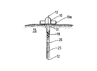
Figure la shows the transducer ~0 and compressor 10
sonically coupled to a target body lS. An ultrasonic
pulse 18 is shown propagating within sonic beam 20 toward
an echo source 2S on beam axie 12. As the pulse 18
propagates through the target 15, corresponding echoes
are generated and arrival times noted at the transducer
aperture 11. The combination of all echoes generated
from reflections within the beam 20 is the echo sequence
or A-line corresponding to pulse 18. ~ ~-
A radio frequency ("RF") signal plot of the A-line
acquired from pulse 18 is shown in Fig. lb. The
amplitude of the signal in volts is plotted against echo
arrival times in microseconds (~s). Later arrival times
-17-
210278~
correspond to progressively deeper regions within the
target body 15. An echo segment or echo wavelet 30,
within a chosen arrival time window, is selected as a
reference. The time window may be selected based on
anatomical data from ultrasound imaging, or may be
arbitrary, e.g., every x micro seconds. The echo segment
or wavelet 30 originates from the echo source 2S.
Figure 2a shows the transducer 10 and compressor 10
being translated along axis 12 to impart a small
compre~-ion (-Yl) to the ti~sue. ~fter the tran~ducer 10
and compressor lOa compress the target body 15, a second
pulse 22 is emitted and the corresponding A-line segment
i~ acquired from a desired depth within the ti~sue.
Fig. 2b shows an RF plot pairing a typical pre-
compre~on A-line, corre~ponding to pulse 13, and a
post-compression A-line, corresponding to pulse 22. The
echo segment or wavelet 32 associated with a given echo
source and pulse 22 is time shifted with respect to the
~ame segment of wavelet 30 associiated with the same echo
source and pre-compression pul~e 1~. The time ~hifted
wavelet 32 may be trasked within the selected time window
u~ing ~tandard pattern matching techniques. The window
~elected must be such that the wavelet of interest will
not be shifted out of the window. This selection may
involve the size of the window or the positioning of the
window. The window selected should reveal both wavelets
or echo segments. The arrival time of echo segment or
wavelet 32 is prior to that of echo segment or wavelet 30
above, since the distance between aperture 11 and feature
25 was shortened by the compression ~Yl-
Fig. 2c shows the cross-correlation function between
the pre- and post-compression A-lines shown in Fig. 2b.
. ' .. ' . ' ' . , '. . ' '.'.,', . ' : ~' ' . . ' ' ' ~' '' : . ' ' ' ' .
-18-
21027g3
In a preferred method of elastography, a transducer
and compressor are positioned on or otherwi~e coupled to
a target tissue and advanced axially toward the target to
compress the target. Alternatively, elastography may be
practiced by retracting a transducer and compres~or from
a previously compressed position. Further, in both
methods the transducer may alone serve as the compressor.
Since the relatively large size of the compressor
precludes penetration of the tissue, small tissue
dioplacement~ occur instead. A pulse iB emitted from the
transducer prior to the displacement, and a first echo
sequence received in response to the pulse is recorded.
Following displacement, a second pulse is emitted and a
second echo sequence is recorded in response to
transmission. Next, a comparison of the waveforms is
made to reveal a decreasing displacement of the tissue - -
structure with depth. The decrea~e will generally be ~ ~-
asymptotic in character.
In the foregoing method, a single compre~sion of a
homogenous target body has been de~cribed. It will be
apparent, however, that other condition~ may be employed.
Thus~ multiple compressions, repetitive or real time
compreseions, varying waveforms and other signal sources,
such as array transducers, may be used. These signal
~ources, for example may be non-repetitive and may
generate spike-like signals.
Further, an internal source of compression, alone or
in conjunction with an external compressor, may be used.
In the case of an internal source of compression, such as
the heart or arteries, the tissue of interest should
preferably be located sufficiently distant from the
internal source of compression such that stress caused by
the internal source of compression, even while changing
as a function of time, remains substantially uniform
` -19-
210278~
throughout the tissue of interest. A transducer may then
be sonically coupled to the target body, preferably with
a compliant body of known uniform elasticity sonically
coupled between the transducer and the target body, such
that the tissue adjacent to the transducer compresses
against the compliant body and transducer as the tissue
is compressed by the internal source of compression, and
decreases in stress against the transducQr and compliant
body a~ the stress caused by internal source of
compression decreases in the tissue of interest. The
strain in the compliant body and tissue of interest may
then be sonically mea~ured using the method described
herein, and the stress against the compliant body -
determined from elasticity and strain measurements for
the compliant body. Finally, this stress may be assumed
to be the level of stress throughout the tissue of
interest, and used with the measured strains to determine
the compres~ibility profile in the tissue of interest.
In tissue that is not homogeneous, the shifting of
tissue in various segments will differ. For example, if
a ~egm~nt of tissue is less compressible than the overall
ti~sue containing the segment, the tissue in the segment
will compress or strain less than if the segment of
ti~sue were of the ~ame compressibility as the tissue as
a whole. Alternatively, when a segment is more
compressible than the tissue as a whole, the segment will
compress or strain more than if the segment were of the
same compressibility as other segments. The presence of
a strain "defect," or segment of different
compressibility, along the compression axis in a target
body influences all other strains along that axis,
increasing or decreasing the otherwise proportional
change in strain with depth along the axis. In this way
a strain "defect" is said to be "smeared" along the axis.
For this reason, it may be preferable to convert the -~
. ", . . .. : ,,.. ~. - - . : ~
-- -- --
-20-
210278.5
strain profiles into elastic modulus profiles. Since the
elastic modulus is a basic tissue property, it may be
ultimately a more reliable parameter. In any event, it
i8 possible to obtain useful images from strain or
S elastic modulus data.
In order to illustrate these principles, it is
convenient to consider a simple one-dimensional cascaded
spring system, where the spring constants repre~ent the
elastic moduli of tissue regions. We assume that all
three springs are equal and are of length e, and that
oach spring represents the behavior of a cylindrical
tissue element with unit cross section. If the top of
the first tissue element is compressed by an axial
downward force such that the overall length of the system
is reduced by (2~y), then a simple statics calculation
shows that each and every spring will shri~k by ~ =
2~y/3. If we define the strain of each spring = ~l/l, it
is clear then that the strain is constant for all
springs, and is equal to 2~y/3~.
Where the center spring has been replaced by an
infinitely stiff spring, i.e. E = ~, the total
displacement is taken up by two outer springs only.
Thus, the strain in the two outer springs will increase
to ~y/~.
It is evident from this example that a strain
profile is dependent on the initial compression and on
the number and stiffness of all springs. A given local
measured value of the strain is influenced by the elastic
propertie.s of elements located elsewhere along the axis
of compression. For these reasons it appears that while
strain profiling may be useful for imaging, it may be of
limited use for quantitative estimation of local tissue
elasticity.
-21-
210278~
If instead of imparting a known displacement a known
stress is applied, it becomes possible to estimate the
elastic modulus of each component in this system of
springs, since the stress remains constant with depth in
this one dimensional system. In this case, the
measurable strain in each spring and the known stress on
each spring may be used to construct an elastic modulus
profile along the compression axis. Such a profile would
be independent of the initial compression, and the
interdependence among the component springs would
disappear.
-.'.~
Further, the stress applied to the target body may
be measured ultrasonically by interposing an anterior
compliant standoff layer which has a known value of E,
and which allows the free passage of ultraRonic waves.
The simultaneous measurement of the strain in this layer
allows the computation of the stress acting on the
target. This layer may consist of compressible or
compliant material such as rubber, sponge, gels, etc.
The material should be compressible and provide for an
ultrasonic transmi~ion path to the tissue. The ~aterial
may be echogenic, but it is not necessary.
2S In the more realistic three-dimensional case, one
would expect that the applied stress would not be
constant along the axis of compression. The reason for
this lies in the fact that stresses along transverse
spring~ become important, and since their vertical force
components are a function of the displacement which in
turn is a function of depth, the resultant forces along
the compression axis vary with depth. On the other hand,
enlarging the area of the compressor, the transverse
springs that are actually stretched, and hence contribute
to the depth dependent stress field, become less
important and the applied stress field becomes more
-22-
2lo278s
uniform. Experiments have confirmed that larger
compre~sors cau~e more uniform axial stre~s fields.
In elastography, however, the velocities of sound in -~
different seg~ents or layers are used, together with time
measurements, to calculate distances within the target
body. More specifically, elastograms are based on time
shift differences among segments of ultrasound A-lines
and preferably, when based on more than about 64 data
points, rely heavily on cross-correlation computations.
The use of cross-correlation analysis for time shift
estimation derives from Fourier theory, and is well known
in the art. In recent years a number of industrial and
medical applications have utilized cross-correlation
analysis for time shift measurements. The application of
ultrasonic correlation techniques to the measurement of
flow velocity of coal slurries has been described.
Similarly, an ultrasonic correlation flowmeter for pulp
~uspen~ion has been proposed. In the medical field, a
number of publications describe the measurement of blood
velocity profiles using one-dimensional and two-
dimensional correlators, as well as applications of
cross-correlation measurements for tissue motion
evaluations, described above.
The generation of an elastogram involves a pairwise
evaluation of the time shift between congruent segments
in an A-line pair, preferably by means of cross-
correlation techniques. The linear cross-correlation of
segment pairs may be computed using FFTs (fast Fourier
transforms). The temporal location of the maximum peak
of the cross-correlation function may be used to estimate
the time shift between the data in the two segments.
However, the time shift differences among segments
of an ultrasound A-line may also be evaluated by using a
210278a
least-means-square match analysis, which is al80 well
known in the art, or by manually measuring the
difforences between A-lines on a display or picture,
in~tead of by cross-correlation. When there are less
than about 64 data points being analyzed, a time domain
computation, such as a least-means-square match, may
often take less time than a fourier domain cro~s-
correlation computation will take to determine the time
shift~. Further, a least-mean~-square match analyei~ may
be computed for a limited number of time-lags, where the
approximate time shift is known, while cross-correlation
using an FFT computation must analyze the entire data
~eguence. Thus, a least-means-sguare match may be faster
than cross-correlation where the approximate time-shift
i~ known, allowing a time-shift determination to be made
~y matching only a portion of the eGho sequences.
By way of illustration only, one approach to
elastography could involve the derivation of an
elastogram from a strain image created from 40-60 A-line
pair~ obtained with a 1-2 mm lateral translation of the
tran~ducer between pairs. An A-line pair consist~ of the
original A-line which is obtained with the transducer
~lightly pre-compressing the target in order to a~ure
good contact, and a compressed A-line which is obtained
after axially compressing the target an additional ~z.
The compressed A-line would be shorter than the original
A-line by 2~z/c, where c is the speed of sound in the
target. The length of the A-line pair is taken to be
that of the original A-line; zeros are appended to the
compressed A-line. These A-lines would be obtained from
a 12 cm total depth in the target, and divided into 40-60
overlapping 4 mm segments obtained every one or two mm.
The data acquisition, and therefore the time scale,
is relative to the face of the transducer. Thus one can
-24
2102785
observe that the relative shift of the signal at the
beginning of an A-line pair is very small, whereas
towards the end it is significant. In general, the time
shift of the compressed A-line relative to the
uncompressed A-line would increase from 0 to a maximum of
2~z/C.
In general, the precision of the time shift Qstimate
improves with increasing segment size. However, it i8
typically better to ~eep the segment size small to
improve the axial resolution of the estimate.
Additionally, because of the relative compression and the
resultant progressive distortion of the data within a
segment pair, the cross-correlation estimate may
deteriorate with increasing segment size. This, in turn
may degrade the precision of the estimate. Thu~, there
are two competing mechanisms that affect the precision of
a time shift estimate as a function of the segment size.
Although this trade-off has not been studied in depth, it
has been observed that a segment size of about 4 mm with
about 3 mm overlap between segments leads to strain data
wh~ch may result in reasonable images with about 1 mm
axial resolution.
The resolution of a measured time shift may be
bounded by the sampling period at which the data is
digitized. To improve the resolution, some interpolation
algorithms have been proposed. For example, a quadratic
interpolation algorithm has been shown to be effective
and it is simple to implement. See, Foster et al., "Flow
Velocity Profile Via Time-Domain Correlation," IEEE
Trans. Ultrason. Ferroel. Freq. Control, Vol. 37, No. 2,
164-174 (1990); See also, Boucher et al . , "A Method of
Discrete Implementation of Generalized Cross-Correlator,"
IEEE Transactions: Acoustics, Speech and Signal
Processing, Vol. ASSP-29, No. 3 (June 1981). This
-,-
-25-
210278~
algorithm first fits a second-order polynomial which
passes through the peak sample value of the cross
correlation and its two neighbors using the Lagrange
polynomial interpolation. Then it analytically locates
S the peak of the fitted polynomial, assigning that
tomporal value to the improved time ~hift e~timate.
Returning to the illustration, after proces~ing one
A-line pair a SQt of time ~hifts, tl through t60, may be
obtained. The corresponding strain profile may then be
defined by the relationship
t~tl-tl Equation7
where sl is the strain estimate for segment pair i, and
where ~x i8 an axial increment.
The process may then be repeated for all A-line
pair~, re~ulting in an array of ~train data. These
value~ may then be scaled and assigned to an intensity
for display, e.g., an intensity varying within 256 grey
scale levels. Due to the large dynamic range of some
strain data, contrast stretching may be applied in order
to observe variation in particular strain ranges. For
example, 256 grey scale levels may be assigned to a user
specified strain range, thus stretching the contrast in
that region.
In general, elastography contemplates sonically
coupling an ultrasonic source to a target body;
energizing the ultrasonic source to emit a first
ultrasonic signal or pulse of ultrasonic energy from the
source along an axis into the target body; detecting from
-26-
21027~
a region within the target body a first echo sequence
including a plurality of echo segments resulting from the
first transmitted signal; displacing the target body
along the axis while maintaining coupling between the
ultrasonic source and the target body; energizing the
ultra~onic source to emit a second ultrasonic signal
alonq the axis into the target body; and detecting from
the region within the target body a second echo sequence
including a plurality of echo ~egments resulting from the
second transmitted signal; and measuring the differential
displacement of the echo segments. A plurality of first
ultrasonic signals or pulses of ultrasonic energy may be
emitted and a plurality of first echo seguences detected
before compressing the target body. Then a plurality of
second signals and pulses are emitted along a plurality
of parallel paths and a plurality of second echo
sequences are detected.
In one embodiment of elastography, a transducer i8
the ultra~onic source and is sonically coupled to direct
an ultrasonic signal or pulse of ultrasonic energy into
the tissue along a radiation axis such that movement of
the transducer along the axis effects a change in
compression of the tissue.
In a preferred embodiment of elastography, the
ultrasonic source is a transducer sonically coupled to a
tissue of interest. A first pulse of ultrasonic energy
is emitted along a path into the target body and the
arrival of a first echo sequence (A-line) including one
or more echo segments is detected from regions within the
tissue along the path resulting from the first pulse of
ultrasonic energy. Thereafter, compression is changed
within the tissue along the path. The compression change
may be accomplished by transaxially moving the transducer
along the path to compress or displace a proximal region
~ -27
2102785
of the tissue. A second pulse is emitted, and the
arrival of a second echo sequence including one or more
echo segments common to the first echo sequence is
detected in response to the second pulse. The
differential displacements of at least one echo ~egment
are measured. The echo sequences detected are from
coumon regions within the tissue.
A comparison of the first and second echo seguences
or waveforms with intervening compression reveal- a
generally decreasing displacement of tissue structures
with depth. In a homogeneous medium, the rate of
decrease will tend to be asymptotic. Of particular
interest is the differential displacement per unit length
- i.e., strain. In a homogeneous compressible medium,
the strain will tend to be constant along the axis of
compression. In a non-homogeneous medium, the strain
varies along the axis of compression.
The strain of a tissue may be calculated using the
arrival times of first and second echo sequences from
proximal and di~tal features in a target body -- i.e.,
tissue -- u~ing the following equation:
(tlg - tlA) - (t2g ~ t2A)
(t1B ~ tlA) Equation 8
t1A = arrival time of a first echo sequence from a
proximal feature;
t1B = arrival time of a first echo sequence from a
distal feature;
t2A = arrival time of a second echo sequence from a
proximal feature; and
t2B = arriva' time of a second echo sequence from a ~ -
distal feature.
The arrival times of the echo segments from a common
point detected in response to a first and second pulse of
ultrasonic energy are compared. The common points may be
. ~ ~ . , . !, .~ . -; ~ ' .:~' ` ' ' , '
-28-
210278~
found in feature~ occurring within the echo signal. The
time shifting of the two echo ~egments is used to
determine compressibility.
Thus, if no change in arrival time has occurred with
an intervening compressive force, it follow~ that a
t~rget body has not baen comprQ~sed along the travel path
leading to the source of the echo segments. On the other
hand, if the arrival time of the second echo segment is
~all-r than tha arrlval time of tho flrst echo ego-nt,
it is clear that compression has occurred and that the
target body is compre~sible. Moreover, the difference in
arrival times, taken together with other available data,
makes it possible to quantify the compressibility of the
target body.
In another embodiment of elastography, body segments
which extend along the transmission path of the
ultrasonic pulse6 are selected within a target body and
separate fir~t and second echo segment~ detected from
within each body segment. Thus, a series of first and
second echo segments is detected for the body segments
~elocted for interrogation. Preferably, the echo
~ognonts are detected from the proximal and distal ends
of body segments relative to the ultrasonic source.
Mea~urement of the time shifts of echo segments in the
first and second echo sequences which correspond to the
proximal and distal ends of each body segment are then ~ ~-
made. By studying the time shifts, it becomes possible
30 to determine whether changes in compressibility occur ` ~-
along the ultrasonic beam within the target body.
A preferred embodiment of elastography involves (1)
sonically coupling a material with a known Elastic
Modulus and speed of sound to the surface of the target
body; (2) emitting a first pulse of ultrasonic energy ~;
~; ~
-29-
2102785
along a path through the material into the target body;
(3) detecting a first echo seguence including a plurality
of echo segments, from within the target body re~ulting
from the first pulse; (4) forcing the material against
5 the target body sufficiently to di~place the target body -
whilo maintaining acoustic coupling botween the material
and the target body; (S) emitting a second pulee of
ultrasonic energy along the path through the material
into the target body; and (6) detecting a second echo
~quonce including a plurality of echo segments common to
the first echo sequence, resulting from the second pulse.
The presence of the material with a known Young's modulus
and speed of sound makes it possible to determine the
Young's modulus of the target body. If the target body,
itself, has multiple layers, it also becomes possible to
determine the Young's moduli of the individual layers.
The ~pplication of Young's modulus to these matters is
explained later in this description.
At thi~ point it is worth noting that elastography
takes advantage of the acoustical properties of
physically compressible or displaceable materials. These
materials -- for example, animal or human tissues --
ofton contain a large number of acoustic ~scatterers".
The scatterers, being small compared to the wavelength of
the eound frequencies involved, tend to reflect incident
sound energy in all directions. For example, in
homogeneous tissue regions, scatterers may comprise a
collection of nearly identical reticulated cells. A
particular arrangement of scatterers will shift in
response to axial forces from the transducer, changing
the time an echo is received from the arrangement. The
echoes received from the various arrangements of
scatterers form an echo sequence. A selected echo
segment or wavelet of the reflected RF signal corresponds
to a particular echo source within the tissue along the
j .. .. ,, ., , . ~, . ~ ~,. .. . . . . .. .. .. . . .. .
-30-
2102785
beam axis of the transducer. Time shifts in the echo
segment or wavelet are examined to measure
compressibilities of tissue regions. It is important
that the ~hape of the echo segment or wavelet not change
significantly, due to compression, such that
identification of the wavelet is not possible, and that
the ~ignals not be decorrelated beyond an acceptable
range. The time shift can be determined by analyzing the
data in a computer or by a visual examination, but the
analy~is will generally be easier with a computer.
Studying an internal region of the human body is
accomplished by sonically coupling an ultrasonic
transducer to the body so as to emit an ultrasonic signal
along an axis into the region, and such that movement of
the transducer along the axis relative to the region will
change the compression of the body between the transducer
and the region; energizing the transducer to emit a first
signal along the axis into the body and the region;
detecting the arrival at the transducer of a plurality of
spaced echo segments resulting from the first signal and
coming from the region; moving the transducer along the
axis relative to the region sufficient to change the
compression of the body between the transducer and the
region while maintaining said sonic coupling; energizing
the transducer to emit a second signal along the axis
into the body and said region; detecting the arrival at
the transducer of each echo segment resulting from the
second signal; and determining the strains produced in
segments of the region between the pairs of echo
segments. ~ ~ ;
Elastography is of particular interest in
interrogating organic tissue, especially human and other
animal tissue. Thus, as a transducer is pressed against
such a material, scatterers in a region within the
210278~ ~:
material are displaced from one position to another. For
elastic materials, release of the pressure enables the
scatterers to return to their original position. A
principal object of such interrogation is to use echo
signals from the tissue in strain studies which may
reveal the presence of abnormalities. In general, when
employing a transdu¢er to transmit signals into a living
body, care should be taken to coordinate the transducer
sound signals with naturally occurring movements. Thus,
in the human body, the transducer should normally be
activated at times which will minimize interference by
movements of structures such as the pumping of the heart
or pulsation of an artery. It should be noted, however,
that it may be possible to use such movements, where the
stress and strain resulting from such movements may be
determined, either in place of or in conjunction with an -~
external source of compression, in the practice of
elastography and of the invention.
It will be noted that the transducers employed in
elastography need not be in direct contact with the
materials to which they are applied. It is necessary,
however, that transducers be sonically coupled to the
materials in a manner such that movement of the
transducers will result in displacement of the materials.
Sonic coupling methods and agents are well known in the
art.
It will be also noted that a material may be
displaced according to elastography either (a) by
advancing a transducer against a compressible elastic
material to increase compression, or (b) by retracting a
transducer from a compressed position within the
material. Changing compression means compressing or
decompressing the target body.
-32-
210278~
As noted above, it is not necessary that an echo
from a discrete feature in a tissue or other compressible
material be employed. It is sufficient that an
identifiable echo segment be present in the echo signal
resulting from a transmittal signal. Even though the
physical features within a material responsible for a
selected echo segment may not be clearly known, the
selected echo segment is an adequate reference for the
purposes of elastography. Thus, the compression of a
material and signal travel times determined before and
after such compression may be based upon comparison of ;
time shifts in the echo segments. Similarly, the
recovery of an elastic material from an initially -
compressed condition and the signal travel times before
15 and after such recovery or decompression may be based ~ ~i
upon comparisons of time shifts in the echo segment.
Elastography may also be employed for estimating
compressibility or compliance in targets having multiple
20 layers. It will be noted that the terms ~ ;`
~compressibility" and "compliance" in the present context
have generally similar connotations. In any event, the ~;~
compressibility in each of the progressively deeper
layers is estimated by employing the same technigues
25 discussed above. For example, the compressibility may be -
e~timated in each layer fro~ only two echo 3equences
along the axis of radiation. The echo sequence may be
divided into echo segments corresponding to the layers.
Thus, imaging of the compressibility parameter in a plane
or volume of a target body may also be accomplished by
appropriate lateral translation of the transducers.
Referring now to Figs. 3a and 3b, another preferred
embodiment of elastography can be illustrated. In this
embodiment, the variation in stress based on depth is
determined by application of an appropriate formula
-33-
21027~
descrlbing stress as a function of other known
quantities, such as depth and the radius of the
compressor.
It is known that the behavior of axial stress under
some compressors may be analytically estimated. A
solution for the axial stress under a circular compressor
ha~ been analytically derived, by ex*ension of a solution
- to the Bous~inesq problem, in Saad~, Elasticity, Theory
and Applications, Ch. 14 (Pergamon Press, NY, 1974),
viz., ~ :
(Z) = (~(a~ ' z~)3a ~' Equation9
where o(z) is the stress in the axial direction (where a
negative value indicates an upward stress), o(0) is the
uniformly distributed applied stress (where the total
load is ~a20(0)), a is the radius of the circular
compressor, and z is the axial distance). Equation 9 may
be rewritten as
- ~:
I [ [ Z ] l I
which emphasizes the fact that the stress profile is
dependent only on the dimensionless ratio (z/a).
Fig. 3a illustrates a varying decrease in axial
stress. The strain observed (line 1) in a foam phantom
-34-
210278~
exhibits a varying decrease as a function of depth when a
127~m circular compressor has been utilized. Thi~
observed strain corresponded WQll with the analytically
e~timated strain (line 2), derived by dividing the
elastic modulus into the analytically determined
variation in stress. 8ecause the elastic modulus was
approximately the same throughout the foam phantom, the
values along lines 1 and 2 are in approximately direct
proportion to the corresponding value~ for stress as a
10 function of distance. -
Fig. 3b illu~trates a plot of analytically derived
stress profiles for circular compressors of varying size.
The normalized stress (¦o(z)/o(O)¦) decreases rapidly for
small compressors, attaining a relatively constant yet
small value at a shallow depth. On the other hand, the
stress profiles of larger compressors tend to drop
progressively much more slowly. Fig. 3c further i~
illustrates the normalized stress in dB as a function of
the quantity (z/a), the ratio of the axial distance from
the compressor and the radius of the circular compressor.
It may be observed that only modest reductions in stress
are encountered for values of (z/a) that are smaller than
or equal to 1.
Formulas such as tho~e derived by Saad~ for circular
compressors may be derived for more complicated shapes
(some of which have been derived in Saada, supra, Ch.
14). On and off axis, the stress distribution for a
given compressor may also be experimentally derived. A
preferred experimental method for deriving this stress
distribution is illustrated in Fig. 4a. A transducer 301
with aperture 303 is sonically coupled to a target body
300 of known elasticity, preferably with an elasticity
similar to that of the types of tissue that the
compressor will be used with, opposite the compressor
r~
-35-
21027~5
305. An A-scan is then taken of the target body along
sonic path 307. The compressor 305 is then compressed a
known distance ~y, a second A-scan taken, and the stress
profile determined from the elasticity and measured
strain along the A-scan. After the stress distribution
is determined along one A-scan, the compressor 305 is
lifted from target body 300, moved laterally a known
distance, and placed in contact with the target body 300
again. The stress distribution is then determined along
the new (relative to the compressor) sonic path according
to the same method as for the first sonic path. This
process is repeated until a desired number of stress
distributions have been obtained. Finally, a three-
dimensional stress distribution for the compressor 30S is
ostimated ba~ed on the measured stress distributions.
Fig. 4b illustrates what a cross-sectional plot of such
an estimated stress distribution might look like in a
target body. The curves represent stress distribution
isobars, with the value of the isobars being relative to
the stress a(0) of the region in the target body
immediately adjacent to the compressor 305.
In another embodiment, the target body 300 may also
be moved, by known amounts, relative to transducer 301
and compressor 305. ~y moving the target body 300
certain distortions may be minimized, ~uch as di~tortions
that might result from repetitively measuring strain
along a sonic path where fixed scatterers exist along
that sonic path. In yet another embodiment, an array of
transducers is used as both the compressor and transducer
to interrogate itself, the array being fired both before
and after compression, yielding measurements of the
strain (and thus stress) distributions along the
different sonic paths.
--36--
210278~
According to a presently preferred embodiment of
elastography, after the stress distribution has been 3
determined by one of the methods described above it is
storQd, such as by any conventional means like electronic
5 or magnetic media or computer memory, for later recall.
The stress distribution is subsequently recalled,
following an elastographic measurement of a desired
target body using the compressor 305. The appropriate
amount of stress for each echo sequence segment of
10 interest i8 then determined, by matching an echo sequence
segment~s known position relative to the compressor with
the value for stress at the same relative position for
the stress distribution. The compressibility of each
segment may then be determined by applying the
15 appropriate value of stress to the strain of the segment,
where the strain may be determined from the measured time
shifts in the echo sequences according to the
elastographic method described above and the applied
strQss may be measured from a compliant layer in front of
20 the transducer 301.
To further explain this preferred embodiment,
reference i6 made to Fig. 5a, showing a phantom
consisting of two triangular foam pieces joined along a
25 diagonal seam. This phantom was obtained by diagonally
cutting a ~quare foam block with a porosity of - 20 ppi,
and then tightly joining the cut pieces. However, as
seen in Fig. 5a, the diagonal seam is not visible when a
B-scan was used (incidentally further showing the
30 limitations of prior art B-scans as compared to
elastograms).
Fig. 5c shows an elastogram corresponding to the B-
scan of Fig. 5b, but without any depth correction.
35 Conventionally, the lighter shaded segments of the
elastogram represent areas of higher compressibility, and
3 ~
-37-
210278~
the darker segments represent areas of lower
compressibility. Clearly seen running diagonally through
the elastogram is the diagonal seam. Further, this seam
correctly shows up as a region of higher compre~sibility,
because the surface along the seam contains a large
number of open foam reticules which would be more
compressible than the intact closed ones. While this
elastogram shows surprising differentiation when compared
with the B-scan of F~g. 5b, it is limited by the
increasingly darker shading (representing decreasing
compressibility) that occurs ~or segments more distant
from the compressor and transducer. This effect arises,
as previously noted, due to the assumption of uniform
stress distribution made in earlier methods of
lS ela~tography.
Fig. Sd shows an elastogram corresponding to Figs.
5b and 5c using the depth-correction method described
above. Because this method accounts for and factors the
variations in stress as a function of depth with the
measured strain, the decreasing compres6ibility seen in
Fig. 5c no longer appears in Fig. 5d. Rather, the
elastogram of Fig. 5d shows relatively uniform values for
compressibility across all depths, as one would expect to
find for a body composed of the same material.
A method is also provided, in a presently preferred
embodiment of elastography, to correct for artifacts and
image deterioration due to aberrations in the target
body. Almost all ultrasonic imaging suffers from such
artifacts and image deterioration, particularly from
aberrations at the body wall. Interspersed layers of fat
and muscle, having differing sonic velocities, may
interfere with focusing and proper registration of
ultrasonic beams.
-38-
210278~
A presently preferred method of correcting for any ~-~
such distortions follows an initial determination,
according to the elastographic methods already described,
of the compressibility of each segment of interest in the
target body. These compressibility profiles are then
used to identify regions having differing sonic
velocities. This identification is preferably computed
from the compressibility profiles, but it may be done
manually from an elastogram or, when identifying the
boundaries of such regions, by a standard B-scan. For
example, when dealing with a human body wall it is known
that fat is much softer than muscle; this difference
should show up clearly on compressibility profile~ or
elastoqrams, and either one may be used to identify the
fatty versu~ muscle regions.
Once the regions are identified, a sound "speed map"
of the regions of interost is calculated by ascribing the
appropriate sonic velocities to the identified regions.
For example, it is further known that one may generally
ascribe a sonic velocity of about 1450 m/s to fatty
regions and about 1580 m/s to muscle. Having identified
which regions are fatty and which muscle, and the
boundaries of thesa regions along each echo sequence of
interest, a map of the varying sonic velocities along
.each sonic beam in an array of beams is made. Once the
speed map is determined, the times required to traverse
the segments along each sonic beam may be calculated and
summed, yielding the time required for each echo sequence
to traverse the body wall. The appropriate time delays,
necessary to correct for variations in echo sequence
travel time along different sonic beams through the same
distance, are then determined. Finally, these delays are
applied to correct each echo sequence and the derived
compressibility profiles and elastograms of the target
body.
-39-
21027g~
While this method has particular application with
respect to correcting distortion and shift~ incurred by
aberration at a body wall, it should be recognized that
it Day al80 be applied to other areas of target bodies,
particularly where the varying regions of sonic velocity
can be readily identified by initial compressibility
profiles or elastograms. This method may be used to
clarify both elastograms and sonograms.
,
Referring now to Fig. 6, an apparatus is shown
schematically for determining compressibility of a target
body 20~ comprising a rigid frame 199; a motor 200
attached to the frame 199; an axial member 201 having a
first and second end, the first end being coupled to the
rigid frame 199, and the second end being coupled to the
motor 200 such that the axial position of the axial
member 201 and rigid frame 199 may be varied by operating
the motor 200; and an ultrasonic source 202 mounted on
the rigid frame 199. The ultrasonic source 202 has a
surface 212 capable of being sonically coupled to the
target body 20~. The target body 20~ rests on a support
215.
The ultrasonic source 202 may be a single transducer
or a transducer array. The axial member 201 may be a
worm gear.
The top surface of a layer 203 with a known elastic
modulus and speed of sound may be coupled to the
ultrasonic source's 202 lower surface 212. The bottom
surface of the layer 203 is coupled to the target body
20~.
The apparatus may also contain a data storage medium
connected to the transducer for storing signals from the
transducer. The movement of the axial member 201 may be
-40-
210278~
controlled in precise amounts by ~sQ~7~ ~otor controller
205 connected to the motor 200, such that operation of
the motor 200 moves the axial member 201 in precise
amounts.
A transmitter 206 may be connected to the ultrasonic
~ource 202 to energize the ultrasonic source 202. A
receiver 207 may also be connected to the ultrasonic
source 202 such that signals generated by the ultrasonic
source 202 in reeponse to echo sequences are transmitted
to the receiver 207. A digitizer 209 may be connected to
the receiver 207 to convert analog signals into numerical .
data. Furthermore, a cross-correlator 210 may be .~:
connected to the digitizer 209. A computer 208 may be . :~
connected to the transmitter 206 such that the computer
208 is capable of triggering the transmitter 206. Also, :
the cross-correlator 210 may be connected to the computer
208 such that data may be received by the computer 208. -
In a preferred embodiment, the cross-correlation may be
accomplishsd by using a software program instead of a
hardware cross-correlator 210, such that the cross- : :~
correlation algorithms discussed above may be
implemQnted, by any well-known ~rogramming technique.
The computer 208 may be programmed to convert the numeric
data, representing echo sequences, into strain or.
compressibility data or into a strain or compressibility .
profile. Images of the strain profile and the
compressibility profile may be displayed on a monitor 211
connected to the computer 208.
It will be noted that the motor controller 20S and
motor 200 may be rigidly connected to additional
structure, such that ultrasonic source 202 may only be
moved as the axial number 201 or additional structure is
moved. It will also be noted that the portion of the
apparatus including the motor controller 205, motor 200,
-41-
210278~
axial nu~ber 201, rigid frame 199 and ultrasonic source
202 may be hand held, where the motor 200 may ~till
axially ~ove the axial member 201 and ultra~onic source
202 while said portion of the apparatus is being held in
a hand against the target body 20~.
Further, instead of being in a handheld device, the -
ultrasonic source 202 may be enclosed in any convenient
means, such as a balloon, for insertion inside target
bodiss. This latter apparatus would have particular
application in examinations for diseases such as prostate
cancer, which may not appear clearly on normal sonograms.
In such an application, the ultrasonic source 202 may be
attached to a flexible control cable and surrounded by a
balloon. Ths balloon and ultrasonic source 202 may then
be inserted into the rectum, with the flexible control
cable connecting the ultrasonic ~ource to the portion of
the apparatus remaining outside of the body. A fluid may
then be used to inflate the balloon inside the rectum,
and to further inflate the balloon to compress against
the walls of the intestinal tract. A means for rotating
the ultrasonic source 202 inside the balloon may also be
usQd for positioning the ultrasonic source to insonify
particular tissues of interest, such as the prostate,
within the body. Pre- and post-compres6ion echo
sequences may then be made and analyzed by the computer
to determine the strain profile for the tissue of
interest. Further, a compliant body of known elasticity
may be disposed such that it is compressed between the
balloon and the intestinal wall, allowing the stress from
the compression to be estimated and a compressibility
profile to be determined.
Althouqh elastography has been described in relation
to clinical diagnosis above, this should be understood
not to be a limiting factor on the utility of
-42-
210278~
elastography. For example, elastography may be used in
forensics, tissue characterization studies, veterinary
medicine, laboratory experiments, and industrial
applications. Also, the present technigue~ may be
employed to any materials that are capable of being
physically compressed or displaced; that i~, a material
which i8 internally displaceable in response to pressure
applied to the material.
The various aspects of elastography will appear more
specifically in the following examples that are purely
illustrative and should not be construed to limit the
scope of the invention. These examples are based on
experiments performed to corroborate the basic
elastographic method. All experiments were performed in
a 120 gallon water tank on synthetic foam blocks and
tissues ~n v~tro. The experimental setup included a
system controlled by a Compag 386 computer via an IEEE
488 bus. A stepper motor controller (made by Superior
Electric Co.) enabled transducer movements in steps of
2.5 microns. A transmitter (made by Metrotek Corp.) was
used to shock excite the transducer. The received signal
was amplified by an input protected, TGC controllable
amplifier, and fed into an 8 bit digitizer operating at
50 MHz (made by LeCroy Corp.). The Compaq computer was
used to compute the digitized data, using a program,
written using ordinary program techniques, which included
routines which substantially implemented the cross-
correlation algorithm disclosed in Boucher et al., supra.
A NEC monitor with a 256 gray shade scale display was
used to display the A-lines, B-lines and elastograms.
All experiments were performed using a 2.25 Mhz, 19 mm
diameter transducer focused from 7-19 cm.
-43-
21027~
EXAMP~E 1: Measurement of elastic modulu~ of foam ~locks
Three types of reticulated open cell polyester foam
samples were cut into 14 X 14 X S cm blocks. Type I was
a black foam with a porosity of ~ 80 ppi (pores per
inch). Type II was a fellow foam with a porosity of ~ 30
ppi. Type III was a coarse black foam with a porosity of
~ 20 ppi. The foam blocks were immersed in a beaker
containing distilled water and a small amount of a
surfactant (by Bath-kleer, Instrumentation Laboratories,
Lexington, MA). The foam blocks were then degassed under
laboratory vacuum (~ 0.5 bar) for approximately 30
minutes, and then transferred to a larqe water tank
maintained at 21 + 1 C. The elastic modulus of each
foam block was determined by uniformly loading the top of
the foam with 6 premeasured lead weights with masses of
50, 100, 150, 200 250, and 300 grams. The resultant
compression of the foam was measured from the difference
betweQn the times of flight of an ultrasonic pulse to and
from a reference plane before and after loading. The
elastic modulus of each foam block was calculated from
the mean of their respective stress/strain data obtained
for the above mentioned loads.
The elastic moduli values were calculated for
compression only and not for expansion. The resulting
mean value of the elastic moduli for foam Type I was -
23 kPa; for foam Type II it was -38 Kpa; and for foam
Type II it was - 21 kPa. The standard deviation in these
measurements was on the order of + 20%. The inverse
elastic moduli values in the three foam blocks were
0.043, 0.026, and 0.048 kPa~l, respectively.
Example 2: Measurement of axial stress uniformity
A block of Type III foam was degassed and immersed
in the water tank at room temperature. An annular
plexiglas plate was attached to the transducer. The flat ~ -~
- -44-
210278~
surface of the transducer aperture formed a part of the
compressor, whose size could be changed by using a
differsnt size annulus. Annuli were made with outer
diameters (o.d.) of 44, 89 and 127 mm. For each annulu~,
the strain profile in the foam in response to a 1 mm
compression was measured.
As mentioned earlier, the extension of the 1-
dimensional model to the 3-dimen~ional ca~e involves
assumption of uniform axial stress field. HOWQVer, this
assumption is strictly valid only for infinitely large
compressors. The theoretical behavior of the axial
stress as a function of the quantity (z/a), the ratio
between the axial distance from the compressor aperture
and the radius of the circular compressor, has been
di~cussed above. This behavior is likely to become
important for large values of (z/a). But, for ratios of
z/a S 1, a relatively modest and gradual decline in the
axial stress occurs, which could be either ignored or
corrected for, as needed.
Since it was not feasible to make direct localized
stre6s measurements, the axial distribution of strain was
measured instead and assumed to be proportional to
stress. The results of the axial strain in foam ~ype III
18 shown in Fig. 3a for a 127 mm compressor. Similar
axial strains were taken for 44 and 89 mm compressors.
From these experiments it became clear that the general
behavior of the strain follows the theoretical
predictions reasonably well.
Example 3: Elastoaraphv in phantoms
Three foam phantoms were used to demonstrate the
capabilities of elastography. The first phantom
consisted of an 11 degree, 140 mm long wedge of foam Type
I surrounded by blocks of foam Type II. The bottom part
-45- ~
210278~
of the phantom was made of foam Type I. The second
phantom consisted of two nearly identical triangular foam
pieces obtained by diagonally cutting a square foam block
of Type III and then tightly re~oining the cut pieces.
The third phantom consisted of a 38 mm thick horizontal
layer of foam Type I embedded between two foam blocks of
Type II.
The elastography experiments were conducted by using
the 127 mm o.d. annulus. The compressor was put in
contact with the phantom and one A-line was taken. The
compressor was then moved axially downward by 1.00 mm,
and a second A-line was taken. The compressor was then
lifted by several millimeters to clear the phantom, and
moved laterally by 1 or 2 mm. The process was then
repeated, until 40-60 A-line pairs were collected from a
40-120 mm wide region.
The A-line pairs were then cross-correlated and a
strain image of the wedge phantom made, in which the
wedge was clearly visualized. A "B-scann was also
constructed from the same raw data, but this B-scan
yielded speckle and poor visibility of the wedge. Next,
an elastogram of the wedge was derived from the strain
image. The gray levels in the elastogram were calibrated
in kPa~l units, and were derived from the strain image by
estimating the average strain in the first 5 mm of the
known anterior foam material. This strain was multiplied
by the known elastic modulus of the first layer, and the
resulting stress was assumed to be the stress applied to
the system.
Similarly, a strain image of the second phantom,
consisting of two triangular foam pieces and shown in
Fig. 5a, was made. The seam between the foam pieces was
clearly visible. The "B-scan" derived from the same
-46-
210278~ ~:
data, shown in Fig. 5b, produced an image where the seam
in the foam was completely invisible. Fig. 5c shows the
corrsspondinq elastogram, and Fig 5d shows the
elastogram after the application of correction for depth
dependent stress.
The phantom images reveal several interesting and
potentially useful characteristics of elastography. The
strain image of the second phantom demonstrated that the
background texture of the image tends to be much more
uniform than a "B-scan" due to the apparent lack or
diminution of speckle. Speckle is a known artifact which
is present in all ultrasound B-scans, and which limits
the attainable image quality. This strain image also
demonstrated the sensitivity of elastography, where the
thin region along the cut surfaces of the two foam blocks
~hows up clearly against the background. Evidently,
cutting the foam results in severed and disrupted foam
reticules. As a result, the region near the cut surface
would be expected to contain a large percentage of open
reticules which would be more compressible than the
intact closQd ones. By comparison, ths HB-scan~ image of
the same structure shown in Fig. 5b is dominated by
speckle and does not show the seam in the foam block,
since the backscatter characteristics of the seam remain
unchanged. Another result is the apparent good lateral
resolution along the whole range from the face of the
transducer, as demonstrated by the relatively uniform
thin line representing the image of the seam. Since the -
transducer focal region extended from 7 to 19 cm, part of
the phantom was in the near zone of the transducer and
thus a significant broadening of the seam image would
have been expected at close ranges. Fig. 5¢ shows a
quantitative elastogram, where the slight vertical
35 streaks are artifacts due to uncertainty in the ~ ~-
estimation of the strain in the proximal 5 mm foam layer.
-47-
210278~
Fig. 5d shows the effect of the theoretical correction
for circular compressors, based on Saada's derivation,
di~cus~ed above.
The strain image of the wedge phantom showed that
the compre3sibility in the wedge wao higher than that in
the ~urrounding material. The increa~ed comprQs~ibility
along the cut wedges of the foam pieces was al80
de~onstrated as an enhanced outline of the wedge. The
corresponding "B-scan" showed a ~ottled appearance of the
wedge, which was again dominated by speckle. It i8 worth
noting that, in general, the wedge need not be visible on
the ~B-scan" in order to give a good elastogram (as was
seen by the case of the diagonal seam in the second
phantom). The fact that the backscatter from the wedge
material is higher than that of the surround was
fortuitous. The corre~ponding elastogram of the wedge
de~onstrated the ability of the technique to gsnerate
quantitative images of the elastic modulus distribution
in the target. Similar images may be especially useful
for diffuse disease in humans, where hardening or
softening of whole organs would result in overall
qu~ntitative brightness changes in the image. The strain
image of the third phantom showed a clear delineation and
25 an excellent ability to visualize a 6 dB change in the -~
elastic modulus of the soft middle layer, reduction of
~peckle, and the cut edge softening e~fect. The
corresponding "B-scan" showed speckle and poor visibility
of the layer.
Exam~le 4: Elastoara~hy in a bacon sla~ ~ ~
A commercial vacuum-packed slab of bacon was ~-
tested in the water tank at 30 +0.5 C. The transducer
was used along with an 89 mm annulus. Slight
precompression was used in order to ensure right contact
betwaen the compressor and the top surface of the slab.
-48-
210278~
This was followed by a 0.5 mm compression along 40
parallel axial directions laterally separated by 1 mm.
The resulting echo seguences were converted into digital
data, processed by the computer, and displayed on the NEC
monitor both as a standard 8-mode image and as a strain
image. 8ecause of the relatively small z/a ratio (Sl),
no correction for depth dependent stress di~tribution was
applied.
The images of the bacon slab demonstrated that the
principles of elastography can be practiced on biological
tissues as well. Bacon is a good example, since fat is
known to be generally softer than muscle. The strain
image showed at least two dark (hard) layers in the
proximal half of the image which probably correspond to
the muscle layers in the specimen. The distal half of
the image tQnds to indicate softer fatty structures. It
was noted, however, that different fat layers in the slab
appeared to possess varying degrees of compliance. The
corresponding B-mode image was guite difficult to
interpret.
These experiments are illustrative of, and should
not be taken as limitations on, an apparatus and method
of elastography. Thus~ for example, it is noted that 8-
bit digitizers are not reguired in elastography, as
signals with more or less than 8 bits may be used.
Similarly, digitizers may operate at freguencie~ other
than 50 MHz, and have been operated as high as 200 MHz
with elastography, with the well-known trade-off of more
precision in the data versus larger sample data sizes as
the freguency increases. Further, transducers other than
2.25 NHz transducers have been utilized, and in
particular, 3.5 MHz and 5 Mhz transducers have been
utilized and observed to yield more precise data than the
2.25 MHz transducer used in the above experiments.
-49-
2~0278~
It will be recognized that elastography may be
practiced and modified in many ways. For example, it is
well known that ultrasonic transducers are available in
matchsd sets wherein a plurality of matched transducers
are as~embled side-by-side in a single head. It is
contemplated that such multi-channel arrays may be
coupled to an animal tissue or other compressible solid
material, and that multiple ultrasonic signals may
thereby be transmitted into the material simultaneously
along an array of radiation axes. Thus, an entire
section of the material may be examined by using such an
array. Images of strain and/or elastic modulus may be
made.
It will also be recognized that one transducer may
be u~ed as a transmitter and that one or more transducers
may be off~et from the transmitter and used as receivers.
While elastography has been shown in connection with
certain presently preferred embodiments thereof, those
skilled in the art will recognize that many modifications
may be made therein without departing from the true
spirit and scope of the invention. Accordingly, it is
intended that the following claims cover all equivalent
modifications and variations as fall within the spirit
: ~
and scope of the invention. ~ ~