Note: Descriptions are shown in the official language in which they were submitted.
Y ~) g2/~S318 2 1 0 g g 5 8 PC~/US92/0170S
1181S OF C~ 8WlFAC~ R~:C~PTOR TARGETISD IIO~CV~F8
FOR ~1~ ~ OF VI~ DI81~ 8
Backaround of the Invention
SThis invention relates to the treat~ent of
di~ea~es associated with viral infection.
Hu~an i~unodeficiency virus (HIV), the etiologic
agent of acguired i~ounodeficiency syndro~e (AIDS~ a
retrovirus which ~electively infect~ certain i~une
~0 syste~ cells, including T4 (CD4~) ly~phocyt~s and CD4
cells of the ~onocyte/~acrophage lineage. In itQ
advanoed stages, HIV infection causes immune ~y~tem
failure and renders the victim susceptible to
opportunistic infec~tions and neoplasms. In the absehce
~S of effective treatment, the mortality rate for AIDS
patients approaches 100% (Fauci, Science 239:617, 1988).
T4 lymphocytes play a central role in ~any i~mune
system functions, and the cytopathic effect of HIV on
infected T4 lymphocytes is thought to be responsibl~ for
20 the devastating effect of HIV infection. Acti~e viral
replication usually leads to the death of the host cell.
However, in certain host cells the virus does not
immediately replicate, and these cells become chronically
or latently infected (Fauci, supr~). Chronically
2S infected cells ~xpr~ss viral proteins at a low level;
latently infected cells have an integrated provirus but
do not express viral proteins. Although viral
replication does not occur in chronically or latently
infected cells until the cells are activated, these cells
30 can servQ as a reservoir of HIV in the body.
Previou~ approaches to ~lowing the progres~ion of
HIV infection include the U8Q of antibodie~ and antibody-
like ~ol-cules directed asainst HIV coat proteins, drugs
that inhibit viral replication, and cytotoxins target~d
3S to infected cells that express HIV encoded proteins.
YO 92/lS318 PCI'/US92/0170S
'- ` 2104g58
- 2 -
Cytotoxic hybrid proteins composed of a cytotoxin
fused to part of the CD4 receptor have been proposed a~ a
way to destroy cells expressing HIV encoded proteins.
This approach relies on the fact that the HIV envelope
5 protein, gpl20, recognizes the CD4 receptor, which is
pre~ent on T4 lymphocytes and certain cells of the
monocyte/~acrophage lineage. Thus, a soluble derivative
of CD4 might be usQd to target a cytotoxin to HIV
infected cells that expre~s surface gpl20. ~his approach
10 is likely to be ~ost effective against productively
infected cells in which HIV is replicating; under these
circu~stance~ there is likely to be ~ignificant
expre~sion of gpl20 on the cell's surface. Chaudhary et
al. ~N~tur~ 335:369, 1988) found that administraticn of a
~S CD4-Pseudomon~s exotoxin hybrid protein to z lymphocytic
cell line chronically infected with HIV cau~es a decrease
in overall protein synthesis. Till et al. (Science
242:1166, 1988) found that a CD4-ricin A fusion protein
decreases DNA Rynthesis in cultures of chronically
20 infected H9 cells. In a vari~tion of this ~tr~tegy,
Capon et al. ~Nature 337:529, 1989) designed a hybrid
protein composed of soluble CD4 and the constant region
of an antibody. This molecule is designed to direct
immune ~ystem response to HIV ~nd the HIV coat protein,
25 gp120. Another molecule of this general type has been
shown to activate complement (Traunecker et al., Na~ure
339:78, 1989).
Human T-lymphotropic retrovirus type I (HTLV-I) is
as ociated with adult T-cell leukemia and may play a role
30 in other diseases including tropical spastic parapares~ 8
and HTLV-I associated myelopathy.
Epstein-Barr virus (EBV) is a B lymphotropic human
herpes virus. The ma~ori~y of people infected with EBV
develop infectious ~ononucleosis. EBV is also associated
~s wi~:h African Burkitt's ly~pho~a, Im~plastic
~ 92/1S318 2 1 0 ~ 9 5 8 PCT/US92/017~
- 3 -
nasopharyngeal carcinoma, and, in immunocompromised
individuals, lymphocytic (usually B-cell) lympho~s.
~u~ry of th~ç lnvention
In general the invention features a method for
5 treating a patient infected with a virus; the ~ethod
include~ administering to the patient a ~olecule which is
capable of ~pecifically binding to a proteinaceous cell
receptor expressed on a cell of the patient and which
contributes to the disease state of the patient, the
10 ~olecule being capable of decreasing the viability of the
cell. ~y ~cell receptor~ is me~nt a molecule which i8
encoded by cellular DNA, binds a ligand, and is expressed
80 that at least a portion of the molecule is exposed on
the cell ~urface. By Uspecifically binding" is ~eaht
lS that the molecule does not substantially bind to other
cell receptors or cell surface proteins. By ~reduces
viability" is meant kills or interferes with
proliferation. By "ligand" is meant a molecule which is
capable of binding to a protein.
In preferred embodiments, the virus is~human
i~unodeficiency virus; the virus is HTLV-I; the virus is
EBV; the proteinaceous cell receptor is the high affinity '
interleukin-2 receptor; the molecule Xills cells bearing
the cell receptor; the molecule is a hybrid molecule
2S which includes a first and a second portion joined
together covalently, the first portion includes a
~olecule capable of decreasing cell viability and the
~econd portion includes a molecule capable of
~pecifically binding to the cell receptor. In more
30 preferred embodiments, the second portion of the hybrid
molecule includes all or a binding portion of an antibody
~pecific for the cell receptor; the second portion of the
hybrid ~olecule includes all or a binding portion of a
ligand for the cell receptor. By a "binding portion~ is
3S ~eant a portion capable of specifically binding to a cell
W09~1S318 PCT/US9V017~
2104~58
- 4 -
receptor. In still more preferred embodiments, the
ligand i8 a growth factor; the ligand is an ~nterleukin.
In yet more preferred embodiments, the interleukin is
interleukin-4; the interleukin is interleukin-6.
S In a preferred embodiment, the first portion of
the hybrid molecule includes a cytotoxin. In a ~ore
preferred embodiment, the cytotoxin is a fragment of a
peptide toxin which is enzymatically active but which
does not po~ses~ generalized eukaryotic receptor binding
~0 activity. In an even more preferred embodiment, the
frag~ent of a peptide toxin includes fragment A of
diphtheria toxin and enough of fragment B of diphtheria
toxin to form a pore in a cell membrane. In ~till aore
preferred embodiments, the molecule is DAB~6IL-2; the
~S molecule is DAB389IL-4; the molecule is DAB389IL-6.
In a preferred embodiment, the molecule includes
all or a binding portion of an antibody specific for the
cell receptor. In a more preferred embodiment, the
antibody is a complement activating antibody.
~0 The invention provides a method for tr~ating viral
disea~es. The method eliminates or neutralizes cells
which bear a cell surface receptor which is induced
following viral infection of the patient; this is
~ccomplished by targeting a molecule (e.g., a cytotoxin
2S or lyt~c ~ntibcdy) to that receptor. ~he cells targeted
can be either infected or uninfected cells of the patient
who is infected with the virus. Viruses can cause
infected cells to express a cell surface receptor; in
some ca~es, the viral disease causes some uninfected
30 cells to express certain receptors, and this expression
may contribute to the pathophysiology of the viral
di~ea~e. Virally induced expression of a receptor a
particular cell ~ncludes: expression by a cell (whether
infected or uninfected) of a receptor which would
3S noraally never be expressed by that cell ~or cell type);
.
~ 92/lS318 PCT/USg~017~
210g9~i8
-- S --
expression by a cell (whether infected or uninfected) of
a r~ceptor which can normally be expressed by that cell
(or cell type) but which is normally expres~ed only under
certain circumstances; and virally associated expression
S of a roceptor by a cell which expression i~ significantly
higher than the expres~ion by an unaffected cell. For
example, HTLV-I infected T-lymphocytes per istently
express the hiqh-affinity interleukin-2 receptor;
norJally this receptor is expressed by T-lymphocytes only
~0 after cell activation. In other instances, including HIV
infection itself, productive viral replication does not
occur until a circumstantial event triggers or ~arkedly
elevates expression of a receptor in association with a
~tate of cellular activation. The method of the
lS invention is applicable to this situation as well. To
summarize, virally associated expression of a cell
receptor includes: any expression of a cell receptor that
is induced, directly or indirectly, by the virus; cell
receptor expression which follows infection with the
20 virus; and cell receptor expression which cont~ibutes to
the disease state.
The method of the invention can be used to treat
HIV infection by killing or neutralizing HIV-infected and
harmful uninfected cells that express the high affinity
~5 interleukin-2 receptor (IL-2R). In vitro experiments
(~ee below) have demonstrated that the high affinity IL-
2 receptor ifi expressed on the surface of HIV-infected
cells prior to expression of HIV encoded proteins, and
the method of the invention is thus useful for destroying
30 HIV infected lymphocytes and monocytes/macroph~ges both
before and after viral replication has occurred.
Accordingly, the method of the invention is capable of
eliminating HIV-infected cells at an early stage, prior
to the production and release of ~ature virus. The
3S method of the invention can eliminate chronically and
WO g2~15318 PCr/uss2/ol7os
210~958
latently infected cells that express the high affinity
IL-2~. 8ec~use the met~od of the invention targets a
protein encoded by cellular DNA rather th~n an HIV-
encoded protein, the method is useful for treating
5 patients infected with any strain of HIV. HIV ifi known
to undergo rapid mutation particularly in the sequences
encoding the coat protein and the reverse transcriptase.
The method of the invention will be effective against HIV
regardless of the exact structure of the coat protein or
~0 the reverse traniscriptase and thus should be effective
ag~inst ~11 HIV variants. This is import~nt becau~e the
HIV in a given patient is likely to consist of a cohort
of viral species. The method of the invention will be
effective against all forms of HIV (e.g., HIV-l and HIV-
15 2) providing that following infection, expression of acell fiurface receptor, such as IL-2R, is induced.
Infection by EBV or HTLV-I is also thought to
cause expression of the interleukin-2 receptor on the
surfaces of cells which would otherwise not express that
20 receptor. Thus, the method of the invention m~y be used
for treatment of diseases caused by infection with EBV or
HTLV~
Generally, the method of the invention will be
- useful for treatment of any viral infection which is
2S associated with the induction of expression of a cell
surface receptor. In addition to eliminating or
neutr~lizing virally infected cells, the method of the
will be useful for treatment of virally caused diseases
in which virally induced expression of a cell surface
30 receptor contributes to the course of the infection or to
the pathophysiology of the disease. Although the
interleukin-2 receptor is suggested as a target in the
exampleis below, the method of the invention can be used
to target cells bearing other virally induced receptors
~ ~92/lS3l8 PCT/USsV017~
210~958
(e.g., the interleukin-4 receptor or the interleukin-6
receptor).
~V and Immune Cell Activation
I une cell activation and accompanying cytokine
S production play an extremely i~portant role in the
progres~ion of HIV infection and in the pathologic
effects of HIV infection.
Activation of HIV infected cells has been shown to
be i~portant for the establishmQnt of a productive
10 infection. The activation signals which have been ~hown
to be capable of inducing cell activation and virus
production include: phorbol esters, W irradiation,
antiqens, mitogens (e.g., phytohemagglutinin), and
cytokines cuch as tumor necrosis factor ~, tu~or necrosi~
15 factor ~, granulocyte-macrophage colony-sti~ulating
factor, and interleukin-6 (see McCune, Cell 64:351, 1991
for a review). Resting T cells, which represent the vast
majority of T-cells in vivo, can bind and take up HIV;
these cells begin to synthesize viral DNA, but they fail
20 to convert HIV genomic RNA to full length doub~e-~tranded
DNA (Zack et al., Cell 61:213, ~990). Thus, infection of
resting cells ~ay lead to an eclip~ed or latent viral
state which can be converted to a productive ~iral-
infection only after cell activation. In other ca~es,
2S the infected cell may be briefly activated, allowing
viral integration and render the cell fully permi~sive
for HIV infection. This cell can then cycle back to a
resting ~tate, possibly allowing the establishment of a
second latent infection. ~n v~tro studies have
30 demonstrated that infected cells may remain in a latent
~tate in which the viral genome is integrated, but viral
tran~crlption occur~ at a very low l~vel (Po~cantz et
al., C~ll 61:1271, 1990). Viral RNA production in such
lat ntly ~n~ected cells is dramatically increased by cell
35 acti~ation. Cell activation triggers high level
. .
~- ~0 g2/lS318 2 1 0 4 9 5 8 PCr'US92~0l70S
expre~sion of virally encoded proteins and viral
replication. High level expression and viral replication
leads to a burst of virion release and host cell death.
The fact that mitogen- and antigen-induced cell
S activation induces HIV expression in T-lymphocytes and
monocyte /macrophages has led to the hypothesi~ that
cytokines play an important role in the activation of
HIV. A protein, NF-~B, whose expression is induced
durinq normal immune cell activation, may be re~ponsible
10 for the enhanced HIV expres~ion which accompanies cell
activation. This protein binds to sequences pre~ent in
the HIV enhancer and, along with other transcription
factors, contributes to the activation of HIV expression
(Lenardo et al., C~ll 58:227, 1989; Bohnlein et al., Cell
lS 53:827, 1988) and to the activation of I~-2R expression
(see below). It appears that normal immune cell
activation and accompanying cytokine production may
contribute to the progression of HIV infection at several
points including: establishment of HIV infection,
20 enhancement of viral protein expression and replication
in productively infected cells, and escape from latency.
Accordingly, administration of immunostimulants to
patients suffering from HIV-induced Lm~unosuppression
may, paradoxically, be detrimental since such agents may
25 induce activation and proliferation of resting T-
lymphocytes and monocytestmacrophages and thus enhance
viral spread.
NIV may itself initiate immune cell activation and
interleukin-2 receptor expression. In vitro experiments
30 have demonstrated th~t exposure of CD4+ lymphocytes or
CD4+ cells of the monocyte/macrophage lineage to HIV or
purified gp120 results in cell activation and expre~sion
of the pSS ~ubunit of the high-affinity interl~ukin-2
receptor, a molecule which is not normally expressed on
3S the surface of resting cells (Kornfeld et al., N~ture
~vog~lS318 PCT/US9~0l~
210~958
g
35:445, 1988; Allen et al. J. Clin . Invest . 85: 192,
l9iO). These result~ are consi~tent with the observQd
increase in ~urface IL-2 receptor on the ~onocytes o~ HIV
infected patients (Allen et al., supra) and with the
S ob~erved increase in soluble IL-2R in the ~eru~ of HIV
infected individuals (Prince et al., J. Im~munol.
140:1139, 1988; Sethi et al., ~mmunol. Lett. 13:179,
1986). Thi~ HIV-triggered expression of surface p55
occurs prior to expression of HIV encoded proteins
(Kornfeld et al., supr~; Allen et al. supr~; Fields et
al ., N~ture 333:278, 1988; Wahl et al ., Proc. N~tl . Ac~d.
Sci. DSA 86:621, 1989). Since purified gpl20 can
activate uninfected cells, soluble gpl20 ~Ry activate
latently infected cells and initiate a productive
lS infection. In addition, soluble gpl20 ~ay activate
uninfected cells thereby producing both a favorable
environ~ent for subsequent infection of those cells and
~ore of the cytokines and inflammatory medi~tors which
contribute to the generalized immunological dysfunction
20 observed during the course of AlDS. It is no~ known to
what extent exposure to HIV or soluble gpl20 activates
ly~phocytes and monocytes/macrophages in vivo.
The method of the invention can eliminate ar
neutralize any cell which inappropriately exprQsses the
2S high affinity ~L-2R as a result of ~IV infection or
contact with HIV or soluble gpliO. Resting lymphocytes
and ~onocytes/macrophages do not express the high
affinity IL-2R, and thus are unaffected.
Cytokines may ~lso play a more indirect role ~n
30 the pathology of HIV infection. As described above,
expos~re of lymphocytes and monocytes/m~crophage~ ~oluble
gpl20 ~ay induce i~mune cell activation (Xornfeld et al.,
supr~; Allen et al., sup~). While these cells are not
infected, their activation may adversely affect immune
3S ~y~te~ function. Activated cells undergo pre~ture
~'~92/lS318 PCT/US92/0170~
- 210~9S8
-- 10 --
differentiation and thus become refractory to ~ctivation
by a ~econd immunologic stimulus. This phenomQnon ha~
been propo~ed as an explanation for the fact that the
many of the i~mune cells of HIV infected individuals are
S phenotypioally differentiated but functionally impaired
(Allen et al., ~upr~). Furtber, the cytokines and
infla~atory ~ediators produced by activated cells ~ay
have adverse ~etabolic and immunological effects.
The i~portance of cytokines in the progre~ion of
lO HIV infection and in the pathologic effects of HIV
infoction suggests that it may be possible to treat HIV
infection by interfering with cytokine action, for
example, by blocking cytokine receptors. Such an
approach can interfere with both HIV infection and the
lS pathological effects of excess cytokine expression. This
approach can be used on its own or to supplement
receptor-targeted cytotoxin mediated elimination or
neutralization of cells bearing a virally induc~d
receptor.
Described in detail below is an approach to
treatinq HIV infection by targeting molecules to cells
beAring the interleukin-2 receptor. However, the method
of the invention i8 not limited to the targeting of cells
bearing thi8 receptor~ In treatment of HIV infection it
25 may be important to target cells bearing other receptors
. (Q . g ., the interleukin-6 receptor or the interleukin-4
receptor). For example, if B-cell hyperactivation
contributes to the progression of HIV (Amadori et al.,
Immunol Today 11:374, 1990), drugs which target cytokine
30 receptors pre~ent on activated B-cells may indirectly
assist in controlling HIV infection. In addition, to the
extent that hyperactivation of immune colls contributes
to the disruption of normal immune function ob~erved in
HIV infection, ~olecules targeted to activated cells via
3S a cytokine roceptor ~ay provide a valuable therapeutic
W092/15318 PCT/US92/01705
.. . ~
210~958
11 --
Approach even when the cell targeted is not itself
infected. For example, the method of the invention may
be useful for treatment of AIDS-related psoriasis.
Psoriasis is characterized by the hyperproliferation of
S epidernal cells, and IL-2R i8 known to be pre~ent in the
der~is of psoriatic plaques (Gottlieb et al., J. Am.
Ac~d. Derm. 18:6, 1988). Since the epider~is is a part
of the irmune system and expresses a full comple~ent of
cytokines, ~any of which (e.g., IL-2, TNF and GM-CSF) are
lO thought to play a role in psoriasis (Duvic, J. Invest.
Derm. 95:38S, l990), the receptors for these cytokines
~ay provide useful targets according to the ~ethod of the
invention. The method of the invention will also be
useful for treating some neoplasms that arise in the
~5 later stage~ of HIV infection and which involve IL-2R
hearing cells (e.g., Kaposi's sarcoma, and AIDS
associated lymphomas).
R~V. E~V and Immune Cell Activation
HTLV-I infection of T-lymphocytes is generally
20 associated with persistent expression of the high
affinity interleukin-2 receptor. This is in contrast to
the transient expression of the high affinity
interleukin-2 observed after normal cell activation.
Persistent expression of the high affinity interleukin-2
2S receptor is thought to be mediated by the HTLY-I t~t-I
gene product which induces expression of NF-~B, a
powerful tr~nscriptional activator. NF-~B in turn
stimulates the expression of the p55 subunit of the
interleukin-2 receptor. This subunit in combination with
30 the p70 subunit, which is constitutively expres~ed, forms
the high affinity interleukin-2 receptor. HTLY-I
infected patients have a high degree of spontan-ous
ly~phocyte proliferation, ~onsistent with an antigen-
independent increa~e in interleukin-2 receptor
3S expression. Further, persistent expression of the high
.0 g2/lS318 P~r/usg2/ol7os
210~8
- 12 -
affinity receptor IL-2R may contribute to HTLV-I ~ediated
cell transformation. Infection by HTLV-II, a related
retrovirus also causes an increase in antigen-independent
lymphocyte proliferation and may also be associated with
5 an increase in interleukin-2 receptor expression. Thus,
IL-2R targeted ~olecules will be useful for treatment of
HTLV-I or HTLV-II infection.
As outlined above, NF-~B plays a role in the
pathology of both HIV and HTLV-I expression; For
10 example, the HIV enhancer has two binding sites for NF-
~B, ~nd induction of NF-~B induces viral tran~cription.
Since NF-~B i8 activated by many of the same signals
which activate T-cells, including mitogens, phorbol
esters, antibodies against cell surface markess which
lS mimic physiologic T cell activation, and activators of
protein kinase C, NF-~B may play an important role in the
course of HIV infection. Other viruses, including
cytomegalovirus have NF-~B binding sites within their
enhancers. Further, cytomegalovirus and hepatitis virus
20 B, like HTLV-I, encode trans-activators which induce NF-
~B expression. A number of viruses induce NF-~B
expression, and any such virus may also induce expression '
of interleukin-2 receptor on the surface of cQlls w~ich
they inf~ct. $o the extent which expression of the
25 r~ceptor (or some other receptor) is induced, diseases
caused by such viruses may be treated by the method of
the invention.
EBV infection is known to induce expression of the
high-affinity interleukin-2 receptor on infected cells.
30 Accordingly the method of the invention can be used to
kill or neutralize EBV infected cells.
Other features and advantages of the invention
will be apparent from the following description of the
preferred embodiments thereof, and from the cl~ims.
WO92/15318PCT/US92/017~
210~958
- 13 -
Detailed Description
The drawing~ will fir~t briefly be describ d.
Drawin~s
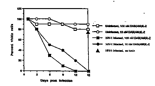
Fig. l is a graphical representation of the effect
S of DAB~6IL-2 on viability of both HIV-infected and
uninfected CD4+ T cell~. The percentage o~ viable cell~
is pre~ented as a function of the number of day~ post
infection.
Fig. 2 is a representation of the results of a
lO SDS-PAGE analy~is of proteins in HIV-infected and
uninfected CD4+ T cells.
Fig. 3 is a graphical representation of the effect
of DAB~6IL-2 on reverse transcriptase activity present in
HIV infected and uninfected monocytes. Rever~e
lS tran~cripta~e activity (cpm) is presented as a function
of DAB~6IL-2 concentration (nM) for uninfected (open
;~ bars) and HIV-l (filled bars) infected cells.
Molçs~lLs Useful in_~he Method of the Inven~o~
In general, there are three ways in which the
20 molecule~ u~eful in the invention can act: (l) the
molecule can kill a cell because the molecule ha~ a
cytotoxic do~ain; (2) thQ molecule (an antibody) can
cau~e cell ly8i~ by inducing complement; and ~3) the
molecule can block binding or uptake of receptor's~
25 ligand. In all three cases the molecule must be targeted
to receptor bearing cells; this is accomplished by
including the receptor's ligand (or a portion or
derivative thereof) or an anti-receptor antibody as part
of the molecule.
Interleukin-2 receptor targeted molecules useful
for treatment o~ HIV infection provide examples of each
of the~e three approaches. A fusion molecule wh~ch
includ~s the IL-2 receptor binding portion of IL-2 and a
cytotoxin can be used to kill NIV infected activated
35 lyophocyte~ and ~onocytes/macrophages. Thi~ molecule can
~92/lS318 2 1 0 ~ 9 5 8 PcT/us92/017os
also kill uninfected IL-2 receptor-bearing cells. Such
uninfected cell8 may contribute to the disease state.
Similarly, the second type of molecule described above, a
complement fixing antibody, in this instance directed
S against the IL-2 receptor, can eliminate infected and
uninfected, IL-2 receptor-bearing cells in a patient
infected with HIV. In this example, the third type of
molecule could be a molecule that blocks binding o~ IL-2
to its receptor. This molecule would prevent infected
lO cell~ from receiving a proliferation signal from IL-2 and
thus could ~low the spread of HIV infection.
Molecules useful for kill1ng or neutralizing IL-
2R bearing cells of HIV infected individuals can take a
number of forms. When IL-2 itself is the targeting-
lS agent, the molecule can be a cytotoxic hybrid molecule inwhich IL-2 is fu~ed to a toxin molecule, preferably ~
polypeptide toxin. Derivatives of IL-2 which bind to IL-
2R, lack IL-2 activity and block binding and/or uptake of
bona fide IL-2 are useful in the method of the invention
20 because they will prevent IL-2-induced prolife~ation of
IL-2R bearing cells. When an anti-IL-2R antibody is the
targeting agent, a cytotoxic hybrid molecule can be
for~ed by fusing ~ll or part of the antibody to a
cytotoxin. The effectiveness of such an antibody/toxin
2S hybrid, like that of an IL-2/toxin hybrid, depends on the
hybrid molecule being takan up by cells to whic~ it
b~nds. Anti-IL-2R antibodies which block binding and/or
uptake of IL-2 are also useful in the method of the
invention. Lytic anti-IL-2R antibodies are useful in the
30 invention because they can direct complement against IL-
2R-bearing cells and thus cause their lysis.
Some the molecules can be hybrid molecules formed
by the fusion of all or pzrt of two or more molecules.
The hybrid molecule can be a hybrid protein encoded by a
3S recombinant DNA molecule, in which case the two domains
WO9~lS318 PCT/US92/017~
` 2104~58
- lS -
are joined (directly or throuqh an intermediary do~ain)
by a peptide bond. Alternatively, two domains can be
produced separately and joined by a covalent bond in a
separate chemical linkage step. In some cases, the
S cytotoxic do~ain of a bybrid molecule may itcel~ be
derived from two separate molecules.
Interleukir~-2 as a Taraetina Agent
IL-2 or any IL-2 receptor binding derivative
thereof can be u~ed as a targeting agent for a cytotoxin.
10 The DNA and amino acid seguences of IL-2 are known
~Tadat~ugu et al., N~ture 302:305, 1983), and its
structure has been predicted by x-ray crystallography
(Brandhuber et al., Science 238:1707, 1987). Analysis of
genetically engineered variants of IL-2 has provided some
lS infor~ation concerning which residues are important for
IL-2R binding (Collins et al., Proc. N~tl. Ac~d. Sci. USA
85:7709, 1988) and bioactivity (Cohen et al. Science
234:349, 1989; Collins et al., supr~). Variants of IL-2
which are useful in the invention include deletion
20 mutants (Genbauffe et al., USSN 388,557, hereby
incorporated by reference) which lack one or ~ore amino
acid residues in the region between residue 74 and
residue 79 (numbering according to Williams et al., Nucl.
Ac~ds Res. 16:1045, 1988). These mutants effectively
2S target toxins to IL-2R-bearing cells (Genbauffe et al.,
supr~). Generally, IL-2 variants useful for targeting a
cytotoxin must efficiently bind IL-2R and be endocytosed~
The ability of various derivatives to bind to the IL-2
receptor can be tested with an IL-2R binding assay
30 described below.
In designing molecules targeted to cells bearing
the IL-2 receptor it must be recognized tAat the IL-2
receptor, like other receptors, has several forms; and it
may be desirable to target cells bearing one for~ and not
3S anotAer. me human interleukin-2 receptor Aas a high-,
~ 92/15318 PCT/VS92/017~
210~S8
- 16 -
an intermediate-, and a low-affinity form. The high
affinity receptor has an apparent Xd of -10 lOM and i8
composed of two subunits, p55 and p75 (also called p70).
When expressed on the cell surface, both the p7s and ps5
S subunits are capable of binding IL-2. The p75 subunit
corresponds to the intermediate affinity receptor (K~ -
8.2 x 10 lM), and pS5 subunit corresponds to the low
affinity receptor (Kd - 1-3 x 10 ~M). The p75 subunit is
expressed on the surface of resting T cell~, natural
10 killer cells monocytes/macrophages, and lymphokine-
activated killer (~AX) cell precursors, while the high
affinity receptor is expressed on activated T- and B-
cells.
In the method of the invention it may be desirable
~S to tarjet only cells bearing the high affinity receptor.
In these circu~stances useful molecules will eliminate or
neutralize cel}s bearing the high affinity IL-2 receptor
at a concentration which leaves cells bearing the
intermediate or low affinity receptor largely unaffected.
20 When the molecule, like IL-2 itself, has affinity for all
three classes of IL-2 receptor, selectivity can be
accomplished by administering the molecule at a
concentration which does not permit ~ignificant bin*ing
to cells bearing lower affinity receptors. A hybrid
25 molecule ~ay have altered receptor affinities compared to
IL-2. Such hybrid molecules may be more or less
~elective for cells bearing the high affinity IL-2
receptor. Por example, cells bearing the high-affinity
receptor are SoO-lOoO times more sensitive to DAB~86IL-2,
30 a fusion protein consisting of part of diphtheria toxin
and part of IL-2, than are cells bearing the
intermediate- affinity receptor (Waters et al., Eur. J.
Immunol. 20:785, 1990).
A cytotoxin can be attached to an IL-2 derivative
3S in a nu~ber of ways. Preferably, an IL-2/toxin hybrid is
WOg~153~8 PCT/US92/017~
~ 210~9S8
- 17 -
~ hybrid protein produced by the expression of a fused
gene. Alternatively, the eytotoxin and the IL-2
derivative ean ~e produeed separately and later eoupled
by ~eans of a non-peptide covalent bond. Linkage methods
S are de~eribed below.
I~erl~gki~-4 as a Taraeti~g Ag~nt
Interleukin-4 (IL-4) is a eytokine whieh aet~ on a
variety of eell types. Its reeeptor is expre~ed on a
nu~ber of eell types, ineluding CD4+ T eells and
10 ~onoeytes. IL-4 ean aet as a T eell growth faetor and it
i8 thought to have an influenee on IL-2 indueed
lymphoeyte proliferation.
A eytotoxin direeted against IL-4 reeeptor-bearing
eells ~ay enhanee the effeetiveness of moleeules direeted
lS against IL-2R-bearing eells. The protein and DNA
sequenee of IL-4 are known (Lee et al., J. Biol. C`lem.
263:10817, 1988). IL-4 ean be u~ed to ereate hybrid IL-
4/toxin ~oleeules similar to IL-2/toxin hybrid ~oleeules.
20 Monoelonal Antibodies as Taraetina Aaents
Monoelonal antibodies direeted against IL-2R, IL-
4R or any eell reeeptor of ehoiee ean be used to direet
toxins to eells bearing that reeeptor. The~e antibodies
or antibody fragoents ean be fusea to a eytotoxin either
2S by virtue of the toxin and the antibody being eneoded by
a fused gene whieh eneodes a hybrid protein ~ol~eule, or
by ~eans of a non-pepti~e eovalent bond whieh i8 u~ed to
join separately produeed ligand and toxin moleeules.
Several u~eful toxins are deseribed below.
Antibody/toxin hybrids ean be tested for their
ability to kill reeeptor bearing eells using a toxieity
assay similar to that whieh is deseribed below for IL-2R
bearing eell~.
ToxiJIs
- ~ g2/1S318 PCr/11S92/01705
21049S8
- ~8 -
The toxin molecules useful in the method of the
invention are preferably toxins, such as peptide toxins,
which are ~ignificantly cytotoxic only when present
intracellularly. Of course, under these circumstances
5 the ~olecule must be able to enter a cell bearing the
targeted receptor. This ability depends on the nature of
the molecule and the nature of the cell receptor. For
~xa~ple, cell receptors which naturally allow uptake of a
l~gand are likely to provide a means for a molecule which
10 includes a toxin to enter a cell bearing that receptor.
Preferably, a peptide toxin is fused to an IL-2R binding
domain by producing a recombinant DNA molecule which
encodes a hybrid protein molecule. Such an approach
ensures consistency of composition.
lS Many peptide toxins have a generalized eukaryotic
receptor binding domain; in these instances the toxin
must be modified to prevent intoxication of non-receptor
bearing cells. Any such modifications must be made in a
~anner wbich preserves the cytotoxic functions of the
20 molecule (see U.S. Department of Health and Human
Services, U.S. Serial No. 401,412). Potentially useful
toxins include, but ~re not limited to: cholera toxin,
ricin, 0-Shiga-like toxin (SLT-I, SLT-II, SL~ IIv),-LT
toxin, C3 toxin, Shiga toxin, pertussis toxin, tetanus
25 toxin, Ps~ud~mon~s exotoxin, alorin, saposin, modeccin,
and gelanin.
DiDhtheria Toxin-based Molecules
Diphtheria toxin can be used to produce molecules
useful in the method of the invention. Diphtheria toxin,
30 whose sequence is known, is described in detail in Murphy
U.S. Patent 4,675,382, hereby incorporated by reference.
The natural diphtheria toxin molecule secreted by
Coryn~b~cterium diphtheriae consists of several
functional domains which can be characteriied, starting
3S at the amino terminal end of the molecule, as
WO9~lS3l8 P~T/US92/017~
~ .
2104~S8
-- 19 --
enzymatically-active Fragment A (amino acids Glyl -
Argl~3) and Fragment B (amino acids Serl9~ - Ser53s), which
includes a translocation domain and a generalized cell
binding domain (amino acid resldues 475 through 535).
The process by which diphtheria toxin intoxicate~
sensitivQ eukaryotic cells involves at least the
following steps: (i) the binding domain of diphtheria
toxin binds to specific receptors on the ~urf~ce of a
sensitive cell; (ii) while bound to its receptor, the
10 toxin ~olecule is internalized into an endocytic vesicle;
(iii) either prior to internalization, or within the
endocytic vesicle, the toxin molecule undergoes a
proteolytic cleavage between fragments A and B; (iv) as
the pH of the endocytic vesicle decrease~ to below 6, the
15 toxin crosses the endosomal membrane, facilitating the
delivery o~ Fragment A into the cytosol; (v) the
catalytic activity of Fragment A ~i.e., the nicotinamide
adenine dinucleotide - dependent adenosine di~phosphate
(ADP) ribosylation of the eukaryotic protein synthesis
20 factor termed ~Elongation Factor 2n~ causes the death of
the intoxicated cell. It is apparent that a single
~olecule of Fragment A introduced into the cytosol is
sufficient to block down the cell's protein synthesis
~achinery and kill the cell. The mechanism of cell
25 killing by Pseudo~on~s exotoxin A, and possibly by
certain other naturally-occurring toxins, is very
similar.
DAB~86IL-2, a fusion protein in which the receptor
binding domain of diphtheria toxin has been replaced by a
30 portion of hu~an IL-2 (Williams et al., J. B~ol. Chem.
3S:20673, 1990; see also Williams et al., Protein Eng.
1:493, 1987), is an example of a molecule useful in the
~ethod of the invention. This molecule selecti~ely kills
IL-2R-expre~sing tumor cells and lymphocytes ~Waters et
35 ~ ur. J. Immunol. 20:785, 1990; ~iyokawa et al.,
- '092/lS318 PCT/US92/017~
21049~8
- 20 -
Cancer Res. 49:4042, 1989). Because of its ability to
kill activated lymphocytes, DAB~86IL-2 has been used to
control graft rejection (Pankewycz et al.,
Transpl~ntation 47:318, 1989; Kickman et al.,
S Transplant~tlon 47:327, 1989) and to treat certain
autoimmune disorders (Forte et al., 2nd Internation~l
Symposium on ~mmunotoxins, 1990).
DAB~86IL-2 is a chimeric molecule consisting of
Met followed by ~mino acid residues 1 through ~85 of the
1~ mature diphtheria toxin fused to amino acid residues 2
through 133 of IL-2. Thus, DAB~6IL-2 includes all of
diphtheria toxin fragment A, which encodes the
enzymatically active portion of the molecule, and a
portion of fragment B. The portion of fragment B present
15 in DAB~6IL-2 does not include the generalized receptor
binding domain but does include the translocation domain
which facilitates delivery of the enzymatically active
portion into the cytosol.
Experimental Methods
Presented below are experiments which demonstrate
that DAB~86IL-2 can kill HIV-l infected T cells,
selectively elimin~te HIV-l infected cells from mixed
cultures of infected and uninfected T cells, and reduce
HIV-l replication in cultures of infected monocytes.
25 DAB~a6IL-2 is ~lao shown to inhibit production of viral
proteins in cultures of infected T ce~ls and to block
product~on of infectious HIV-l in cultures of infected T
cells.
Other molecules targeted to IL-2R may be scxeened
30 using the methods described below.
P~ 1L~ DAB~6~
DAB~6IL-2 was produced in E. coli harboring the
DAB~6IL-2 encoding plasmid, pDW24 (Willi~ms et al., J.
Biol. Chem. 265:20673, 1990, except ampr is replaced by
3S kanr). The protein was purified by immunoaffinity
WO92/lS318 rCT/US9~0l705
210~958
- 21 -
chromatography and high pressure liquid chromatoaraphy
(Williams et al., supra).
Rillina of HIV-1 Infected T Cells by DAB~86~Lc~
DAB~86IL-2 at 10 8M or 10 9M destroyed HIV-l
S infected T cell~ while having ~lmost no effect on
uninfected T cells. CD4+ T cells were prepared from the
peripheral blood of HIV-l negative donors by negative
~election to remove B cells, macrophages, natural killer
cells, and CD8+ T cells. B cells and macrophages were
10 removed by passage over glass wool. CD8+ and CD16~ cells
were removed using anti-CD8 (OKT8, American Type Culture
Collection, Rockville, MD) and anti-CD16 (Leu lla,
Bectin-Dickinson, Mountain View, CA) monoclonal antibody
coated magnetic beads by the method of Haregewoin et al.
~S (N~tur~ 340:309, 1989). Cells were cultured at 2 x
10~/ml in RPMI 1640 (GIBCO/BRL, Bethesda, MD) and 10~
bovine calf serum (GIBCO/BRL, Bethesda, ND) supplemented
with lymphocult-T (Boehringer-Mannheim Biochemicals,
Indianapoliæ, IN; corresponds to -10 9N IL-2). Infections
20 were performed by incubation with ~TLVIII~ (prepared from
filtered supernatants of infected H9 cells, A$DS Research
and Reference Reagent Program NIAID, National Institutes
of Health, Bethesda, MD) for 1 hr at a multiplicity of
infection of 10 (cal~brated by li~iting dilution of'~H9
2S cell~). DAB~86IL-2 (10 7M or 10 ~M) was added on days one
and three post-infection. Cell cultures were split twice
we~kly, ~nd vi~bility was determined by ~ trypan blue dye
exclusion assay (Xruse et al., eds. Tissue Culture :
Methods and Applications, Academic Press, 1989)
Referring to FIG. 1, treatment of uninfected cells-
with 10 ~ (open circles) or 10 7M (open squares)
DAB~6IL-2 only transiently ~mpaired proliferation of T
cells in response to 10 9M IL-2. Further, cultures of
un~nfected T cells treated with DAB~86IL-2 and untreated
3S uninfected T cells achieve similar cell densities after
~ g2/lS318 2 1 0 9 9 5 8 PCT/US~2/017~
- 22 -
two weeks of culturing. In contrast, HIV-l infected cells
were eliminated by incubation with 10 ~M (filled circles)
or 10 7M (filled squares) DAB~86IL-2. This toxic effect
occurs despite the fact that the cells are cultured in
S the presence of 10-9M IL-2 which i5 reguired for cell
viability and which has a 10- to 100- fold higher
affinity for IL-2R than does DAB~8~IL-2 (Waters et al.,
~upra). Infected cells which were not treated with
Daa~ 2 were >75% viable after two weeks, demonstrating
10 that the reduction in viability of DAB~86IL-2 exposed
cells i8 a specific effect and is not due to viral
replication.
Selec~ive Killina of HIV-1 Infected T Cell by DAa~86IL-2
DAB~6IL-2 p~evented production of tbe HIV-l
lS encoded proteins, gpl20, p55, and p24, in mixed cultures
of HIV-l infected and uninfected T cells.
CD4l T cells prepared and infected as described
above were incubated overnight in RPNI 1640 medium
containing 10% bovine calf se`rum and wasbed three times
20 with the same medium prior to addition of uninfected
cells from the same donor. Infected T cells (10~) and
uninfected T cells (107) were cultured in the presence of
10 ~ -2 (Lymphocult T) with or without 10 8 M DAB~8~IL-
2 at 10~ cells/~l. Every two days cells were pelleted,
25 washed twice, and resuspended in fresh IL-2 containing
media. D~B~86IL-2 was added to the treated cultures on
days 1 and 3 and was washed out 24 hourF la~er. The
untreated cultures were washed according to the same
~chedule but were not exposed to DAB~6IL-2. Two weeks
30 after infection, the cells were labelled overnight with
3sS in methionine-free media (GIaCO/BRL, B~thesda, ND).
Proteins were immunoprecipitated (Coligan et al, eds.,
Current Protocols in Immunology, John Wiley & Sons, New
York, 1991) with either anti-HIV globulin (NIAID) or the
W092/~S3l8 PCT/US92/01705
210~958
- 23 -
framework anti-MHC class I antibody (W6t32).
Immunoprecipit~ted proteins were separated on SDS g-16
and visualized by autoradiography.
Referrin~ to FIG. 2, the proteins in lanes 2-4
S were i~munoprecipitated using anti-HIV antibody; the
proteins in lanes 6-9 were immunoprecipitated using anti-
MHC class I ~ntibody; lane 1 has size markers; and the
arrows along the right ~ide indicate the expected
po~itions of HIV-l ~ncoded proteins gpl20, p55 and p24.
Io The HIV-l encoded proteins gpl60, p55 and p24 were
pre~ent in untreated mixed culture (lane 4), but could
not be detected in the DAB~86IL-2 treated culture (lane
S). A~ expected, normal NHC class I proteins were
detected in both the treated (lane 9) and untreated (lane
lS 8) ~ixed T cell cultures. Unmixed cultures of both
uninfected cells (lanes 2 and 6) and infected cells
(lanes 3 and 7) ~erved as controls. Cell viability was
>95% for both treated and untreated mixed cultures,
indicating that the cytotoxic action of DAB~86IL-2 was
20 limited to infected cells.
DAB~86IL-2 treatment of mixed cultures of infected
and uninfected T cells completely eliminated production
of HIV-l p24 protein. Thls result was demonstrated by
means of a sensitive ELISA assay.
2S Treated ~nd untreated mixed cultures of infected
and uninfected CD4~ T cells were prepared as described
above. Cells were pelleted and resuspended twice weekly,
and the culture supernatants were assayed for the
pre~ence of p24 two days later using an ELISA assay
(Abbott, Chicago, IL).
Referring to Table 1, in untreated mixed ~ultures,
the level of p24 increased steadily for at least 18 days
post-infection. In contrast, p24 was undetectable in
mixed cultures which were treated with 10-SM DAB~6IL-2.
'`'~92/lS318 PCT/US92/017~
210'1958
- 2~ -
This experiment demonstrates that DAB,86IL-2 can bloc~ all
production of an HIV-l encoded protein.
T~bl- 1S
Incubation of ~ixed T Cell Cultures with DAB~6IL-2
S Elim~nates Production of p24 Antigen
D~ Int-ct-d litV-1 In~t~ x d ~ d T C ll.~ultur-~
Cultu d ~ T~ Cultur-~ ~ D~486IL-2'
-
0 6 0 0 0
9 0 2S6I O O
~' 12 0 >500 J9 0
O >SOO - 29~ 0
t~ O >SOO >500 0
;~ lS
;~ I Picogr_ ot p24 in th~ u~#~t nt t~ t~-r c-ll ~hin~.
21o~ D~4~6IL~2 ~dd on ~ys 1 ~
~'
na~ IL-2 Prevents HIV-l Infection
Treatment of mixed cultures of infected~and
20 uninfected T cell with DAB~86IL-2 prevented the production
of infectious HIV. This was demonstr~ted by co~paring
the p24 produced in a co-culture of H9 tu~or cells ~nd
~: cells from a DAB~86IL-2 treated mixed infected and
uninfected T cell culture with that produced by a co-
2S culture of H9 tumor cells and cells from an untreated
mixed infected and uninfected T cell culture. Despite
the fact that H9 cells can be readily infected by HIV, no
HIV p24 could be detected in the co-culture supernatant
if the mixed infected and uninfected T cells h~d bsen
30 treated with DA~86IL-2 at least three days prior to their
addition to H9 cells.
CD4+ T cells were purified, infected with HIV-1,
washed, and then mixed with uninfected cells at a ratio
of 1:10 as described above. Cells were cultured in 10-9M
.
~ <``~
~!~0 92/1S318 PCI'/US92/0~
210~958
- 2s -
IL-2 with or without 10 8M DAB~86IL-2 (~dded on days 1 ~nd
3). On days 0, 1, 6 and 9, 5 x10⁵ T cells wore
collected, washed ~nd co-cultured with 5 xl0S uninfected
H9 tu~or cells (NIAID). Co-cultures were incubated for 6
S days prior to ~easure~ent of the p24 present in the co-
culture supernatant. p24 was measured by the ELISA a~say
described above.
~bl- 2s
Incubation of T Cell Cultures with DAB~86IL-2
Eliminates Infectious Virus
C ll Cu~tur~ D~U61~-2D y ot T cel~ cu~twe p241
lS po~tllIV~1 inf~ct~on ~y o o- eo-
cultur~
Unlnf et-d T C ll~ 0
20 IllV-1 int cted T cell~ 0 >S00
T C~ xtwe --~- >500
T C~ll ~ixture 10~ 0 ~ >S00
T Cel~ ~ixture ---- 1 >S00
T C ~ xtur~ 10~ 1 >S00
T C-l~ ~ixtur- ---- ~ >500
T C-~ xture 10~ 6 0
T C~ xture ---- 9 >500
T C ll ~1xt~re 10~ 9
~ ~ured in pico~r~
~ eferring to Table 2, when the mixed infected and
uninfected T cell culture was not treated with DAB~IL-
2, p24 W~8 produced in the co-culture supernatant. This
3S re~ult i~plies that infectious HIV was produced in the
untreated mixed cell culture. In contrast, when cells
fro~ a DAB~TL-2 treated mixed infected and uninfected T
cell culture were added to ~9 tumor cells 3 or 9 days
.- ~n s2/ls3ls PCT/US92/0170S
2104958
- 26 -
after the second addition of DAB~86IL-2, no p24 could be
detected in the co-culture supernatant. This re~ult
implies that DAB~86IL-2 treatment was able to clear the
mixed infected and uninfected cell culture of infectious
S HIV. DAB~6IL-2 treated cultures responded normally to
IL-2 and phytohemagglutinin indicating that the tre~tment
is not generally toxic to T cells.
~2 EliF~n~Ç~LH¦_-l Replication i~ Mo~oçy~çs
Treatment with DAB~86IL-2 blocked HIV-l
replication in monocytes as judged by a reverse
transcriptase assay. Aliquots of monocytes (107)
purified according to Wahl et al. (Cell Immunol. 85:3553,
1984) were suspended in 1 ml of primary macrophage
culture supernatant containing HIV-l/HTLV-IIIJ~_L (2.5
xlO~ cpm reverse transcriptase activity). As a control,
aliquots of monocytes were suspended in RPMI 1640
containing 10% fetal calf serum (FCS, GIBCOIBRL).
Infected and control cultures were incubated for 1 h at
37C w~th intermittent gentle agitation. The cells were
then washed with RPNI 1640 plus 10% FCS, resuspended in
RPNI 1640 containing 10% FCS, antibiotics, and glutamine,
plated (2 x10⁶) on chamber slides and incubated at 37C.
DAB~6IL-2 (10 8 to 10-1OM) was added beginning on d~y 3
(when IL-2R is expected to be present on infected
monocytes) and every third day thereafter. Supernatants
from the infected and control monocyte cultures were
assayed directly for reverse transcriptase activity by a
modification of the method of Spira et al. (J. Clin .
Microbiol. 25:97, 1987). Briefly, 15 ~1 aliquots of
supernatant were removed from each culture, 5 ~1 of a
buffer containing 30% glycerol and 0.5% Triton x-100 was
added to each aliquot in order to solubilize any virus
present. 25 ~1 of ~ reaction mix containing S ~Ci of 3H-
thymidine triphosphate t20 Ci/mmol; Du Pont NEN, Boston,
MA), 0.45 ~g poly rA oligo (12-18 bases long; Pharmacia,
~92/lS318 2 1 0 ~ 9 S 8 PCT/US92/Ot7~
- 27 -
Piicataway, NJ), and lOmMi MgC12 was added to each test
~liquot. The re~ctions were incubated at 37^C for 2.5 h,
and the reaction products were eth~nol precipitated.
Referring to FIG. 3, 10 ~M DAB~IL-2 virtually
S eli~inated reverse transcriptase activity ~n HIV-l
infected monocytes (solid bars). Lower DAB~6IL-2
concentrations inhibited reverse transcriptase activity
to a l~sser extent. DAB~86IL-2 did not effect the
background ~ea~urement o~ reverse transcriptase activity
in uninfected cells (open bars). Further, adherent
~onocyte/macrophages remaining in the DaB~86IL-2 treated
cultures were viable and morphologically
indistinguishable from untreated cells.
Use for DAB~86 L-2 for Treatment of Kaposi's S~rcoma
Certain epidermoid cancers and ~arcomas can also
express functional IL-2 receptors. Accordingly,
molecules targeted to cells ~earing the IL-2 receptor can
be u~ed for treat~ent of cancers and sarco~as a~sociated
with ~iral diseases. IL-2 recepto~s were de~onstrated on
Kaposi's sarcoma cells using 8-D phycoerythrin labelled
monoclonal anti-TAC (anti-pS5) antibody. Two patients
with advanced AIDS and disseminated, chemotherapy
resistant Xaposi's sarcoma underwent one course of 5
doses of DAB~86IL-2 administered as a daily 90 ~in
intravenous infusion. Both patients had an approximately
30~ regression in their skin lesions.
Pre~aration of DAB 9IL-4
38
A synthetic gene encoding human interleukin-4 was
synthesized (Milligen/Biosearch 7500 DNA synthesizer).
The IL-4 sequence (Yodota et al., Proc N~t'l AC~d sci.
USA, 83:58994, 1986) was modified to incorporate E. coli-
preferred codon usage (deBoer et al., in ~xi~izing Gene
Erpression , Reznikioff et al., eds., 1986, Butterworths,
Boston), and restriction endonuclease cleavage sites were
added to facilitate subsequent cloning steps. I~-4
~ g2/lS318 2 1 ~ ~ 9 5 8 PCT/US92/017~
- 28 -
coding sequence (Hisl to Serl29) was inserted into pDW27
plasmid. pDW27 is derived from pDW24 (Willi~ms et ~1.,
J. Biol. Chem. 265:11885, 1990) by deleting DNA
corresponding to amino acids 388 to 485 of n~tive
diphtheria toxin.
S~ L~ 3~9IL-4
The ~bility of DAB38~IL-4 to reduce vi~bility of
v~rious cell types w~s me~sured using an inhibition of
protein synthesis ~s~y; the results of thi~ ~ss~y ~re
presented in Table 3. ICSo (M) is the concentration of
DAB3~9IL-4 sequired for a 50% decrease in proteln
synthesis. The r~t, Con A-activ~ted, normal ~plenic
ly~phocytes were f~r less sensitive to DAB3~9IL-4 th~n ~ny
of the other cells or cell lines. Since the r~t
interleukin-4 receptor does not bind human interleukin-
4, this result demonstrates the specificity of DAB3~9IL-
4. The~e rat cells are sensitive to a diphtheria
toxin/rat interleukin-2 hybrid molecule.
` S~bl- 3s
- 20 DAB ~IL-4 Sensitivity of Normal and Neopl~stic Cells ~nd
Cel~ Lines
C-tl or C~lt Lln Cl~c1~icetion ICsO (1~)
25 _ _
T c 11 orl~n
NUT 102/6TC Nu~n, CTCL, NTLV-I ~ 2.9 X 10 1~
C9t~PL Nu~n, NTLV-I I, tr~-o~ed 6.3 X lo ll
30 ~ e ll ori~in
N~J i Nu~n, Nuclti tt 'c l~_ EBV~ ~.2 X 10 10
~t~nel~e~
un~ Nu~n, hictiocytic ly~pha~ 2.0 X 10 9
~C
PN~ et1vot~d T eellc Nu~n 1.6 X lo 10
~prl_t
Con ~- ct1v t-d nor_l
e T e ttc t >107
W092/15318 rcs/usg2/ol7~
21019~8
- 29 -
~xç~ation of DAB38~ 6
A synthetic gene encoding human interleukin-6 was
synthesized (Milligen/Biosearch 7soo DNA synthesizer).
The IL-6 ~equence (Revel et al., EPA 8611404.9) was
nodified to incorporato E. Coli preferred codon u~age
(deBoer et al., supr~)~ and restriction endonuclea~e
cleavage site~ were added to facilitate subseguent
cloning steps. The entire IL-6 coding ~equence was
inserted into pDW27 plasmid as described above for
DAB3~9IL-4.
Mixe~ Toxinæ
The cytotoxic portion of some molecules useful in
the invention can be provided by a mixed toxin molecule.
A ~ixed toxin molecule is a molecule derived from two
different polypeptide toxins. Generally, as discussed
above in connection with diphtheria toxin, polypeptide
toxins have, in addition to the domain responsible for
generalized eukaryotic cell binding, an enzymatically
active domain and a translocation domain. The binding
and translocation domains are required for cell
r-cognition and toxin entry respectively. The
enzy~atically active domain is the domain responsible for
cytotoxic activity once the molecule is inside a cell.
Naturally-occurring proteins which are known to
have ~ tr~nslocation domain include diphtheria toxin,
Ps~udo~on~s exotoxin A, and pos~ibly other peptide
toxins. The translocation domains of diphtheria toxin
and P~eudomonas exotoxin A are well characterized (Qee,
e.g., Hoch et al., Proc . Natl . Acad . Sci . USA 82:1692,
1985; Colombatti et al., J. Biol. Chem. 261:3030, 1986;
and Deleers et~al., FEBS Lett. 160:82, 1983), and the
existence and loc~tion of such a domain in other
molecules ~ay be determined by nethods such as those
. `"') 92/lS318 PCr/USg2/01705
210~958
- 30 -
employed by Hwang et al. Cell 48:129, 1987); and Gray et
al. Proc. N~tl. Ac~d. Sci. USA 81:2645, 1984).
One useful IL-2/mixed toxin hybrid molecule i~
formed by fusing the enzymatically active A subunit of E.
5 col~ Shiga-like toxin (Calderwood et al., Proc. N~tl.
Ac~d. Sci . USA 84:4364, 1987) to the translocation domain
(~ino acid residues 202 through 460) of diphtheria
toxin, and to IL-2. This three-part hybrid ~olecule,
S~T-A/DTB'lIL-2, is useful in the method of the
invention in the same way as DAB~86IL-2 described above.
Tbe I~-2 portion of the three-part hybrid cau~es the
molecule to attach specifically to IL-2R-bearing cells,
and the diphtheria toxin translocation portion acts to
insert the enzymatically active A subunit of the Shiga-
like toxin into the targeted cell. The enzymaticallyactive portion of Shiga-like toxin, like diphtheria
toxin, ~cts on the protein synthesis machinery of the
cell to prevent protein synthesis, thus killing the cell.
The difference between these two types of hybrid toxins
is the nature of their enzymatic àctivities: the
enzymatic portion of DAB,86IL-2 catalyzes the ADP-
ribosylation by nicotinamide adenine dinucleotide of
Elongation F~etor 2, thereby inactivating this factor
which is necessary for p_otein synthesis, while the
enzymatic portion of SLT-A/DTB'/IL-2 is a ribonucle~se
capable of cleaving ribosomal RNA at a critical site,
thereby inactivating the ribosome. SLT-A/D~B'/IL-2
hybrid would therefore be useful as a treatment for the
same indications as DAB~86IL-2, and could be substituted
or used in conjunction with it if, for example, a
patient's activated T-cells develop a resistance to
DAB~6IL-2 .
Linkaae of Toxins to Bindina Liaands
The binding ligand and the cytotoxin of useful
hybrid molecules can be linked in several ways. If the
WOg2/1S318 PCT/US92/017~
21049S8
- 31 -
hybrid molecule is produced by expression of a fused
gene, a peptide bond serves as the link between the
cytotoxin and the binding ligand. Alternatively, the
toxin and the binding ligand can be produced ~eparately
and later coupled by means of a non-peptide covalent
bond.
For ex~ple, the covalent linkaqe ~ay take ~he
for~ of a disulfide bond. In this case, if the binding
ligand i8 a protein, e.g., IL-2, the DNA encoding IL-2
can be engineered to contain an extra cy~teine codon as
de~cribed in Murphy et al. V.S. Serial No. 313,599,
hereby incorporated by reference. The cysteine ~ust be
po~itioned so as to not interfere with the IL-2R binding
activity of the molecule. For example, the cysteine
codon can be inserted just upstream of the DNA encoding
Pro2 of the mature form of IL-2. The toxin mol~cule must
be derivatized with a sulfhydryl group reactive with the
cysteine the modified IL-2. In the case of a peptide
toxin this can be accomplished by inserting a cysteine
codon into the DNA sequence encoding the toxin.
Alternatively, a sulfhydryl group, either by it~elf or as
part of a cysteine residue, can be introduced using solid.
pha~e polypeptide techniques. Por example, the
introduction of sulfbydryl groups into peptides i8
2S described by Hiskey (Peptid~s 3:137, 1981).
Derivatization can also be carr~ed out according to the
method described for the derivatization of a peptide
hormone in Bacha et al. U.S. Patent No. 4,468,382, hereby
incorporated by reference. The introduction of
sulfhydryl groups into proteins is described in M~asen et
al. (Eur. J. Biochom . 134:32, 1983). once the correct
~ulfhydryl groups are present, the cytotoxin and IL-2R
binding ligand are purified, both sulfur groups are
reduced; cytotoxin and ligand are mixed;, (in a ratio of
~bout 1:5 to 1:20) and disulfide bond formation iB
.- W092/15318 PCT/US92/017~
210~958
- 32 -
allowed to proceed to completion (generally 20 to 30
minutes) at room temperature. The mixture is then
dialyzed again~t phosphate buffered saline to remove
unreacted ligand and toxin molecules. Sephadex
chro atography or the like is then carried out to
separate on the basis of size the desired toxin-ligand
con~ugates from toxin-toxin and ligand-ligand conjugates.
Assavs for IL-2 Rece~tor Binding and IL-4 Receptor
Bindina
The IL-2R binding ability of various molecules can
be ~easured using an IL-2R assay for high affinity (Ju et
al., J. Biol . Chem. 262:5723, 1987) or inter~ediate
affinity receptors (Rob et al., Proc . Natl . Ac~d . Sci .
USA 84: 2002, 1987). The IL-4R binding activity of
various molecules can be measured using the assay
de~cribed by Park et al. (J. Exp. Med . 166:176, 1984) or
the as~ay described by Foxwell et al. (Eur. J. Immunol.
19:1637, 1989).
As~ays for Toxicity
Moleculeæ of the invention (both antibodies and
hybrid molecules) can be screened for the ability to
decrease viability of cells bearing the targeted receptor~
by means of assays such as t~ose described below.
Toxicity towardæ IL-2R bearing cells can be tested
as follows. Cultured HUT 102/6TG (Tsudo et al., Proc.
N~tl. Ac~d. Sci. USA 83: 9694, 1986) or YT2C2 (Teshigiwari
et al., J. Exp. Ned . 165:223, 1987) cells are maintained
in RPMI 1640 medium (Gibco, Grand Island, NY)
supplemented with 25 mM HEPES (pH 7.4), 2mM l-glutamine,
100 U/ml penicillin, 100 ~g/ml streptomycin, and 10
fetal calf serum (Hazelton, Lenexa, XS). Cells Are
seeded in 96-well V-bottomed plates (Linbro-Flow
Laboratories, McLean, VA) at a concentration of 1 x 10⁵
per well in complete medium. Putative toxin~ are added
to varying concentrations (10 12M to 10-~M) and the
WO92/15318 PCT/VS9V017~
.
2104958
- 33 -
cultures are incubated for 18 hrs. at 37C in a 5% C02
atmosphere. Following incubation, the plates are
centrifuged for 5 min. at 170 x g, and the medium removed
and replaced with 100 ~1 leucine-free medium ~MEM, Gibco)
containing 8 ~Ci/ml (3H-leucine; New England Nuclear,
Boston, MA). After an additional 90 min. at 37C, the
plate~ are centrifuged for 5 min. at 170 x g, the medium
i5 removed, and the cells are collected on glass fiber
filters using a cell harvester (Skatron, Sterling, VA).
Filters are washed, dried, and counted according to
standard methods. Cells cultured with medium alone serve
as the control.
Toxicity towards cells bearing IL-4R may be tested
by an assay similar to that described above for IL-2R
bearing cells, but utilizing N~A144 cells (Rabin et al.
J. Immunol. 127:1852,1981) or HUT 102/6TG cells, seeded
; at 1 x 10⁵ cells per well and incubated for 40 hours.
Therapy
Generally, the molecules of the invention will be
administered by intravenous infusion. They ~ay also be
administered subcutaneously. Dosages of molecules useful
in the methods of the invention will vary, depending on ,
factors ~ucb as whether the substance is a cytotoxin, a
lytic antibody, or an cell receptor blocking molecule.
In the case of toxic molecules the extent of cell uptake
is an important factor; less permeable molecules must be
admin~stered at a higher dose.
More than 60 patients have received D~B~86IL-2 in
Phase I/II clinical protocols. The molecule ig well
tolerated with the maximum tolerated dose (MID)
established by transient asymptomatic hepatic
transaminasQ elevations i~ about 30% of patients treated
at the MTD. Anti-tumor effects have been sc~ne in
approxi~ately 40S of patients; responses were noted in B-
cell leukemias and lymphomas, cutaneous T-cell lymphoma
.
~92/lS318 2 1 0 ~ 9 S ~ PCT/US92/017~
- 3~ -
and Hodgkin's disease (LeMaistre et al., Blood
360a:abstract 1429, 1990; Woodworth et al., Fourth
Int~rnation~l Conference on Human Retrovirology, 1991).
Serum concentrations of 10 8M DAB~86IL-2 have been ~afely
achieved in patients with IL-2 receptor expres~ing
malignancies. Significant anti-tumor effects have been
observed in highly refractory leukemia/lymphoma p~tients
and these effects have occurred despite the presence of
elevated soluble IL-2R levels in all patiente. Thi~
observation is consistent with data which suggest that
soluble IL-2R does not interfere with binding of IL-2 to
the high affinity interleukin-2 receptor. Ani~al and
hu~an studies h~ve demonstrated that DAB~86IL-2 has no
general immunosuppressive effect (LeMai~tre et al.,
15 supr~ ; Woodworth et al., supra).
Experiments indicate that binding and
internalization of DAB~86IL-2 by cells bearing the high
affinity IL-2 receptor occurs within 30 minutes of
exposure, resulting in maximal inhibition of protein
synthesis within several hours. Therefore, the molecule
should be effective even if the serum half-life is rather
short.
For DAB~86IL-2 a typical course of therapy ~ight
be 0.025 to 0.3 mg/kg/day for 10 to 30 days. This course
of treatment can be repeated several times to provide
effective therapy.
Otber Embodiments
The molecules described above act to decrease cell
viability by directing a cytotoxin (or a lytic antibody)
to a targeted cell. Also useful in the method of the
invention are ~olecules which interfere with the targeted
cell's ability to utilize a cytokine.
Derivatives of IL-2 or other cytokines which block
utilization of endogenous cytokine are u~eful for
- ` ~92/15318 PCT/US92/01~
2104~S8
- 3s -
preventing proliferation of targeted cells. For example,
activated cells deprived of IL-2 f~il to proliferate and,
in the absence of the essential anabolic stimulus
provided by IL-2, will eventually die. With regard to
HIV infection, if utilization of IL-2 is prevented, the
infected process will be disrupted. The ability of a
given IL-2 derivative to interfere with IL-2 function can
be te~ted in an IL-2 bioactivity assay such as the one
described by Ju et al. (J. B~ol. Chem. 262:S723, 1987).
Hybrid molecules in which the toxin has been rendered
inactive can be also used to block a cytokine receptor.
A non-toxic mutant diphtheria toxin molecule has been
de~cribed (Uchida et al. J. Biol . Chem . 248:3838, 1973),
and this molecule can be used to produce a non-toxic IL-
2/diphtheria toxin hybrid. See Svrluga et al. U.S.Serial No. 590,113, hereby incorporated by reference, for
an example of such a hybrid molecule.
Monoclonal antibodies can be used to kill or
neutralize cytokine receptor-bearing cells in a number of
ways. Ac described above, anti-cytokine receptor
antibodies fused to a toxin molecule can be used to
deliver the toxin to receptor-bearing cells. Lytic anti-'
cytokine receptor antibodies can themselves kill cytokine
receptor-bearing cells by acti~at~ng complement. For
example, monoclonal antibodies which activate complement
can be used to de~troy IL-2R-bearing cells. Complement
inducing antibodies are generally those of the IgGl,
IgG2, IgG3, and IgM isotypes. Monoclonal anti-IL-2R
antibodies can be screened for those able to activate
complement using a complement-dependent cytotoxicity
test, as follows.
Human T-lymphocytes and EBV transformed B-
lymphocytes are labeled with 51Cr sodiu~ chro~ate and used
as target cells; these cells are incubated with hybridoma
culture supernatants and with complement, and then the
WO9~lS318 PCT/VSg2/017~
210495~
- 36 -
supernatants are collected and counted with a ga~ma
counter. Tho~e supernatants exhibiting toxicity against
activated T-lymphocytes, but not resting T- or B-
lymphocytes, are selected (described in detail in by
Leonard et al., Proc. N~tl. AC~d . sci . USA 80:695~,
1983). The de~ired anti-~L-2 receptor antibody is
purified from the supernatants using conventional
~ethods. The specificity of the antibody can be
d~onstrated by chowing that the activity is blocked by
exogenous IL-2.
Also useful are antibodies which block binding
and/or uptake of a cytokine. For example, monoclonal
antibodies which interfere with the binding and/or uptake
of I~-2 are useful in the method of the invention because
IL-2R bearing cells deprived of IL-2 fail to proliferate.
Blocking monoclonal antibodies (and other blocking
molecules) can be tested for their ability to interfere
with IL-2 bioactivity using the method of Ju et
al.,(supr~) . Generally, assays useful for blocking
molecules will be competitive binding assay~ which
measure the ability of the molecule being to interfere
with binding of one or more of the receptor' 8 natural
ligands.
Monoclonal antibodies useful in the ~ethod of the
2S invention can be ~ade by immunizing mice with human IL-
2R~ T-lymphocytes, fusing the murine splenocytes with
appropriate myeloma cells, and screening the antibodies
produced by the resultant hybridoma lines for the
requisite IL-2R binding properties by means of an ELISA
assay. Antibody production and screeniny can be
performed according to Uchiyama et al. (J. Immunol.
126:1393, 1981). Alternatively, useful antibodies may be
isolated fro~ a combinatorial library produced by the
~ethod of Huse et al. (Science 246:1275, i989).
WO92/1s3~8 PCT/US9~01705
21099~
- 37 -
The invention can employ not only intact
monoclonal or polyclonal antibodies, but also an
i~munologically-active antibody fragment, for example, a
Fab or (Fab)~ fragment; an antibody heavy chain, an
antibody light chain; a genetically ngineered ~ingle-
chain Fv ~olecule (~adner et al., U.S. Patent No.
4,946,778); or a chimeric antibody, for exa~ple, an
antibody which contains the binding ~pecificity of a
murine antibody, but in which the remaining portions are
of hu~an origin.
What is claimed is: