Note: Descriptions are shown in the official language in which they were submitted.
48284CAN9A
TROCAR
Technical Field
The present invention is directed to trocars
for inserting an access tube through an abdominal wall,
and more particularly to a trocar having an improved
obturator and trigger.
Backgrounr,~the Invent~~
An increasing number of abdominal surgical
procedures are being performed with laparoscopic
techniques in order to avoid a large skin incision.
Typically in laparoscopic surgery, a special needle (a
needle similar to the pneumoneedle described in U.S.
Patent No. 4,808,168 is inserted through the skin, and
used to inflate the abdominal cavity with an
insufflating gas such as carbon dioxide (C0,). Once the
abdomen is adequately dilated, the needle is removed
2o and a rigid access tube or cannula with a diameter
larger than the pneumoneedle (for example 5, 10 or il
mm) is passed through the skin in the same location.
The access tube provides access for
laparoscopes or other laparoscopic surgical tools such
as the stapler described in U.S. Patent No. 5,040,715
or the surgical clip appliers described in U.S. Patent
No.'s 5,084,057 and 5,100,420. To drive the access
tube through the skin, the surgeon places a trocar in
the lumen of the access tubs to provide a sharp,
1~ading edge for cutting tissue.
Th~ art is replete with trocar d~via~a,
including those shown in U.S. Patent Nos. 4,535,773,
4,601,710, 4,654,030, 4,902,280 arid 4,931,042. Those
trocars typically comprise an obturator with cutting
surtaxes for penetrating the skin, and a spring-loaded
protective sleeve that surrounds the obturator. As
-1-
these trocar devices are urged through the skin,
friction with the skin causes the protective sleeve to
slide proximally (rearwardly). After the access tube
has penetrated through the skin, there is no longer
friction between the protective sleeve and the skin,
and the spring is designed to urge the protective
sleeve distally (forwardly) to cover the cutting
surfaces. Some of those trocars lock the prot~:ctive
sleeve in the forward position to reduce the risk of
to accidental puncture of the underlying organs.
These prior art trocars rely on a similar
principle of operation: The friction or drag on the
protective sleeve as the trocar is advanced through the
skin pushes the protective sleeve back (proximally) to
expose the cutting surfaces. Once the access tube has
penetrated the skin, the drag on the protective sleeve
is reduced and the sleeve. accelerates distally
(forwardly) under the bias of the spring to cover the
cutting surfaces.
Figure 1 illustrates a portion of a typical
prior art trocar similar to the lOmm Auto Suture
Surgiport T.M., generally available from U.S. Surgical
of Norwalk, Conn. That trocar includes an access tube
1, an obturator 2 and a shield 3. The shield 3 is
biased distally to cover the obturator 2. The shield 3
comprises a generally cylindrical tube with a slightly
rounded or angled end portion 4.
Existing trocars such as the trocar shown in
Figure 1 encounter prablems because a significant
amount of force usually must be applied to penetrat~
the skin (particularly the tough fae~oia). As a result
of the significant insertion force, the trocar may
continue to advance toward the underlying organs after
it has penetrated the skin. Thus, the protective
sleeve must "catch up'° to the moving trocar point
b~fore the trocar reaches the underlying organs.
-2-
Figure 2 illustrates another prior art
trocar. This Figure generally illustrates a portion of
trocar that is currently being sold in the United
States under the name 10/11 mm Endopath~ (generally
available from Ethicon of Somerville, N.J). U.S. Patent
No. 5,066,288 to Deniega et a1. describes a trocar
similar to the trocar shown in Figure 2. That trocar
includes an access tube 5, an obturator 6 and a shield
7. The shield 7 is biased distally to cover the
obturator 6. Unlike other trocars, the shield 7 of the
trocar shown in Figure 2 includes a bullet shaped end
portion 8 comprising three semicircular lobes 9.
U.S. Patent No. 5,066,288 states that the
trocar restricts tissue trauma. However, like the
trocar shown in Figure 1, trocars similar to those
shown in Figure 2 also encounter problems because a
significant amount of force is nevertheless required to
penetrate the skin (particularly the tough fascia).
Again, as a result of the significant insertion force,
the obturator may continue to advance toward the
underlying organs even after it has penetrated the
skin.
Figures 3 and 4 illustrate yet another trocar
similar to the trocar described in U.S. Patent No.
4,654,030 to Mall. That trocar includes an access tube
(not shown), an obturator 10 and shield 11 that is
biased distally to cover the obturator 10. The
obturator has a triangular bas~ 12, and three generally
equilateral triangular surfaces 13.
The shi~ld 11 comprises three parabolically
shaped bevels 14 which form a triangular shaped opening
15. The parabolically shaped bevels 14 intersect at
three edges 16. While U.S. Patent 4,654,030 states
that the trocar shown in Figures 3 and 4 markedly
reduces the force required to insert the trocar into
_3_
body Cavities, the trocar shown in Figures ~ and 4 is
believed to suffer from several drawbacks including:
(1) the shield 11 is believed to concentrate tissue
trauma generally at the edges 16 during insertion into
the body cavity resulting in undesirable tissue trauma
at the incision site, (2) the shield 11 (particularly
the edges 16) may become caught on tissue which
restricts movement of the shield 11 relative tR;o
obturator l0, which is particularly undesirable after
the obturator has pierced the abdominal wall; and (3)
the edges 16 of the shield 11 may be relatively sharp
and may expose the underlying organs to damage from
contact with the edges 16 of the shield 11 itself.
U.S. Patent No. 5,152,754 discloses a trocar
comprising an obturator which retracts relative to the
access tube just after the dbturator pierces the tissue
defining the body cavity.
Bri ef Describti ~r~ of the Irwo_~nt~ i can
According to the present invention there is
provided a trocar having an improved obturator and
trigger which (1) affords an acceptable amount of force
required to insert the trocar into the abdominal
cavity, (2) restricts force concentrations (and tissue
trauma) at the incision site due to the shape of the
trigger; (3) obscures any potential sharp edges on the
trigger to restrict the chances of the trigger catching
tissue or other structures which may inhibit the
trigger s movement relative to the accees~ tube after
the trigger has penetrated into the bady cavity; (4)
re~tricta damage to underlying organs due to th~a shape
of the trigger; and (5) resists binding or rotation of
the obturator relative to thg trigger.
According to the present invention, there is
provided an improved trocar assembly having a novel
obturator and trigger. The trocar is placed in the
-4-
~~~~~vw
lumen of a cannula to facilitate insertion of the
cannula through tissue defining a body cavity.
The trocar comprises a handle, and an
elongate obturator which extends from the handle. The
direction of elongation of the obturator and its center
define a longitudinal axis. The obturator comprises
cutting surfaces for cutting and penetrating the tissue
defining the body cavity, and a trigger having a
plurality of tissue cams. Each tissue cam comprises an
1o inner surface and an outer, generally planar surface
situated at an angle relative to the longitudinal axis.
Each tissue cam also has a distal end portion that is
arcuate about an axis normal to the outer surface.
Each of the distal end portions of the
trigger has a distal most point. The distal end
portions of the tissue cams intersect at edge portions.
The distal most points of the distal end portions are
spaced distally from the edge portions.
Optionally, the trocar includes a mechanism
for restricting rotation of the obturator relative to
the trigger. Such a mechanism may comprise the trigger
having a decent member, and the obturator having a
generally cylindrical base portion having a chamfered
edge that engages the detent member.
Preferably, the trigger is mounted around the
obturator. The trigger. is adapted to move
longitudinally and axially movement relative to the
obturator. The trigger retracts proximally relative to
the obturator as the trocar is advanced through the
3o tissue defining the body cavity. The trigger advances
distally after the cannula has penetrated through the
tissue defining the body cavity.
Also, preferably, the trocar includes a
mechanism for retracting the obturator proximally
relative to the cannula after the obturator has cut the
tissue defining the body cavity. More preferably, the
_5_
CA 02107852 2004-03-03
79237-6
obturator retracts proximally after the trigger advances
distally.
Alternatively but not preferably, the present
invention may comprise a trocar comprising an obturator
defining a longitudinal axis and a protective sleeve having
a plurality of tissue cams. The protective sleeve's tissue
cams each have an inner surface and an outer, generally
planar surface situated at an angle relative to the
longitudinal axis. Each of the protective sleeve's tissue
cams also have a distal end portion that is arcuate about an
axis normal to the outer surface. Preferably, the distal end
portions of the tissue cams intersect at edge portions.
Distal most points of the distal end portions are spaced
distally from the edge portions of the protective sleeve.
In accordance with another aspect of the present
invention, there is provided a trocar for placement in the
lumen of a cannula to facilitate insertion of the cannula
through tissue defining a body cavity, the cannula having a
distal end, said trocar comprising: a handle, an elongate
obturator extending from said handle and defining a
longitudinal axis, said obturator comprising cutting
surfaces for cutting the tissue defining the body cavity, a
protective sleeve having portions adapted to project beyond
the distal end of the cannula, said protective sleeve having
a plurality of tissue cams, each tissue cam comprising an
inner surface and an outer, substantially planar surface
situated at an angle relative to said longitudinal axis, and
a distal end portion, and wherein each of said distal end
portions has a distal most point, said distal end portions
of said tissue cams intersect at edge portions, said distal
most points of said distal end portions are spaced distally
from said edge portions, and each of said distal end
-6-
CA 02107852 2004-03-03
79237-6
portions are arcuate about an axis normal to the outer
surf ace .
Brief Description of the Drawings
The present invention will be further described
with reference to the accompanying drawing wherein like
reference numerals refer to like parts in the several views,
and wherein:
Figure 1 is a partial side view of a prior art
trocar illustrating penetration of tissue by the trocar;
Figure 2 is a partial side view of a second,
bullet nosed prior art trocar illustrating penetration of
tissue by the trocar;
Figure 3 is a perspective view of a third prior
art trocar obturator having portions broken away to show
details;
Figure 4 is a perspective view of the obturator of
Figure 3 assembled with a sleeve in a retracted position;
Figure 5 is a perspective view of a trocar
assembly according to the present invention;
-6a-
Figures 5 through 8 sequentially illustrate
the operation of the trocar of Figure 5 wherein:
Figure 6 is a sectional view illustrating the
relative positions of the obturator and trigger after a
button of the trocar has been pressed and just prior to
insertion into the abdominal cavity;
Figure ? is a sectional view illustrating the
relative positions of the obturator and triggex as the
trocar just begins to pierce the tissue defining the
body cavity;
Figure 8 is a sectional view illustrating the
relative positions of the obturator and trigger after
the obturator has pierced the tissue defining the body
cavity and after the obturator has retracted relative
to the trocar handle;
Figure 9 is an enlarged end view of the
trocar assembly of Figure 5;
Figure to is a sectional view of the trocar
assembly of Figure 5 taken approximately along lines
10-10 of Figure 5 and with portions broken away to
emphasize detail; and
Figure 11 is an enlarged perspective view of
the novel trigger forming a portion of the present
invention having portions braken away to emphasize
details.
net~~~ed Description of the Prpf~~gd Embodiments
Referring now to Figures 5 through 11 of the
drawing there is shown an embodiment of a trocar device
or assembly generally designated by ref~rence~ character
200. The trocar 200 is similar to the trocar described
in U.S. Patent No. 5,152,754. Features of the trocar
200 that are similar to the features of the trocar
described in U.S. Patent No. 5,152,754 have been given
the same reference character to which the suffix °B~~
has been added.
_7_
The trocar 200 includes the novel obturator
and trigger according to the present invention. The
trocar 200 facilitates insertion of a cannula through
tissue defining a body cavity, for example inserting an
access tube 1028 through an abdominal wall 201. The
access tube 1028 comprises a cannula 104B that has an
enlarged fixture 1068 at its proximal end similar to
the fixture 106 described in U.S. Patent No. 5,152,754.
There is an opening 1108 at the proximal end of the
chamber similar to the opening 110 in U.S. Patent No.
5,152,754.
A trap door valve member 112B similar to trap
door valve member 112 (in U.S. Patent No. 5,152,754) is
also present to close the opening 1108. The valve
member 112B is operated by a pushbutton 1168 similar to
the pushbutton 116 (again further shown in U.S. Patent
NO. 5,152,754).
The trocar 200 has a stopcock (not shown) by
which pressurized gas (insufflating gas) can be
provided to the chamber to maintain the gas pressure in
the body cavity, and thereby keep the cavity inflated
to facilitate the surgical procedure.
The trocar 200 comprises a handle or housing
1268 that is releasably attached to the fixture 106B so
that after the trocar 200 is used to insert the access
tubs 102B in the tissue defining the body cavity, the
trocar 20o can be removed leaving the access tube 1028.
The tube 1028 can then be used to introduce surgical
instruments into the body cavity.
The proximal end of th~ fixture 1068 haa~ a
funnel-shaped extension 1288, with an enlarged rim
1308. Resilient fingers axe also present and have a
shoulder to engage the rim 130B and attach the handle
1268 to the fixtur~ 1068. The sides of the handle have
two pivotally mounted buttons 1388 which operate
_g_
similar to the buttons 138 described in U.S. Patent No.
5,152,754.
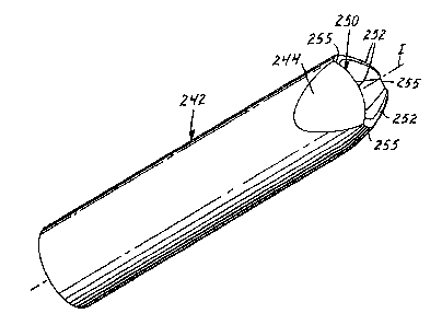
The trocar 200 also includes an obturator 240
extending from the distal end of the handle 1268. Note
Figures Gas. The direction of elongation of the
obturator 240 and its center defines an imaginary,
longitudinal axis I. The trocar also includes trigger
242 mounted around the obturator 240 for axial movement
relative to the obturator 240. The trigger 242 is
preferably mounted so that it can retract proximally
relative to the obturator 240 in response to drag from
the tissue 201 defining the body cavity as it is
advancing through the tissue 201. The trigger can then
advance distally after the cannula or access tube 1028
has penetrated through the tissue 201 and reduced the
drag on the trigger 242.
The obturator 240 comprises proximal 204 and
distal 205 end portions. The distal end portion 205
comprises a base part 206 and also has generally planar
surfaces 207 (preferably three) intersecting to form
cutting edges or surfaces (again preferably three) and
a point 208 at the distal most end of the obtu~rator.
The axis I is generally parallel to the direct~.on of
elongation of the obturator 240 and passes through
point 208.
Trigger 242 has proximal and distal end
portions with the proximal end portion attached to
trocar adapter 211. Unlike trigger 142 shown in U.S.
Patent No. 5,152,754, the distal ~nd of th~ trigger 242
comprises three tissue cams 244. Each of the tissue
cams 244 has an inner surface 245 and a planar outer
surface 246 situated generally at an angle relative to
the longitudinal axis I. Preferably, the included angle
between the outer surface 246 and the axis I is between
about 10 degrees and about 40 degrees. Mast
preferably, the angle is about 15 degrees.
_g_
The cams 244 are believed to beneficially
reduce the insertion force required to insert the
trocar 200 through the abdominal tissue 201. The cams
244 each include a navel arcuate end portion 250 that
is arcuate about an axis td normal to the outer surface
246 (See Figure 8).
For example, when the trocar 200 is used to
insert a cannula 1048 having an lumen interior diameter
of about 1o millimeters, the radius of curvature of the
1o arcuate end portion 250 should be between about 0.18
inches and about 0.32 inches, and preferably
approximately 0.25 inches.
The distal end portions 250 each have a
distal most point 252. The distal end portions 250 of
the cams 244 intersect at edge portions 255. The
distal most points 252 of the distal end portions 250
are spaced distally (relative to the fixture 1068) from
the edge portions 255.
While not desiring to be bound by any
particular theory, it is believed that the arcuate end
portions 250 distribute the insertion force applied to
the tissue 201 by the trigger 242 more evenly, than for
example, a structure as shown in Figure 4. The arcuate
end portions 250 thus restrict trauma to the tissue
201. Additionally, the arcuate distgl end portions 250
of the trigger 242 axe believed to (1) obscur~ any
potential sharp edges on the trigger 242 (e. g. portions
255) (2) restrict the opportunity for the trigger 242
to catch tissue which may inhibit the trigger s distal
3o movement after it has penetrated into the body cavity;
and (3) restrict damage to underlying organs due to the
blunt shape of the and 250 its~lf. Unlike prior art
trocars, the potentially sharp portions 255 are
obscured by the arauate end portions 250. The arcuate
end portions 250 restrict damage to underlying organs
from contact with the trigger itself.
-10-
G ~? i'9 ,
The trocar 200 optionally includes means for
restricting rotation of the obturator 240 relative to
the trigger 242. That means may comprise the trigger
242 having inner and outer surfaces with the inner
surface having a detent portion 243 (Note Figure 6).
The means also includes the base part 206 of the
obturator provided in a generally cylindrical shape
with a chamfered edge 263 (Note Figure 10) for engaging
the detent member 243 of the trigger 242. The means
l0 described in this paragraph is believed to restrict
binding or rotation of the obturator and/or trigger
relative to the housing of the trocar 200.
The trigger 242 is resiliently biased
distally (relative to the housing or handle 126B). In
the embodiment shown in Figures 5-11, the trigger 242
is biased with a spring (not shown), similar to the
coil spring 146 shown in U.S. Patent No. 5,152,754.
The distal end of the spring (not shown) is supported
on an internal shoulder 148B on the trigger, and its
proximal end engages a raised shoulder on the obturator
(described in more detail below).
The action of the spring (not shown but
similar to spring 146 in U.S. Patent No. 5,152,754)
resiliently biases the trigger 242 distally. However,
the trigger can move proximally against the bias, under
the forces applied by the tissue defining the body
cavity as the trocar 200 is advanced through the tissue
201. The trigger 242 has a °resilient" characteristic
and moves distally when the applied faroes are removed,
as occure~ once the access tube 102B ie ine~rted through
th~ tissue 201 and the trigger 242 is na longer in
frictional contact with the tissue 201.
The trocar 200 also comprises means for
retracting the point 208 on the obturator 240 relative
to the cannula 1048 after the paint has penetrated
-11-
~~d~w~
through the tissue 201, thereby reducing the risk that
the point 208 will cause damage inside the body cavity.
In trocar 200, the retracting means is
triggered upon the distal (forward) movement of the
trigger 242. 6Vhen the novel obturator 240 and trigger
242 are used with the trocar 200, it is important that
the trigger move distally so that the retracting means
of the trocar 200 may operate, The arcuate su~.rface
portions 252 obscure the relatively pointed structures
255 to restrict the chances that the trigger 242 will
catch tissue which might prevent the trigger 242 from
moving distally and "triggering" the retracting means.
Like the embodiment shown in U.S. Patent No.
5,152,754, in the trocar 200, there is a tubular
extension 1508 secured on a shoulder on the proximal
end of the obturator 240. An enlarged head 1528 is
secured onto the proximal end of the extension lSOB.
The extension 1508 has a generally radially extending
flange 1548. The distal surface of the flange 1548
forms the shoulder that engages the proximal and of the
spring (not shown). Preferably four proximall;~
extending resilient fingers 1568 are disposed around
the obturator.
The proximal ends of the fingers 1568 have
beads 1588 that can engage the flange 1548 on the
extension 1508, and thereby hold the obturator against
proximal retraction under the bias of spring (not shown
but similar to spring shown in U.S. Patent No.
5,152,754). The distal ends of th~ fingers 1568 are
anchored to a ring 1578. Tha ring 1578 is supported on
a gen~rally cylindrical base similar to the bas~ 159
shown in U,S. Patent No. 5,152,754.
Before use, as shown in Figure 6, the beads
1588 on the fingers 1568 engage the flange 1548,
holding the spring (not shown) in compression and
holding the obturator 240 from proximal retraction. A
_la-
locking member 1608, slidably mounted on the proximal
portion of the extension member 1508, locks the fingers
1568 in engagement with the flange 154B. The bottom
edge of the locking member 1608 has notches 162H which,
when the locking member 1608 is in its distal most
position, receive and engage the fingers 1568 and hold
the beads 1588 in engagement with the shoulder.
The cylindrical section 1448 of the trigger
242 is sized and positioned to engage the locking
to member 1608, and slide the locking member proximally as
the trigger 242 slides proximally (as occurs as the
trocar 200 is advanced through the tissue 201 and the
friction of the tissue acts against the trigger 242).
As shown in Figures 6-7, when the trigger 242
moves proximally, it pushes the locking member l6oB
proximally. Thereafter, further proximal movement of
the trigger 242 moves the locking member 1608. The
cylindrical section 1448 of the trigger 242 is sized to
engage the fingers 156B, and hold the beads 1588 in
engagement with the flange 1548 after the locking
member 160B has been displaced. Thus, as shown in
Figure 7, when the trigger 242 is in its fully
retracted position, the locking member 1608 has been
moved proximally, and the walls of the cylindrical
section 1448 hold the fingers 156B in engagement with
the flange 1548.
The mechanism of the trocar 200 is now primed
so that any distal advancement of the trigger 242 will
cause the trocar 200 to retract the obturator point
208. Thus, the importance of permitting this distal
advancement is apparent.
When the cannula 1048 of access tube 102B
pierces through the tissue 201, it shields the trigger
242 from contact with the tissue 201, and thus the
tissue 201 no longer exerts frictional force on the
trigger 242. The reduction'in force on the trigger
-13-
allows the trigger 242 to advance distally under the
bias of the spring (not shown). This distal
advancement is believed to be facilitated by the shape
of the cams 244 of the trigger 242.
As the trigger 242 advances, the cylindrical
section 144H releases the fingers 156B. When the
fingers 1568 are released, they spring resiliently
outwardly, and the beads 1588 clear the flange 1548.
This allows the obturator 240 to retract under 'the bias
of the spring (not shown).
As the obturator 240 retracts, it moves the
locking member 1608 with it. Moreover, the cap 1528
protrudes through an opening in the handle 1268
providing a visible signal, as well as a tactile signal
that the obturator point 208 has retracted.
As shown in Figure 8, the point 208 of the
obturator rebracts relative to the cannula 1048 to a
retracted position. Alternatively, the point 208 of
the obturator may be designed to retract completely
Within the lumen of the cannula 1048 so that the tip is
located proximally relative to the distal end of the
cannula 1048. This may conveniently be accomplished by
increasing the length of travel of flange 1548 between
the position shown fn Figure 7 and the position shown
in Figure 8.
Operation
The operation of the trocar will now be
described with reference to Figures 6 through 8 which
sequentially illustrate the operation o! the trigger.
3o The tracar 200 optionally includes a protective cap
(not shown) over the point 208 that is removed prior to
use of the trocar 200. The access tube 1048 is already
installed over the distal end of the trocar 200. The
user grasps the handle of the trocar 200, with the palm
of the hand over the proximal end.
-14-
~~f~°'l'~~%
The trocar 200 is advanced against the tissue
201 defining a body cavity, far example the abdomen.
The arcuate end surfaces 252 are believed to distribute
the insertion force applied by the trigger 242 to the
tissue 201 over a greater area than would, for example,
a flat, planar surface. The shape of the arcuate end
surface 252 is also believed to restrict force
concentrations on the tissue 201 to thereby restrict
tissue trauma.
1o As the trocar 200 is advanced, friction or
drag from the skin urges the trigger 242 proximally.
As the trigger 242 moves proximally, its enlarged
proximal end also moves proximally.
The proximal end of the trigger 242 pushes
the locking member 1608 proximally, releasing the
notches 1628 from their engagement with the fingers
156B, while the enlarged end of cylindrical section
1448 simultaneously moves over the fingers to continue
to hold the beads 1588 in engagement with the shoulder
(Compare Figures 6 and 7). The user continues to
advance the trocar, penetrating the tissue 201.
Once the cannula 1048 has penetrated through
the tissue 201, the drag on the trigger 242 is reduced,
and the spring (not shown] urges the trigger 242
distally. The distal motion of the trigger 242 also
causes the enlarged end to move distally, releasing the
fingers 1568. The fingers 156B are displaced radially
outwardly, releasing beads 1588 from their engagement
with the shoulder. This allows the spring (not c~hownj
3o to expand, pushing the obturator 240 proximally
(Compare Figures 7 and Sj. Thus the point 208 begins
to move proximally, i.e., it r~tracts.
The proximal motion of the point 208 causes
the point to be quickly moved away from the underlying
organs. As the obturator retracts, the proximal end of
the button 1528 projects through the opening in the
-15-
~~.~"1~ ~~
handle, nudging the palm of the user and providing a
positive tactile signal that the trocar has penetrated
the tissue 201. Thus the user will have information as
to when to stop advancing the trocar 200.
The user then grasps the fixture 1068 of the
access tube 1028, and pulls the trocar proximally,
leaving the access tube 1028 in the abdominal tissue
201. As noted above, the access tube preferably has a
trap-door valve that closes the cannula 1048 when the
trocar is withdrawn to prevent the escape of gas from
the abdomen. The trocar can be quickly prepared for
reuse (on the same patient) by pressing the enlarged
end of the button 1528 down through the opening 1788,
until the beads 1588 on the fingers 1568 engage the
shoulder 1448, and the notches 1628 on the locking
member 1608 hold the fingers in place. Another access
tube can be placed over the trocar 200, and the
procedure repeated.
The present invention has been described
above in relation to the trocar 200 which has a means
for retracting the obturator 240 relative to access
tubes 1028. However, alternatively, but not
preferably, the present invention may be used in
conjunction with a trocar with an obturator that
remains relatively stationary With respect to its
cannula. Fox example, the pzesent invention may
comprise a protective sleeve having the shape shown in
Figure 11. The protective sleeve shown in Figure 11
may be used, for ~xample, to r~place th. protective
sleeves ahawn in U.S. Patent No'n. 4,535,773:
4,654,030, 4,931,042, and 5,066,288.
Insertion force tests were conducted using an
obturator and four different shaped structures. The
insertion force for the four structures was separately
measured through two different materials: 1. a 0.125
inch membrane of Neoprene', and 2. a 0.030 inch
-16-
xo rs ~ ,~" ~
fv ~. ~ ~~ ~.
membrane of Polyurethane. The structures were sized to
approximate a protective sleeve or trigger for a lOmm
trocar. The location of insertion of the four tips
into the membranes was randomly assigned.
The first structure (structure A) was a
generally cylindrical shaped structure. The s~acond
structure (structure B) comprised the
trigger/protective sleeve structure according to the
present invention. See Figure 11. The third structure
(structure c) comprised a structure generally as shown
in Figure 4. Finally, the fourth structure (structure
B) comprised a structure generally as shown in Figure
2, and was taken from a lOmm Ethicon Endopath"' trocar,
generally available from Ethicon of Somerville, New
Jersey. Structures A, B and C were constructed from
Lexan HPS ,~1-1125 polycarbonate; generally available
from General electric (GE).
Referring now to Table 1, the obturator tip
shape for use with the above structures was selected as
~ither a triangular (2,3) or triangular, with a flat
edge (1,4j (to prevent rotation of the sleeve r~lative
to the obturator). All of the obturator tips ~~ere
constructed from the same material (ASTM #2024
Aluminum). All of the obturators were sharpened
initially.
-17
i~ ~ ',h'' r-' ca
_i E i-d
I
I
I
a
w
. .
ak
f~.~-~NrINNN tIIe1'Inlllvph00 rlrlf'~IM Mf'~lel'~1p1p1phOt
v . v
H N riMV'.1NM ~-1V'V' ofMV'M N.-1e1V'M Nri'iN
N M .-I
C OCC Cp ~' '
'
rororororor ~~C roroQC.
ro roro roro
x xxx xx xx.c.ass~x
~ ~ v vva~va~ aro~v v vvv vv vvv vv vv
~ a~ c~
roa c caa aa ~a c d a~arm a~d ddd dd dro
a a c
.1~ ~ ~01G!N01 01GiQ1 isHN1.1NH N61L1Nw 61N
y Q1 CI
v ~ ~ ~Hw f~~~ ~a ~
~ H
. d .
v
P
ddC~dd d I 0 000 00 000 00 00
v G
x x xxx zx xz x w www ww www a.w ww
x x z
M O
N O Iff0.
O~ v0 M 0~d'
M
.
.
r-1 M N N 09 0~ COO
O
W
a
W N n ofh
~ M . NV' N N u1
. -11.
N M M M O~D h
OO O off O O OO 1!1 If1.-1
~D
O
MM N ~D h d'C~N O 00M
M
N
NN N N 1(1 If1lf11p
N
O
If1 O Of1 U1 O O O tt110U1
I 0~
M N M1 N .-1 N N .-9.iO
. O
~
N NN N U1 If9 1(1 If1IdfG
H
. CZ
v
ro
'"
The test was performed as follows. An
Instron brand force testing machine (with a 1000 pound
load cell) was selected to measure insertion force.
The force was measured in pounds. The obturator and
sleeve structures (e. g. A-D) were placed in a generally
vertical position and the membrane was placed in a
generally horizontal orientation. A fixture was used
to fix the relative positions of the obturator and the
sleeve structures (A-D). The relative positions of
io obturator and sleeve structures were manually chosen,
but the positions were chosen to approximate the
positions of the structure (A-D) relative to the.
obturator as the trocar is passed through tissue. For
example, the end of the cylindrical shaped structure
i5 (A) was placed just proximal to the end of the cutting
surface of the obturator. Note that no cannula was
used in these tests.
The combination sleeve/obturator assembly was
moved in a direction normal to the surface of the
20 membrane and toward the membrane at a speed of
approximately fifty (50) inches per second. .Average
Peak Insertion Force test results are shown in Table 2.
TABLE 2
25 Average Peak Insertion Forces (lbs)
Shape/Material A B c D
Neoprene 2.25 2.36 3.12 2.93
Polyurethane 8.16 5.85 7.25 8.95
3o The Neoprene and Polyurethane membranes were
selected to approximate tissue. Additionally, the
r~lative positions of the test structures A-D and the
obturator were manually positioned to approximate their
orientation ag a trocar is passed through tissue.
35 However, factors too numerous to list here may affect
-19-
~d ~ v
the actual insertion force for a trocar. Far example,
the spring constant of the trocar, the sharpness of the
obturator, the interaction of the sleeve/obturator and
cannula (note no cannula used in this test), the
friction constant of.the slee~re, and the size of the
obturator may all affect the actual insertian force
encountered by a surgeon.
-20-