Note: Descriptions are shown in the official language in which they were submitted.
..1
1 zloos~o
C O G E O O G N O SUE
The present invention relates to an implant anchoring
element, preferably for bone tissue integration, consisting
of a mainly rotation-symmetric body made of a biocompatible
material and of which one end is designed for supporting of
presthetic appliances, artificial joint components, dental
bridge constructions, artificial teeth etc. and which on
its outer surface is provided with an external screw
thread, as well as a method to apply such an anchoring
element in the maxilla and the zygomatic bone far retention
of a tooth prosthesis or a bridge construction.
The invention can be advantageously used for any
application where a double anchoring of such prosthetic
appliances are desirable with regard to possible defects in
the bone tissue, long bones etc., or in cases where a
double retention is necessary for additional position
fixation of the anchorage, e.g. by the reconstruction of
finger joints, wrist joints etc.
In the following the invention will be described for its
application as a fixture for the retention of artificial
teeth or bridge constructions, whithout limiting its
utilization for this type of use.
UACRGROUND
In general, partially or completely edentolous upper jaws
can be treated by drilling hales in the jawbone and the
consequent insertion of anchoring elements in these holes
by the use of preferably cylindrical fixtures with an outer
thread and to supply them with single-tooth or bridge
prostheses in the latter case with more than one fixture to
21Q9~5U
2
be applied in the bone in order to retain a bridge
construction.
In certain cases when the patient has been edentolous for a
long time and for this reason the jawbone has been partly
resorbed, the dimension and the retantion force of the
jawbone is inadequate for the anchoring of one or several
fixtures. The patient may thus not be successfully treated
with a desirable posthetic appliance, or may not be willing
to accept the risk which in such a case may be
considerable, for loosing the prosthesis or a fractured
jawbone.
SUMMARY OF THE INVENTION
The object of the present invention, in a case previously
described, is to achieve a fixture whereby artifical teeth
or a tooth bridge construction can be successfully retained
in an upper jaw with such properties that it does not
fulfil the dimensioned or retention force demands normally
required for such a procedure. Additionally, the invention
may be utilized for obtaining a fixture to optimize
positioned and retentioned fixation by the reconstruction
of joints, e.g. finger and wristjoints.
This is achieved by the use of the previously described
anchoring element having substantially cylindrical shape
and contralateral ends of different diameters, wherein the
end portion with the smaller diameter is intended to be
inserted into the zygomatic bone and is at least partially
threaded whereas the other end part with the larger
diameter with its at least partly threaded portion is
intended to be retained in the maxillar bone and both
threaded parts having the same pitch.
CA 02109850 2003-08-18
21209-442
3
Both threaded parts may suitably converge with
each other to form a continuous outer thread with different
diameters.
According to a broad aspect, the invention
provides an anchoring element for implantation in tissues,
especially bone tissue, having a substantially rotational
symmetric shape and consisting of a biocompatible material,
which has a supporting end and an insertion end, the
supporting end, being intended to support prostheses,
artificial joint components, tooth bridges or artificial
teeth, and which on an outer surface is provided with
external screw threads, whereby the anchoring element is
essentially cylindrical and has a proximal end and a distal
end, and a proximal end part and a distal end part of
different diameter, whereby the distal end part has a
smaller diameter and is positioned at the insertion end of
the anchoring element and said end parts are at least
partially supplied with a portion of said external screw
threads, the external screw threads being directed towards
the insertion end and having the same pitch on both said
portions, characterized in that the length of the anchoring
element is such that the proximal end part of the anchoring
element is adapted for fixation in the maxilla while the
distal end part is adapted for fixation in the zygomatic
bone.
According to a suitable embodiment of the
invention the anchoring element can have a nonthreaded
middle zone between the threaded portions of the end parts.
According to a further embodiment of the invention
by its use as the mentioned anchoring element for fixation
of artificial teeth or bridge constructions, the anchoring
element is designed to be retained by its tooth-adjacent
CA 02109850 2003-08-18
21209-442
3a
part in the maxilla and its distal part (insertion part) in
the zygomatic, whereby the stress forces in the maxilla are
relieved.
In order to achieve this the length of the
anchoring element is suitably dimensioned for retention in
the maxilla with its tooth-adjacent part and in the
zygomatic with its insertion part and is supplied with means
of retention of prosthetic elements at its tooth-adjacent
end.
It is preferred for the diameter of the fixture to
be larger over a substantial extension from its proximal end
than over a substantial extension from its distal end, the
outer diameter of the external threads in both portions
being different but the distal and proximal external threads
having the same pitch.
It is preferred for the fixation means to comprise
a bore with internal threads and at least one abutment area
for the tooth prosthesis or bridge, and for the bore to be
positioned at an angle in respect of the central axis.
It is furthermore preferred that the angle
comprised between the central axis of the fixture and the
bore axis of the fixation means is between 15° and 60°,
preferably
4 2109850
about 45° or alternatively with a possibility for
adjustment of this angle within a suitable range.
At its distal end the fixture according to the invention
can be provided with a bore and through slits extending
from the portion near the distal end or from that end in
direction of the proximal end. It is also appropriate for
the distal end to be somewhat bevelled. The threads can
have self-tapping design.
According to another preferred aspect of the invention a
non-treaded intermediate portion is arranged be~wween the
proximal and the distal external threads. It is preferred
for the intermediate portion to have the same outer
diameter as the outer diameter of the distal threads. This
intermediate portion preferably is somewhat smaller than
the distance between the sides of the zygomatic bone and
the upper jaw facing each other.
According to a further embodiment a periosteal plate is
placed around the gingival penetration area at the tooth-
adjacent end of the anchoring element, whereby this plate
is porous and is having a thickness of between 0,1 - 0,3 mm
and suitably is supplied with a concentric hole with a
diameter slightly larger than the gratest outer diameter of
the anchoring element.
The present invention is also disclosing a method for
attaching said fixture to the jaw bone and the zygomatic
bone, said method comprising:
(a) making a hole in the jaw bone,
(b) making a hole in the zygomatic bone in line with
the hole in the jaw bone, the hole in the zygomatic
bone being given a smaller diameter than the hole
in the jaw bone, insertion of a fixture with first
/:.",.
210950
external threads adapted to the diameter of the
hole in the zygomatic bone, said first external
threads having substantial extension from the one
end of the fixture, and second external threads
5 adapted to the diameter of the hole in the jaw
bone, said second external threads having
substantial extension between the second end of the
fixture and the first external threads, said
fixture end bordering the first outer threads being
inserted first,
(d) simultaneous screwing-on of the fixture into.both
holes,
(e) orienting the screwed-on fixture by rotating it
around its longitudinal axis, thereby positioning a
bore at the free end of the fixture provided with
inner threads in a tilting position in respect of
the longitudinal axis of the fixture such as to
allow anatomically correct mounting of the dental
prosthesis or bridge,
(f) mounting the dental prosthesis or the bridge on the
fixture.
It is preferred for the screwing-on of the fixture to
comprise self-tapping action.
The above objects, features and and advantages of the
invention will become apparent from the follwing
description and appended claims, taken in conjunction with
the accompanying drawings, without being limited to the
shown embodiments.
zlo9~~0
BRTEF DESCRIPTION OF THE DRAWINGS
In the accompanying drawing:
Fig. 1 shows an anchoring element according to a first
embodiment of the invention in a somewhat lateral
side view,
Fig. 2 shows a schematic view of the orientation of the
anchoring element in relation to the maxilla and
the zygomatic bone, in situ after operation,
Fig. 3 shows an alternative embodiment of the anchoring
element in situ after operation and supplied with a
periosteal plate, and
Fig. 4 a view of the plate shown in Fig. 3.
INSCRIPTION OF PREFERRED EMBODIi~ENTS
In principle the anchoring element shown in the figures is
mainly cylindrical and at one end, the so called supporting
end, supplied with supporting means to receive directly or
indirectly after heeling into the bone, prostheses,
artifical join components, tooth bridges, artificial teeth
etc. Such anchoring elements are previously described, -
cp. US 5,064,425, showing the so called insertion end
supplied with an outer thread, whereby the anchoring
element is screwed into a prefabrcated drilled hole with a
slightly smaller diameter than the outer diameter of the
thread on the anchoring element.
In this case the thread should preferably be self-tapping,
as exemplified in US 5,064,425.
21J9~a0
In order to achieve optimal heeling and osseointegration
with surrounding tissue, the surface of the anchoring
element should preferably be formed with micropits within
the size range of 10 - 1000 nm, as e.g. described in US
4,330,891.
In the enclosed drawings, one of many possible indications
for a suggested anchoring element according to the
invention is illustrated, namely for the retention of
artificial teeth or bridge constructions in partients with
different defects in the jawbone.
By the use of the suggested anchoring element the
possibility exists of a double anchoring in the maxilla and
the zygomatic bone.
The invention may naturally also be utilized for many other
applications, e.g. long bones may be used for such a double
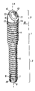
anchoring principle. The anchoring element shown in figure
1 is prepared in one piece of pure titanium and mainly
built up of two adjacent and lined up cylinder-shaped
segments.
The fixture shown in Fig. 1 is manufactured as a single
piece of pure titanium and essentially composed of two
cylindric segments 1, 2 bordering each other and in being
in alignement, wherein the segment proximal to the jaw and
having a diameter larger than that of the segment distal to
the jars 2 is designated by 1.
The outside of the fixture is threaded, except for a
mounting portiom extending from the proximal end of the
fixture and connected to the cylindric proximal segment 7..
The outer threads comprise first threads 4 on the proximal
cylindric segment 1 and second threads 5 on the distal
cylindric segment 2. The pitch is the same for both threads
2109850
4, 5 merging at the border zone between the cylindric
segments. The inner diameter of first threads 4 is somewhat
but not much larger than the outer diameter of the second
threads 5. Threads 4, 5 are self-tapping.
A symmetrically centred bore (not shown) extends from the
distal end 6 and has an extension corresponding to about
half the length of the proximal segment 2. Two through
slits 7, 8 arranged symmetrically in segment 2 and in its
longitudinal direction extend from a plane perpendicular to
the central axis near the distal end & by a length of about
three thread pitches. Slits 7, 8 establish communication
between the outside of proximal segment 5 and the
symmetrically centred bore arranged therein for transport
of bona material removed by ablation. The outside of distal
segment 2 is bevelled (bevelling 16) towards end 6.
The mounting section 3 is contained within a cylindric
chamber with a diameter corresponding to the outer diameter
of cylindric segment 1. The mounting segment 3 comprises a
base portion 9 having the form of a cylindric body
dissected by a plane at an angle of 45° i respect of the
cylinder axis. The circular basis of base portion 9 is
connected to the proximal end of proximal cylindric segment
1 with which it merges. Nearest to the proximal cylindric
segment 1 base portion 9 has an annular flange 14 to which
an annular groove 15 connects in direction of the proximal
end. End face 10 of base portion 9 is defined by the
aforementioned dissecting plane and, at its proximal zone,
smoothly rounded joins the cylinder mantle of base section
9, the beading decreasing gradually towards the distal
portion of the base section. Because of the bevelling the
profile of end Pace 10 is substantially circular. In its
center end face 10 has a bore 12 running at an angle of 45°
in respect of longitudinal axis A for cylinder segments 1
and 2, i.e., for the fixture. At bore 12 base section 9 is
21U9850
9
extended under formation of a frustrum of a cone 11
tapering in direction away from base section 9. Mantle
surface 13 of the cone frustrum 11 and the annular end face
are designed for sealing abutment of a dental prosthesis
5 or bridge (not shown) that can be mounted on the base
portion by screw means.
The .parts of the fixture according to the invention in
contact with living bone tissue have a surface promoting
10 integration, preferably a surface covered by micropits or
similar surface irregularities in the order of 10 - I,000
nm.
Implantation starts by providing in the maxilla a bore with
a diameter corresponding to the inner diameter of threads 4
and at an angle deviating about 45° from the vertical (the
longitudinal axis of the human body in an upright
position), followed by a bore in the zygomatic bone having
a diameter corresponding to the inner diameter of threads 5
and in line with the first hole. Thereupon the fixture is
inserted with its narrow end 6 into the hole in the upper
jaw until, by means of its bevelled portion, it comes into
engagement with the hole arranged in the zygomatic bone.
Thereupon the fixture is screwed on into the hole in the
zygomatic bone in a self-taping manner and, after threads 4
having reached the jaw bone, also simultaneously into the
latter in a self-taping manner. When attaining a sufficient
insertion depth for the fixture, such depth being defined
by the fixture's free end having the correct distance from
the jaw bone, the screwing process is stopped. By fine
tuning, that is, anti-clockwise or clockwise rotation
around ita longitudinal axis, bore 12 provided with
internal threads is brought into correct position for
mounting of the prosthesis, i.e., in a position in which
bore 22 is substantially parallel with the longitudinal
axis of the human body. The position for an implanted
2109850
fixture according to the invention is schematically shown
in Fig. 2; the zygomatic bone has been designated by O and
the maxilla by M.
5 The dental prosthesis or bridge can be mounted after
surgery or after a healing period during which the fixture
is progressively anchored in bane tissue.
The length of the fixture and cylindric sections 1 and 2
10 and the angle of bore 12 in base portion 9 are adapted to
the anatomical requirements of the individual patient. A
limited range of fixtures according to the invention,
including fixtures of varied total length, varied ratio of
length of the cylindric sections andwaried angle of bore
12 in base section 9 in relation to the longitudinal axis
of the fixture, will suffice for covering the needs of most
patients.
It is also possible to produce the fixture in two parts, a
separate base part 9 and a part 1, 2 provided with external
threads. The base part and the part with external threads
can be connected in various ways, for instance, by a
symmetrically centred tap positioned on the end of base
part 9 facing away from bore 12, said tap being arranged
for screwing on into a threaded bore in the free end of
part 1 provided with external threads, both parta otherwise
having planar abutting surfaces (line ~ in Fig. 1 indicates
their position) for abutment against each other.
In a second embodiment of the fixture according to the
invention shown in Fig. 3 only the proximal portion of the
proximal segment 1' has first external threads 4'. The rest
of the proximal segment 1' has a polished surface, i.e.,
essentially the portion positioned upon implantation in the
maxillary sinus 17 between the maxilla M and the zygomatic
bone Z. First external threads ~' thus have an extension
11 210950
essentially corresponding to the depth of the through bore
provided in the maxilla M in preparation for implantation.
The distal segment 2' is provided with corresponding second
external threads 5'.
As is also shown in Fig. 3 this second embodiment can be
provided (as can the first embodiment shown in Fig. 1) with
a periosteal plate or foil 20 for enhancement of
integration of the implant with living tissue in an area
where the distal end zone of distal portion 2' with
mounting portion 3' emerges from the through bore in the
maxilla M. The periosteal plate or foil 20 is a porous disc
of thin pure titanium, preferably from about 0.1 to about
0.3 mm thick and having a concentric hole 21 with a
diameter slightly larger than the largest outer diameter of
the fixture 1', 2', 3', allowing plate or foil 20 to be
fitted like a collar around the part of the fixture
protruding from the maxilla M. Periosteal plate or foil 20
is fitted with its one side against outer surface 19 of the
maxilla M after partial removal of the periosteum 18
(thickness of periosteum 18 is exaggerated in Fig. 3 for
reasons of comprehensiveness) which is then folded back
against the other side of the periosteal plate or foil 20.
The pores (not shown in Fig. 3 and 4) in the periosteal
foil or plate 20 are through pores and provide for
communication between both sides of foil or plate 20: it is
preferred for the pores to have an average diameter of from
about 0.1 to about 0.01 mm. The periosteal foil or plate 20
promotes anchoring of the fixture around its proximal part
and reduces the risk of communication between the oral
cavity 22 and the maxillary sinus 17.