Note: Descriptions are shown in the official language in which they were submitted.
8678z
PS C:spec/
~~
2 1 i ~
1 ENDOSCOPIC SURGICAL PROCEDURE AND
INSTRUMENT FOR IMPLEMENTATION THEREOF
The present invention relates to a method of
implementing an endoscopic surgical procedure on a
5 patient, and more particularly, is directed to a novel
and uni~ue technique of performing a uniportal palmar
subligmentous endoscopic carpal tunnel release.
Moreover, the invention is also directed to the provision
of a unigue endoscopic surgical instrument adapted to be -
10 employed in the implementation of the foregoing method of
endoscopically effecting the carpal tunnel release.
Carpal tunnel syndrome is a numbness in the
thumb, index, middle and ring fingers resulting from
pressure being exerted on the median nerve inside the
15 carpal tunnel, interfering with the function of such
median nerve. This may readily manifest itself as a pain -
radiating as far as the shoulders and neck of the
patient, resulting in impaired grasping ability by the -- 3
hand and loss of sleep. This physical phenomenon is the -
20 result of repetitive work and motions being carried out
with the hand over lengthy periods of time, and is
experienced by more ever younger people.
In essence, the carpal tunnel is formed by an
arch of the eight wrist bones, spanned on its palmar
25 surface by the transverse carpal ligament, the flexor
retinaculum. The carpal tunnel functions as a large ~ ~ -
mechanical pulley to provide the appropriate moment arms
for the digital flexor tendons as they pass through the
tunnel. The tendons can then transmit force out into the
.
.. .
.
- i~ 21~04~
--2--
1 fingers and impart only an appropriate amount of tension
to develop torque at the level of the wrist.
Within the carpal tunnel, these tendons are
lubricated and nourished by two synovial membranes - the -
5 radial and the ulnar bursa. The median nerve also shares
the carpal tunnel, then branches out to provide sensory
innervation to the palmar surfaces of the thumb, index,
long and a portion of the ring finger. In addition, a
small motor branch of the median nerve supplies the
10 thenar muscles, which are responsible for lifting the
thumb into opposition with the fingers. -~
Currently, a considerable array of methods or
surgical techniques, and suitable therewith correlated
surgical instruments, are being employed for purposes of
15 implementing surgical procedures in effectuating carpal
tunnel release in patients, and are generally designed --
for particular and highly specialized applications in -
this medical technology.
The customary procedure in implementing carpal
20 tunnel release has heretofore been the forming of a
lengthy incision, up to 8 cm in length across the palm
from the wrist to the middle thereof, resulting in an
unsightly scar, requiring division of all anatomical
structures between the skin and the flexor retinaculum;
25 i.e. the transverse carpal ligament. This created the
potential for inadvertently cutting or injuring the
palmar cutaneous nerve. Moreover, the patent normally
encountered significant postoperative pain and
discomfort, weakness of grip and pinch strength because
3o of pillar infraction and the excessively lengthy extent
of the incision. Such open surgery not only normally
- - . . .' ' ` ' - .: , '`, . . . :- .
2~ i~451
--3--
1 left the patient with a cosmetically unsightly scar
extending from the wrist to the center of the palm, as
mentioned hereinbefore, but also necessitated a lengthy
and painful convalescence for the patient, whereby this
5 convalescent period frequently caused the hand to be
incapable of any significant physical work or
manipulation for many weeks and even months, thereby
effectively rendering the patient incapable of carrying
out any meaningful work with the operated on hand and
10 resulting in considerable financial losses being
sustained by the patient.
~nong more recent developments and advances in
such surgical procedures, arthroscopic surgery employing ~ ;
the use of endoscopic devices has found widespread
15 application, among others in connection with carpal
tunnel release, in that in comparison with earlier ~ -
customary surgical methods, any incisions necessary for
such endoscopic/arthroscopic surgical procedures have
been considerably reduced in size, thereby alleviating
20 potential postoperative complications and pain . ~-
encountered by the patient, while reducing any scarring
to cosmetically desirable levels. Among various types of
surgical procedures, techniques involving approaches by
means of arthroscopic and endoscopic systems to carpal
25 tunnel surgery have been acknowledged as being superior
in providing significant advances over earlier so-called
open surgical procedures necessitating large incisions.
Such endoscopic surgical procedures Xave found widespread
acceptance in effectuating carpal tunnel release for the
3o purpose of alleviating the symptoms in a patient caused
by carpal tunnel syndrome, also referred to as tardy
-4_ 211~4~
1 median nerve palsy, normally caused by the compression of
the median nerve within the carpal tunnel.
These more recent endoscopic surgical
approaches to remedying varying types of surgical
5 problems afforded desirable alternatives to such earlier
open surgical procedures, and especially when applied to
effectuating carpal tunnel release, have found widespread
favor with surgeons and patients in comparison with the
earlier surgical methods which primarily constituted
10 complex open surgical procedures, and which involved
lengthy and painful postoperative convalescent periods.
Among numerous publications which describe
recent advances in endoscopic surgical methods and
instruments employed in connection therewith,
15 particularly such as may be employable for carpal tunnel
release procedures, there may be found the Agee carpal
tunnel release system as disclosed in Agee, et al. U. S.
Patent Nos. 4,963,147 and 5,089,000, both of which
disclose endoscopic surgical instruments and surgical
20 procedures implemented therewith, which when applied to
carpal tunnel release through an effective severing of
the flexor retinaculum, or transverse carpal ligament,
are adapted to provide relief to the patient. However,
the instrument and methods developed by Agee, et al. as
25 described in those publications, although superior to
open surgery, inhibit readily unobstructed visualization
of the surgical site during the sequence of severing the
flexor retinaculum and do not provide adeguate control in
the manipulation of the instrument so as to reduce the
3o inherent danger of damage to surrounding nerves and
_5_ 2~
1 tissue to an acceptable minimum, and additionally
necessitate the forming of two entry portals or incisions
in the wrist and hand. Moreover, the endoscopic
instruments developed in Agee, et al. are relatively
5 cumbersome and expensive, requiring the surgeon to always
use both of his hands, and necessitate the use of a
swivel cutting blade construction operable independently ~-
of a viewing scope, which does not always provide the
appropriate visualization during cutting of the flexor
10 retinaculum so as to potentially present the danger of
causing damage to adjacent or contiguously located tissue
or nerves relative to the operating site, which could
lead to serious and possibly permanent injury to the
patient.
Another surgical system and instrument -`
providing for an advanced technique over Agee, et al.,
which is particularly adapted for carpal tunnel release
through the intermediary of an endoscopic surgical -
procedure is disclosed in Chow U. S. Patent No.
20 S,029,573. However, in that instance, although setting -
forth a considerable advance over the methodology
disclosed in the Agee, et al. U. S. patents, the surgical ~ -
procedure employed by Chow requires the formation of two
entry and exit portals or incisions, one in the wrist
25 area and one in the palm, and the passage of an
endoscopic medical instrument, such as an obturator
through a considerable length beneath the subcutaneous
areas of the palm of the patient. Again, the necessity
for two widely separated incisions or entry portals, and
3o the requirement for inserting a scope from one end of the
instrument from one portal and with the instrument
, ~- 2110~1
-6-
1 extending outwardly from the other portal or incision,
while surgically severing or cutting through the flexor
retinaculum or transverse carpal ligament from the other
portal or incision, engenders a considerable obstruction :~
5 toward a clear nonproblematic visualization of the
operating site during the severing of the transverse
carpal ligament and, once again, raises the spester of a ~ :
potential risk of causing injury to tissue and nerves
adjacent the operating site, especially such as to the
10 median nerve, which could lead to serious permanent
injury to a patient and possibly reguire additional
corrective surgery necessitating subjecting the entire
surgical or operating site to open surgery. Moreover,
Agee, et al. and Chow require the surgeon to
15 simultaneously employ both hands during the surgical
procedures, thus necessitating the utilization of an
unusually high degree of dexterity in manipulating the
various components of the endoscopic surgical
instruments.
The foregoing limitations and potential
drawbacks which are encountered in the prior art
publications are clearly and ambiguously obviated and
improved upon through the inventive and novel method of
implementing an endoscopic surgical procedure, and the
25 unique and inventive endoscopic surgical instrument
developed for accomplishing this purpose, especially for
the effectuation of a carpal tunnel release; in essence,
the severing of the flexor retinaculum or transverse
carpal ligament through an endoscopic surgical procedure
3o in which there is effected, by means of a uniportal or
2,1 i 0 ~
-7-
1 single incision, a palmar subligmentous endoscopic carpal
tunnel release technique. This sur~ical procedure only
requires the formation of a single and relatively small
entry portal or incision in the palm proximate the distal
5 side of the flexor retinaculum, thereby reducing any
postoperative symptoms of the patient with only a
cosmetically appealing scar formed on the palm, while
eliminating the need for a second portal or incision
proximate the wrist of the patient. Moreover, the -
10 endoscopic instrument employed in implementing the
inventive method utilizes a unique cutting device which
is mounted on a scope insertable through a cannula which
has been initially inserted to extend beneath the flexor
retinaculum from the distal side of the flexor
15 retinaculum or transverse carpal ligament, upon the
formation of a passage beneath the flexor retinaculum, ---
after hyperextending of the hand, by the preceding ~-
insertion and manipulation of a curved dissector.
Thereafter, the dissector is removed and the cannula and -
20 an obturator which is contained therein are inserted
through the incision into the previously formed passage
beneath the flexor retinaculum. The cannula of the
surgical instrument has the obturator withdrawn
therefrom, and in place of the latter, a scope is
25 inserted into the cannula which enables unhindered and
unobstructed visualization of the operating site and of :
the flexor retinaculum.
The scope is then withdrawn from the cannula,
and the same scope or another scope with a cutting blade ~ -
3o mounted at the leading end thereof inserted into and
advanced through the cannula towards the flexor
' .
:: :
8 2 1 ~ 0 ~
1 retinaculum. Severing of the latter is then effected by
the cutting blade while affording an unhindered view of
the operating site through the scope, thereby resultingly
dramatically reducing or even completely eliminating the
5 risk of any injury being sustained by tissue and nerves
in the vicinity of the operating site; for example, such
as the median nerve. This particular unhindered
visualization of the operating site also enables the
surgeon to exercise an improved degree of control over
10 the possibly single-handed manipulation of the endoscopic
instrument and cutting blade.
Pursuant to a feature of the invention, the
guiding member or cannula of the endoscopic instrument,
and which contains the obturator which is initially
15 employed to be advanced beneath the flexor retinaculum or
transverse carpal ligament subsequent to withdrawal of
the curved dissector, is provided with lateral or
sideways wing-like or flange-like protrusions of
curvilinear configurations which, in conjunction with an
20 upwardly curving tip of the obturator projecting
forwardly of the leading end of the cannula, is adapted
to displace any tissue, or such as the media nerve, out
of the path of the obturator and cannula as is being
advanced; in effect, through essentially a sideways or
25 lateral "shoving" action, thereby preventing any
potential damage to such displaced tissue and nerve
during the subsequent cutting procedure by maintaining
such tissue well out of the way. Moreover, the leading
tip of the obturator by being curved slightly upwardly
3o towards the lower surface of the flexor retinaculum is
also adapted to remove or dislocate any possible tissue
9 2 ~
1 or fascia located close to the surface of the flexor
retinaculum and to ensure that the cannula and,
resultingly, the subsequently inserted cutting blade are
located as closely as possible to the flexor retinaculum.
The foregoing inventive concept ensures a -~
simple and extremely efficient endoscopic surgical method
which is particularly adapted, in conjunction with the
use of the novel endoscopic instrument, for the
implementation of a carpal tunnel release through the
10 severing of the flexor retinaculum while producing ~ .
minimal or no postoperative pain and discomfort in the
patient, with a shortened convalescent period and with
the formation only of a small cosmetically attractive
scar on the palm of the patient's hand.
Accordingly, the present invention is directed -
to a novel and unique method of implementing an
endoscopic surgical procedure through a uniportal entry ~.
to an operating site by a novel endoscopic surgical
instrument.
The present invention is more specifically - :
directed to a method of implementing an endoscopic :
surgical procedure at a selected operative site on a .. -.-:
patient; comprising the steps of making an incision on
said patient at a locale proximate said operative site to -
25 establish an entry portal; inserting an elongate : ~ -
insertion member into a longitudinal bore of an elongate
cannular guide member having open proximal and distal
ends and an open slot extending along the length thereof
communicating with said open ends, said elongate
3o insertion member being slidably receivable within said
cannular guide member and being configured so that at . :
' :'~ .'
-10- 2 1 1 0 ~
1 least portions thereof conform with said open distal end
and said open slot of the guide member to form a smooth
exterior surface in combination therewith; introducing a
leading end of the combination of said cannular guide
5 member and the therein inserted insertion member into
said entry portal and advancing said combination a
predetermined distance relative to said operative site;
withdrawing said insertion member while permitting said
cannular guide member to remain in place at said
10 operative site; inserting endoscopic viewing means into
said cannular guide member for direct visualization of
said operative site and the positioning of said guide
member relative to said site; withdrawing said
endoscopic viewing means from said cannular guide member;
15 mounting a surgical instrument on further endoscopic
viewing means proximate the leading end of said viewing
means; inserting said composite endoscopic viewing means
and said surgical instrument into said cannular guide
member such that the surgical instrument protrudes into
20 the open slot in said cannular guide member, and
advancing said composite endoscopic viewing means and
surgical instrument so as to contact tissue at said
operative site with said surgical instrument;
operatively engaging said tissue with said surgical
25 instrument while advancing the latter under direct
visualization through said endoscopic viewing means so as
to perform a desired operative procedure on said tissue;
withdrawing said composite endoscopic viewing means and
surgical instrument from said cannular guide member;
3o withdrawing said cannular guide member through said entry - .
portal and suturing said incision. ` .
.
: .
- . . , .; -
1 The present invention is further directed to
an endoscopic surgical instrument with a novel scope-
mounted cutting or blade element for implementing the
endoscopic surgical procedure pursuant to the invention.
5 More specifically, the present invention is directed to
an instrument for implementing an endoscopic surgical
procedure at a selected operative site on a patient
through an incision on said patient at a locale proximate
said operative site to establish an entry portal; said `
10 instrument comprising an elongate cannular guide member .
including a longitudinal bore having open proximal and
distal ends and an open slot extending along the length
thereof communicating with said open ends; an elongate .
insertion member being slidably receivable within said
15 cannular guide member and being configured so that at
least portions thereof conform with said open distal end ::; - .
and said open slot of the guide member to form a smooth -:-:
exterior surface in combination therewith, said cannular~.
guide member and the inserted insertion member being ~.
20 advanceable a predetermined distance relative to said ~ --
operative site, said insertion member being withdrawable
while permitting said cannular guide member to remain in
place at said operative site; endoscopic viewing means
insertable into said cannular guide member for direct ~ ~:
25 visualization of said operative site and the positioning
of said cannular guide member relative thereto and
thereafter withdrawable from said cannular guide member;
a surgical instrument being mounted on further endoscopic
viewing means proximate the leading end of said further
3o viewing means; said composite further endoscopic viewing
means and surgical instrument being insertable into said
: ~ .
. .
, ~;
, ' , ,'' ''-.~ '
-12- 2 1 ~ 0l
l cannular guide member such that the surgical instrument
protrudes into the open slot in said cannular guide
member enabling advancing said composite further
endoscopic viewing means and surgical instrument so as to
5 contact tissue at said operative site with said surgical
instrument and operatively engaging said tissue with said
surgical instrument under direct visualization through
said further endoscopic viewing means so as to perform a
desired operative procedure on said tissue, said
lO composlte further endoscopic viewing means and surgical
instrument being withdrawable from said cannular guide
member, and said guide member being withdrawable from
said entry portal.
Still further, the present invention is
15 directed to an endoscopic surgical instrument of the type
described, in which a scope which is adapted to be
advanced through a cannula located beneath the flexor
retinaculum has a cutting device mounted thereon to
enable severing of the flexor retinaculum while being
20 able to afford the surgeon an unobstructed visualization
of the operating site.
Reference may now be had to the following
detailed description of preferred embodiments of the
endoscopic surgical instrument constructed pursuant to
25 the invention, and to a surgical procedure for the
effectuation of carpal ligament or tunnel release on a
patient by a transverse severing of the flexor
retinaculum, taken in conjunction with the accompanying
drawings; in which:
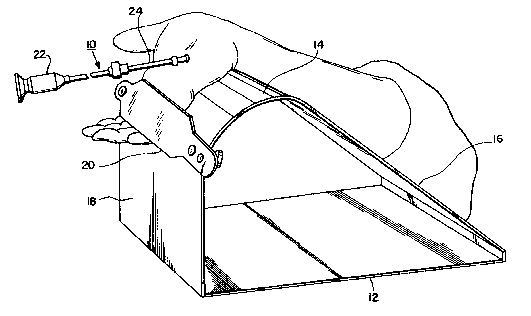
3o Figure l illustrates a generally perspective
view of the hand of the patient in a hyperextended
. .. ., , ~: :
,
.- ,
.~ ` ` ,
: . : . ,
. . . . . ..
. .
-13-
l position during a surgical procedure for effecting2~ ~a~5
ligament release, utilizing the endoscopic surgical
instrument pursuant to the invention;
Figure 2 illustrates a longitudinal top view of
5 a slotted cannula of the endoscopic surgical instrument
pursuant to the invention;
Figure 3 illustrates a sectional view taken
along line 3 - 3 in Fig. 2;
Figure 4 illustrates a top view of the leading
lO end portion of a modified slotted cannula;
Figure 5 illustrates a sectional view taken
along line 5 - 5 in Fig. 4;
Figure 6 illustrates a longitudinal side view
of the leading end of an obturator adapted to be inserted
15 into the slotted cannula of Figs. 2 or 4; -
Figure 7 illustrates a top view of the leading
end of the obturator; ---
Figure 8 illustrates a longitudinal side view
of the endoscopic instrument, showing the scope and - -
20 cutting device mounted on the latter inserted into the -- --
slotted cannula;
Figure 9 illustrates a top view of the leading
section of the endoscopic instrument shown in Fig. 8;
Figure 10 illustrates, on a somewhat enlarged
25 scale, a sectional view of the encircled portion 'A' of
the instrument of Fig. 8;
Figure 11 illustrates a longitudinal side view
of a scope and cutting device or blade mounted thereon ,
prior to the insertion thereof into the slotted cannula;
-14- 21 ~
l Figure 12 illustrates a top view of the leading
end portion of the scope and cutting device of Fig. 11;
and
Figure 13 illustrates the palm portion of the
5 hand of a patient showing the surgical markings applied
thereto prior to implementing the incision for the carpal
tunnel releasing surgery.
Reverting now in more specific detail to the
description of the invention as represented by drawing
lO Figs. 1 through 12, Fig. 1 of the drawings illustrates an
endoscopic system 10 employed for the procedure of
effectuating the surgical release of a transverse carpal
ligament; in essence, the severing of a flexor
retinaculum in order to alleviate the symptoms and
15 debilitating effects of carpal tunnel syndrome.
In this instance, the hand of a patient with
the endoscopic system 10 is supported on a hand rest 12,
which is in the form of a bolster having a curved upper
surface 14 between an inclined or sloping surface 16
20 enabling the lower arm portion of a patient to be
supported thereon, and a vertically depending front
surface 18 with a strap 20 attached thereto for
maintaining the hand of the patient in a hyperextended --
position in readiness for the endoscopic surgical~-~
25 procedure~
As shown in Fig. 1 of the drawings, the
endoscopic instrument 10 which is to be utilized for
effectuating the carpal ligament release; in effect, the
severing or transverse cutting through of the flexor
3o retinaculum, is shown in the operative position thereof
inserted ~brough an incision into the hand of a patient;
. ~ -
.
. ~
2 1 1 0 4 ~
-15-
l with the surgical procedure being set forth in more
specific detail hereinbelow.
Referring to Figs. l through 11 of the
drawings, and particularly Figs. 2 through lO, the
5 endoscopic surgical instrument 10 comprises an
arthroscope 22 which includes a cannula 24 having a ~ ;
through extending longitudinal slot 26 formed therein, -
and a knob or flange-like member 25 at one end thereof, --
as shown specifically in Figs. 2 and 3 of the drawing.
An obturator 28, as in Figs. 6 and 7, is
adapted to be slidably received within the cannula, and
presents a smooth outer surface through the intermediary
of an axial, upstanding rib portion 30 which is ~ -
engageable in close conformance within the longitudinal ~ ;
15 slot of the cannula upon insertion therein. The leading - - ~ -
end of the obturator 28 is a tapered tip portion 32 which
is bent upwardly in a direction towards the longitudinal
rib to impart to the tip a somewhat upward curvature for
a purpose to be described hereinbelow in more extensive
20 detail.
Although the cannula 24, as shown in Figs. 2
and 3, is illustrated as being circular in cross- ~-
sectional configuration along its external surface, ~ -~
pursuant to a modified embodiment, as shown in Figs. 4
25 and 5, at opposite sides of the longitudinal slot 26, the
outer surface of tbe cannula 24 may be equipped with
integrally formed outwardly extending curvilinear flange
portions 36 and 38 so as to essentially form so-called
wings or fins, as described further on hereinbelow.
3o These fin-like wings or flange portions 36 and 38 are
integrally formed with the cannula and are also curved so
.: :
. ..
-16- 2 lt 0
1 that upon insertion of the obturator into the cannula,
the tip end of the obturator essentially forms a smooth
curvature at its juncture with the flanges 36 and 38.
As shown more specifically in Figs. 8 through
5 10, the endoscopic instrument 10 is illustrated in its
condition for cutting through the flexor retinaculum to
effectuate carpal tunnel or ligament release.
Hereby, the arthroscope 22 includes a suitable
knurled knob 40 having an internal threaded portion 42 in
10 a cylindrical extension 44 and a ~apered bore 46 for
receiving a tubular knife or cutting blade holder 48.
The blade or knife holder 48 is adapted to receive a
scope 50 of cylindrical configuration extending
therethrough and lock the latter within the blade holder
15 by simply axially displacing the knurled nut 40 through
threaded interengagement between the internal thread 42
of the nut and an external thread 52 on the blade holder.
This will cause the tapered bore 46 of nut 40 to either - -
compress the slotted portion 54 of the blade holder to
20 clampingly engage the scope 50 or to loosen it so as to
enable axial adjustment thereof relative to the blade
holder.
A scope in the form of a rod member, in the
absence of a blade holder, and which is connected to a
25 video scanner (not shown) is adapted to be inserted
through the cannula for effective visualization of the
operative site.
The scope S0, at the leading end thereof
includes a mounting for a cutting element, such as a flat
3o knife blade 60 having a leading cutting edge 62, and with
the scope 50 having a tapered or angled forward end
-17-
l surface 64 enabling light to be projected against the
cutting device so as to illuminate the region of the
operating site.
The knife blade 60 is adapted to be slid
5 through the cannula 24 while mounted on the scope 50 so -
as to be slidingly engaged within the longitudinal slot
26 of the cannula during the forward advance thereof and `
while severing the flexor retinaculum. Moreover, the
extent of forward advance of the knife blade in the
lO cannula is readily controlled by adjusting the relative
axial positioning of the scope within the tubular blade
holder 48 and thereafter clamping the scope within the
knife holder through activation of the knurled knob 40.
As shown in Figs. 11 and 12 of the drawings,
15 the cutting blade 60 may also be directly mounted on the
holder 48 for the cylindrical scope 50, which has the
distal end thereof provided with the external thread 52
which is engageable with the clamping nut 40, and with
the slotted end portion 54 adapted to be tightened onto
20 the scope. -
The inventive endoscopic surgical procedure for
effecting carpal tunnel release utilizing the novel
uniportal palmar subligmentous endoscopic carpal tunnel
release technique, and employing the novel endoscopic
25 surgical instrument 10 is now described hereinbelow, by
way of example.
Initially, after the hand is prepped, a -
regional anesthesia is applied to the hand of the patient
which is to be sub~ected to the operative procedure.
3o Thereafter, two lines are drawn, one transversely across
the palm from the distal border of the thumb and another
~. p..
21~4~1
-18-
l between the middle and ring fingers of the patient. At
the point of intersection of the lines, and at a
proximity of 1 cm thereto, a 1.5 cm long incision is made
in the thenar crease or in a slight ulnar direction. The
5 incision is deepened to expose the palmar fascia through
the intermediary of blunt scissors in order to avoid
injury to the palmar cutaneous branch of the median
nerve. The distal edge of the flexor retinaculum is
identified and divided for 5 to 6 mm approximately.
lO Throughout this process, the palmar arch and the median
nerve branches are protected. This Dalmar fascia is then
divided longitudinally exposing the flexor retinaculum.
The hand is thereafter placed on the hand rest
or bolster 12, with the forearm to which a tourniquet has
15 been applied being supported on the inclined surface 16.
The wrist is hyperextended in that the hand is positioned
palm facing upwardly on the curved surface 14 with the --
fingers depending forwardly, and then clamped by means of
the strap 20 to the bolster.
In this hyperextended position of the hand, a
curved dissector is inserted through the incision so as
to cause the posterior surface of the flexor retinaculum ~ -~
to be carefully dissected so as to feel the synovial
tissue of the flexor retinaculum. Suitable retractors
25 maintain the incision in an open spread condition. This
enables the open incision or wound to be thoroughly
irrigated.
Thereafter, the curved dissector is withdrawn,
and the cannula 24 with the obturator 28 positioned
3o therein with its tip 32 forwardly extended, is advanced
into the incision along the path previously defined ~y
4 ~ 1
-19-
1 the dissector in close proximity to the internal surface
of the flexor retinaculum. This closeness is enhanced by
the curvature imparted to the tip of the obturator.
Thereafter the obturator 28 is withdrawn while -
5 permitting the cannula 24 to remain in place beneath the
flexor retinaculum, and a scope (without a cutting blade)
is inserted through the cannula 24 to enable thorough
visualization of the posterior surface of the flexor
retinaculum. Hereby, it is important to be able to
10 identlfy the flexor retinaculum endoscopically through
the presence of its transversely oriented fibers. In
the event that the scope ascertains that there is a ~ ~
presence of some synovial tissue obstructing the ~ ~ ~s
visualization of the transverse fibers, either a blunt
15 dissector or a blunt hook may be employed to peel the
thin and generally flimsy synovial lining away from the
flexor retinaculum. Alternatively, if this particular
presence of such tissue is of a substantial nature, the -
cannula 24 is withdrawn, the obturator repositioned
20 therein, and the entire procedure repeated. This must be
implemented until such time as the transverse fibers of
the flexor retinaculum are clearly viewed endoscopically.
Upon the transverse fibers of the flexor
retinaculum beinq clearly identified, the scope is then
25 withdrawn from the cannula 24, and the scope 50 having
the cutting device, consisting of the blade 60 mounted
thereon, is inserted through the cannula 24 and advanced
towards the operating site represented by the transverse
carpal ligament or flexor retinaculum. The angled
3o leading end 64 of the scope 50 on which the cutting blade
60 is mounted enables projection of illuminating light
. ~ . . . . ~- . ` .
21~4~1
-20-
1 against the blade and the surrounding regions of the
operating site so as to constantly afford direct
unobstructed visualization of the operative region during
the carpal ligament releasing procedure.
As the scope and the cutting device or blade 60
mounted thereon is advanced, the cutting edge 62 of the
latter will divide the flexor retinaculum throughout its
transverse width while being maintained under endoscopic
visualization. Upon completion of the severing of the
10 flexor retinaculum, the scope 50 and the thereon mounted
cutting blade 63 are withdrawn from the cannula, and a
scope without a cutting device thereon is reinserted into
the cannula to provide for a viewing of the cut edges of
the flexor retinaculum so as to ensure the complete
15 division thereof and not of to palmar facia. Once the
intactness of the median nerve and surrounding structures
have been verified through suitable rotation of the -~
cannula about its longitudinal axis so as to afford a
broader overview, the entire endoscopic surgical
20 instrument 10 is withdrawn from the operating site out of
the incision.
Prior to closing and suturing the incision, the
wound is again inspected, such as through the insertion
of a blunt dissector, and if satisfactory, the wound is
25 then irrigated and sutured, with a tincture of benzoin
applied thereto, thereafter applying a steristrip and the
hand placed in a soft fluff dressing.
From the foregoing, it becomes readily apparent -
that the inventive surgical procedure, employing only a
3o uniportal or single incision enables the operation to be
implemented much more rapidly than heretofore, while
-21- 21~4~
. ~. . .
l forming only a cosmetically attractive small single scar
in the palm, while extensively reducing the postoperative
recovering period of the patients. In at least one-third
of the patients, no pain was experienced postoperatively,
5 obviating the necessity for any medication.
Moreover, the average length of time
postoperatively for being able to gainfully utilize the
hand and, thereby to return to wor~, was approximately 14 ~ -
days, with executives normally being able to return to -~
10 work at about 7 days subsequent to the operation,
clerical/secretarial staff at approximately 17 days, and
workers involved in heavy physical labor at approximately -~
28 days after surgery. -.
While there has been shown and described what
15 are considered to be preferred embodiments of the
invention, it will, of course, be understood that various
modifications and changes in form or detail could readily ~ -
be made without departing from the spirit of the
invention. It is, therefore, intended that the invention
20 be not limited to the exact form and detail herein shown
and described, nor to anything less than the whole of the ~: :
invention herein disclosed as hereinafter claimed. -
., ~,
-
:
. ~ ~
3o
;