Note: Descriptions are shown in the official language in which they were submitted.
CA 02112297 2004-07-09
rOMPUT~R S zMtJLATION OF T.~TVE ORGAN
HACF~GROUNn o~ THE iNVENTiON
~U. S. Patents 3,453,745 and 3,552,036 disclose
el.eet~onioally operated means to simulate. cotitxol, and modify
ECG signals. The signals are displayed. on standard oaciloacope-
type monitors.' However, these patents only address mans to
simulate ECG signals. . .
U. S. Patent 4,091,549 discloses means to trace heaxt
el2etrieal actipity through speciF~ic points of a heart by means of
illumination of specific parts of an illustration of a heart.
U. S. Pat-ent 4,254,562 discloses means to trace blood~flow
through specific points of a heaxt model or other component of a
living organism. ~ .
2
CA 02112297 2004-03-10
OBJECTS OF THE INVENTION
One object of the invention is to display to an observer a
pictorial image of a heart in a body of a patient generating
EEG, EMG, ECG, or other diagnostic electrical signals a:> these
signals are occurring in the patient. The diagnostic signals
are commonly voltages measured of the heart tissue indicating
expansion and contraction of the heart.
Another object is to provide a three dimensional
interactive view of a heart's operation from electrical
measurement signal type information.
Another object is to provide the operator with a view of an
active, working heart model driven from electrical measurement
signal type information in three dimensions.
Another object is to allow the operator to manipulate
various characteristics and dynamics of the modeled heart or the
signals from an actual or simulated EEG, EMG, ECG, or other
diagnostic electrical measurement signal type monitor anal
observe the results on interactive, three dimensional graphical
models for study, teaching, diagnosis, and research purposes.
Another object is to provide the capability to surgically
or chemically interact with, probe, or explore the object of the
study.
Another object is to provide the capability to do the
foregoing with other parts of the body.
3
211229
SUMMARY OF THE INVENTION
A computer system receives two dimensional slice data of a
heart or other organ to be simulated in three dimensions. It
also receives chemical composition data of the heart or other
organ, and chemical composition data of other parts of the body.
These data are put in the computer memory. Then a Voxel View or
three dimensional volume rendering program forms images of the
organ to be studied. For example, with the heart it generates
images of the atria and ventricle. Diagnostic data obtained
from a patient conveniently with electrical measurement signals
includidng an electro-cardiagram electro-myogram,
electro-encephalogram, and other diagnostic measured electrical
signals obtained from a patient are fed into the system and are
placed in computer memory. Physiological data of the patient,
including the strength, weakness and other parameters of the
organ, is also, supplied into the system. This can be done
manually with a keyboard or mouse, or may be
supplied from a hard disk, a floppy disk or a tape. This is
also fed into memory and is used to modify the three dimensional
image data of the organ.- This data is then synchronized with
the electrical signal diagnostic data. Conveniently the first
derivative of the electrical data signal is taken, and P and Q
waves determined from the derivative. From this information the
organ, including sub-parts, may be simulated. This data may be
fed ip. black and white or preferably in color to a device
which shows the organ for visualization, operation simulation,
or training.
4
CA 02112297 2004-03-10
THE DRAWINGS
Figure 1 is a schematic representation of the three
dimensional organ monitor of the present invention.
Figure 2 is a schematic representation of the conversion
model program used in the present invention.
Figure 3 is a schematic representation of the physiological
model dynamics program used in the present invention.
Figure 4 is a schematic representation of the three'
dimensional graphics program used in the present invention.
Figures 5(i) - 5(r) show a listing of a computer program
for the system according to the present invention.
211227
SUMMARY OF OPERATION
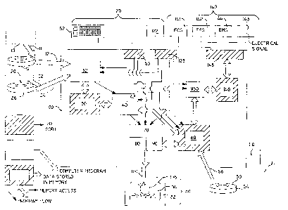
Figure 1 is a block diagram of the overall system. Five
major features are described.
One feature is the interactive devices (120), which may be
hand generated data including keyboard (52), mouse (122), touch
sensitive screen, light pen, or other device. Data may also be
introduced by a voice command signal.
Another feature is the input chemical composition and
dimensional slice data. This may come from several sources,
including Nuclear Magnetic Resonance Imaging Data (11),
Computerized Tomography Data (22), or Interactive Device Generated
Data, which may come from magnetic tape (14), {26), (50) or data
in computer memory or from an appropriate interactive device
(120). Physiological data may be provided through communications link(56
Another feature is the diagnostic input signals (140). These
may be Electrocardiogram (ECG) signals (14i), Electroencephalogram
(EEG), signals (142), Electromyogram (EMG) signals (144), or other
diagnostic data and/or electrical input signals (143).
Another feature is the output device (130), in the preferred
version being a cathode ray tube (CRT) (132).
Another feature is the Graphics Image Generator Computer
System (100). Within this computer system, which may be composed
of several networked computers, several computer programs {32),
(40), (70), and (950) run, accessing several blocks of memory
{30), (60), and (146). These utilize the described input and
output means to graphically display a three dimensional object
that_ref~ects input activity and can be manipulated by the user.
6
211229
DETAILED DESCRIPTION OF PREFERRED EMBODIMENT
In accordance with the present invention, a Nuclear Magnetic
resonance Scanner such as G.E. Medical Systems Model No. MR Signa
Advantage 1.5 Tesla (NMRS) General Electric Co., Milwaukee,
Wisconsin, (10) provides Imaging Data (11) in grey scale two
dimensional slice format. It is used as one input to the Graphics
Image Generator Computer System (100). This data is ready
available and can be gathered from the patient undergoing
diagnosis, or from pre-recorded data from some other NMRS source.
The data may enter the system via a digital data communication
link (12), which enters the Graphics.Image Generator Computer
System (100) through a provided communications port, or read from
a computer memory or magnetic tape (14). This data represents two
dimensional slices of the heart or other object for study which
are used to generate a three dimensional picture.
A computerized Tomography Scanner (20) such as a Siemens
Medical Systems Somatom Plas supplies Scan Data (22) in grey scale
two dimensional slice format is also used as input to this system.
This data is readily available, and can be gathered from the
patient undergoing diagnosis, or from pre-recorded data from some
other CTS source.\ The data may enter the system via a data
communications link _(24) tfirough a provided communications port,
or read from a storage media, such as magnetic tape (26), hard
discs and floppy discs. As an example, the chemical composition
of selected parts of the body may be inputed. Other sources of
chemical composition data may be used as inputs as well.
A three dimensional Volume Rendering program, (32) such as
"Voxel View", which reads, CTS, NMRS, or similar data into memory
(30) is activated in the Image Generator Computer System (100) to
receive the data from communications links (12) and (22). "Voxel
view" is a Registered Trademark of Vital Images, Inc. A brochure
is in the application file, and is available from them at P.O. Box
551, Fairfield, Iowa 52556, (515) 472-7726., The data (30) is then
ready to be manipulated by software (40) running in the Image
Generator Computer System (100). For example, Silicon Graphics
Models Nos. 4D/GTB,
7
211~29~
GTXB, or VGXB process the input data into a format suitable to the
model dynamics methods. The software (40) comprises a conversion
program which puts the data into a format in memory where each
particular part of the heart is identified, so that model dynamics
programs (950) can accurately model each part's reaction to
stimulus data. The software conversion program (40) is
illustrated in Figure 2.
Figure 2 is a block diagram of an example of a simple
conversion program (40) that inputs a three dimensional data block
(30), and outputs two data point arrays, one for atria, and the
other for ventricle points stored in memory (60). The conversion
program is entered through entry point IDENTIFY (410).
First, it finds the centroid of all input data points (30) in
the CENTROID section (420) of the program. It also determines the
centroid of the upper and lower halves of the data. The upper and
lower half centroids will be used as reference points for
compressing and filling these sections.
The next section, DIVIDE (430) positions a dividing plane
passing through the data centroid, dividing it into two halves.
This dividing plane could be user defined. Data above the
dividing plane is considered the atria and below is considered the
ventricle.
The next section, LOAD (440) takes this data and transfers it
to two data point arrays, one for the atria, and the other for the
ventricle, in a memory section (60). The conversion program then
exits (450).
8
CA 02112297 2004-03-10
Hand generated data (50) from a keyboard (52) or stored in
computer memory or stored on_magnetic tape (54), may be fed
directly into the system by communication link (56) to add to,
modify or correct slice shape data chemical composition data and
to input physiological data or that data indicating the relative
strength of various parts of the organ to be simulated. Data
may be modified for simulation or teaching purposes through Link
56. Modified or simulated data may also be introduced through
communications Link 12 and 22.
The data from the conversion program (4) and from hand
generation is stored in residual memory (60) in the Graphics
Image Generator Computer System (100). The image (104) of the
item being studied, in this example a heart (106), is generated
from this set of data. This date (60) is also accessed by the
model dynamics program (950) where it is modified in the
procedure shown in Figure 3 to reflect activity in the item of
study, (here heart 106).
Figure 3 is a block diagram followed by a computer program
of an example of a simple physiological model program for a
dynamic heart representation (950). It is composed of an entry
point (952), an initialization section (954) where memory blocks
are cleared and initialized, and control variables are
initialized. This program will access memory blocks (60) set up
by the conversion program (40) as shown in Figure 2.
The physiological model program enters a loop, beginning
with the Read-Wave section (960) where input signals data (146)
is accessed. The first derivative of the input signal magnitude
with respect to time will indicate whether the signal
9
211229'
is increasing or decreasiryg With each change of direction, ~n
index identifying the current wave sectioh is incremented. This
index identifying the c~.cr~rent wave section must be synchronized
wi th the actual l nput si gnal . and thi s synchrQn l z At l or. l s dope l n
the neXt. SQCtion. .sync-wave t9b2) .
. A f 1 ag l s set when the wav8 l s known tQ be sj~nchroni zc3d arnd
that sect l on l s slGi peed. Otherwi se, the sect l on sWTp ~' es t he
input signal over a period of time. The largest upward spike is
the Q wave, where -~hE? wave magni tude corresponds to ventri c1 ~
compr~essi on . When synchroni zed, the sync -F 1 ag l s set, and thi s
section will not perf4rm again r~nless synchronization is lost,
The neXt section is the update-wave section (96A). It is
performed l f the l nput si gnal and physi of oc~i cal model prpqram are
known to be~ synchron l zed ( l . e. , the sync f 1 act l. s set ) . Thi s
secti on accesses th.e wave secti on l ndex set l n th.e read-mave
section (~6G;. It also accesses the current input signal (14E),
and assigns this magnitude to the currently indexed point in a
mempr~y Section known as the wave form array. It also sets the
wave type far the currently indexed point in th2 wave form array
to the current wave type ~.;si ng the Curr2nt Wave secti on l ndeX se't
in the read-wave section L9603.
.t
ice'
The next secti on l s the Heart-I3ynami cs Secti orr 0966) . it
accesses the memory C60) where the arrays ciescribina floe three
d l mensi onal 1 ocati ons o-f poi nts on the surfa~-~ of Spec l f l c ' con-
tours of the heart ~r~ stored. If fihe Currently indexed point
has ' a "P" wave type., the atr l. ~ l s conpr-essed , and the vex qtr l c 1 a
is f l 1 Ied. A "Qt" wave type wi 3 ~ ~fi 1 l the atr~r.d~ and compress the
ventricle. Compression of points is done by carnpmting a ctampr~s-
211r297
5ion factor, dependent upon the magnitude of thC,input signal.
computing three dimensional point transition using the compres-
lion factor, and modifying the array points containing the three
dimensional location of the points of the section to be compres-
sed. The same.process is d~~ne when filling<a section, the diffe-
rence being, a fill -factor is used instead of a compression
-factor. After the heart-dynamics section (966, the physiologi-
cal model program returns to the read-wave Section- (960) where
the current input signal is again sampled, the direction of
signal change is noted, and the loop is repeated.
A Three Dimensional Graphics Program C7r1), that accesses tfie
da ~ tf~C~) is provided which uses geometric image functions C84>
provided by the Graphics Image Generator Computer System 1100) to~
draw an i m.~ge of the i tem o-f study, and r eT 1 ects the acti vi ty of
the items such as a heart, as modelled bytt-re model dynamics
program (954). Geometric Image Functions (80), are provided to
draw simple shapes or objects. Program t74) use=_ these functions
to produce an i mage of the i t2m of study such as heart C10~~ .
The Three Dimensional Graphics Program (79) is ii3.ustrated in
Figure 4.
Figures 4 is a block diagram of the three dim~.r,sionai grap-
hits programs (74) . It runs in parall=-=1 with ~Fra physiolGgicaJ.
model programs (950) to draw an image of the- it~Yn os study in
~tfii= emoodiment a heart.
The program is entered th,~flugh entry poi-nt 3D1~18L.F_L C3Z0),
anct conti nuea i nto tht: i n i ti al i.zatian secti on C72C~) wfiere control
variables are =_et up. The pr ogr'am ti~en proce~ec~ ~n the, DRAW-
21
2112297
MODEL i73Cr) section, where s~raphics image functions are accessed
to draw the model using simple geomett-it shapes. In this exam-'
ple, . a tr iangular mesh tECh.nique is appropriate, and is fully
documented by Silicon Graphics, makers of the preferred embodi-
mes~t Graphics Image Generator Computer. When the update pass
through the graphics image function=_ is completed, the program
waits t74t3) for the physiological model programs 1954) to signal
tf~e 3D Graphic) Program t70) to update the +display. It then
repeats the DRAW MGDEL secti;=:n 1730) and continues looping.
The i''iachine Operating System (9n3 runs within the Graphics
Image Generator Computer (100) and is the interface between the
user and the graphics Generator Machine. The preferred operating
system is IFiI:t, which is well known, and information is available
in ø,ublications concerning the Graphics Image Generator. For
example, see the IRIX System Library, available from Silicon
Graphics.
The preferred embodiment Graphics Image Generator is the
IFt~S 4Df320VGXB from Silicon uraphics of Shoreline Blvd., P. O.
Box 7311, t"lountain View, CA 94037-2011. It h.as a one mi 11 ion
vectors/second and one million polygons/second capacity. Pubii-
rations ar'A'e available concerning the use o-f this system a:nd or a
found in the InIS 8yste,r. Library, available from SiiiCOn Grap-
hits. In the preferred embodiment it is equipped wit!n anal
input Capacity into'which external signals Such as ECG (I41)~
BEG X142) ~ CMG tI44) ~ =fr other ~l2ctrical 5igna15 (I43) ark
inrsut.
In the prefQr r-~=-d QmbOdiment..graphics Image G==~~matar- Syst2;~~,
tt-:=s preferred Gra.phiGS Irr~ar~e uenera.tor Cc=my_it>=r i~:~ thr IRIS
1~
2112297
4Dl320VC-XH~ arid ail computer programs are executed within it. but
the Graphics Image Generator Computer System may consist of
several computers linked or networked together.
~Pawer for the system is 129 volt house :current Cild) prefe-
rably having surge protection <112) included, is also provided.
Interactive Devices (120) such as a i~eyboard (52) and mouse
C122) are supplied wifh the Graphics Image Geherator Computer
C10U) . 'the prefert-ed devices are a si 1 icon graphi cs l~,eyboard~
model number EO 3410051] Qart number 34097 and a silicon graphic
mouse, model number M4. The keyboard (5Z) connects tv the k,ey-
board fort (53)~ and the mouse (I22) connects t o the mouse port
C 1237 provi ded w~i th the Gr aphi cs Image Gener-atcr Computer t 1 OO) .
These interactive devices (120) interact with all software prog-
rams (32)~ (40), (7Q), and (95~) running in the system.
The l mage o-F the l tem of study l s dl spI ayed on an output
device 0130). In zhe.~referred embodiment a Color CRT Terminal
(1321 such as a Mitsubishi Color Display Model No. HA 3945a
AC124v, bOHz~ l.bA is used.
Input signals (140) from an electrocardiogram tECG 141.)3
e1 ectrcencephal ogram (EEC- 142) ; _ e1 ectromyogram~ (EMC 144) and/or
other electrical input signals (143) are supplied to an analog
input (148) into computer memory (146) o' _ t~h~ Graphics Image!
Cenerato~ (I00>. These inputs may be direct output from' an
actual ECM (141), EE6 (iq2)~ EMC (144), an ECG, EEG, or EMC
Si mui ator or other ei e~-~rz cal l nput si gnal s CI 43~ . TI~~eJe =~.r a
well known diagnostic devices, and techniques fer gEnerating
~u211~297
simulated signals thereof are also well known.
Standard Red-Green-Blue (RGB) connectors (150) are used in the
preferred version to connect the CRT terminal (132) to the Graphics Image
Generator Computer System (1001. These connectors are supplied with the
CRT ( 132) and are conventional.
The Input Data, such as ECG (141 ), EEG (142), EMG (144), or other
electrical input signals (1431 is converted in the Graphics Image Generator
(100) to digital form at a memory location (146) for use in the model dynamics
program (950) by analog input (148).
The Model Dynamics Program (950), such as those for modelling a heart,
use either actual or simulated input signals such a ECG (141 ), EEG (142), EMG
(144), or other electrical input signals (143) to determine the reaction of
the
item of study. It accesses the data (60) for modification to reflect the input
signal dynamics, and the updated data is then displayed in the image (104) via
the three dimensional graphics program (70) accessing the shared image data
in memory (60).
The system described herein consists of input data from various sources
including, but not limited to, NMR data (10), CT data (20), hand generated
data
(50), or other diagnostic data. Also included are appropriate interactive
devices
( 120), such as a keyboard (52), mouse ( 122), or other devices such as touch
sensitive screens, light pens, voice recognition systems, or other compatible
interactive means (not shown).
The Graphics Image Generator Computer System (100) is where the
software programs run, and input data is stored and manipu-
14
r~~2i?297
lated in its memory. The Graphics Image Generator Computer System may
consist of several interconnected computers. The software programs, such as
Volume Rendering type (32), input conversion (40), three dimensional graphics
(70), and physiological model (950), are user activated via appropriate
devices,
including keyboard (52) or mouse (122). The user first activates the Volume
Rendering type input program (32) to bring the NMR ( 10), CT (20) or other
data
source into a computer memory block (30). The user then activates the input
data conversion program (40), which accesses the Volume Rendering type
program output data (30), puts it into a manipulatible form in memory (60) for
the physiological model programs (9501 and three dimensional graphics
programs (701 to access.
After the conversion program (40) is completed, the user activates the
thre dimensional graphics program (70), which accesses the converted data in
memory (60), and generates the image (104), in this case a heart (106). The
three dimensional graphics program (70) continually loops within itself,
accessing the data in memory (60) and updating the display (130) each pass
through until the user interrupts the task. While the Three Dimensional
Graphics Program (70) is running, the physiological model program (950) is
activated to monitor the input signals (140), and modify the data in memory
(60) to reflect the input signals.
Meanwhile, the three dimensional graphics program (70) is accessing the
memory (60) as updated by the physiological model
~~u~112297
program (950) and the resultant change is seen in the display (130).
In this case, the programs (701 and (950) are running in parallel in
separate processors to take advantage of the dual processors in the preferred
embodiment Graphics Image Generator Computer System (100), the IRIS
4D/320VGXB. In accordance with the present invention, the programs could
be further parallelized by using more processors, or may run sequentially if
only
one processor is available. Information on parallelization is available in the
Silicon Graphics' Publication Library, available from Silicon Graphics. Other
processors may be added by interconnecting the system with other computers
to increase processing power, or processors may be replaced as faster ones
become available.
The programs and parameters used in these models may be modified or
updated depending on the intent of the study or diagnoses, and may model a
wide variety of body functions including muscles and nerve signals throughout
the body or bodies. Any object with three dimensional data and knowledge of
its properties available may be modelled by this system.
16
211229'
Another embodiment of the present invention involves the use
of orthoscopic video data or lathroscopic video data which is
utilized to examine through a fiber optic lens and a camera
located outside the body actual physical portions of organs of the
body. These devices are well known in the art and the following
~e example of how and where these devices can be purchased.
if.P. Video; Ki,-scr~:iev Medical Co-p, ~3 S. St. Hopkin~on Mass. 01748.
This data can be inputted into the present invention through
digital data input (143) and then into suitable computer board
(148) where the data is processed.
In addition the physiological model program (950) is modified
to include a suitable program.to control the arthoscopic and
latroscopic data such as PowerSceneTM, a program available from
Cambridge Research Associates, 1430 Springhill Rd., Suite 200,
McClain, VA 22102. This program is particularly adapted to
process arthoscopic and lathroscopic data. This program works
in conjunction with the physiological model program (950).
Thus by running the PowerSceneTM program in conjunction with a
physiological program (950) the arthroscopic and lathroscopic
image is available in the CRT (132) in a manner previously
describe involving memory (60), through the action of three
dimensional graphics program (70). geometric image functions
function (80), and machine operating system (90).
1 '7
211227
Alternatively the arthroscopic or lathroscopic data may be
inputted at (10) providing imaging data (11) which is fed into the
PowerSceneTM program and works in conjunction with the
three-dimensional volume rendering program (32).
The PowerSceneTM program acts in conjunction with the
Voxal View program to develop a three-dimensional image which is fed
into the memory (30).
Software (40) then transmits this image into the three-
dimensional graphics program (70), geometric image function (80),
and the operating system (90) will generate the image to be
provided in CRT (132).
In addition the CRT (132) may be a very small CRT utilized on
an operating physician's helmet so that it can be simultaneously
observed by a physician and utilized during surgical operations.
Such very small CRT's are known in the art, and an example is
Raiser Sim-Eye from Raiser Electro-Optics; 2752 Loker Ave, West;
Carlsbad Calif. 92008.
.f
r-
18
211229'7
r E
L
'; O
U~
U_
cQ
L T
t~
z
O
cEo ' U
ut
o Z UI
a O
O v ~ co ~ ~ I"U_, >
a Z Z Z Q
o _. O ~ ~i y ~I o
F-!~O N ~~>>ZZI
m ~ n U IQ Q Z Z Z
Q = Z ~ tii "~~~0 O ~ Z
_m O Z o '- In ca n
LL! O II II N M 'C II II II N ~ ~ O ~ lL O LiJ ~ z LLI LL O
Q _E Z f- d. ~ ~ N ~ ~ II II II ~ uJ ~ O 11 Q LL (O ~ <n ~ ~ CL ~ 0
~ tJJI 11 II II ~ ~ > > > > >
O ~ ~ S I--~~ (r a Y z tL ~ N ~ ~ ~ ~ N cv J d O ~I ~ Q E a C G a 2 ~I (~ O O
a~muuW ac7~cn~f-~u.~oz p~~~?~~ooc~cJnc~n
19
2112297
N
U
G~
c0
n
c_J
O_
d
N
U_
f0
C
a
0
_O
!Z
vi
vi
~
:C 'C
t
E
~ c ~ tC
tC p
i m C
O O
C c
7 .
E
N.
LL O ~ U m V U
~ N ~ > m
vi Q m ~ v 3 t m m (n
m ~, ~ ~ N ~ co ~ > U
~
~ Z d E m : to
3 3
c
.
a
> ~ u~
>
d > ~ ~ ~ o ~ ~
N Cn V U ~ m : ~ N ?~ ~' N ~I (n
LL ~ U 7 ~ J
I
E
~ O .U ~ ~ ~ m ~
N ~ ~ ~ ~ > U ~
'
o
a v~ 3 ~ D
~ m J
U m - Q . o o CC
c9. ~: ~d ~aQC'dll~>rV~a~~~~ II
> a o ~~ J
~ ~Z
Nh- ~- o~ 3~ m m~ a
N Q _ a D
_
C) J O ~ J ~
C J
z
~
Y m U
(C
~ m
J
o O z
~ ~ ~ ~ U tn U ~ 3 U U Z
a E U
Z o Q cn tip
. . . . J . . . . . w
~ . . . . . . . . . .
.
.
.
2112297
0
U
c>3
C
D
_U
d
H
D
Z
LJJ L1J U
1 j
t- > > >
~ z .
>
aaaQa
Qa
p~ O _ a
O
Q ~N
N U ~
I l Q.~O~V~
Q O tlJ ~ II II II 11
J 1l II II 11
U O cu > L.-.
I(L Q II lt7tV1
~_NvV
~c
O
v '"
Z ttl II ~ J Z a _ ~
>-- ~~ 11 _ O
>Z _
Z Z Z Z Z Z
Z Z
NZ II ~ OOOOOOOO a
O ~ u ~ U ~
! Z Z U U U
~ w o . O U CC
D ~ U U U
O Lu u1 mu um~
u1 l
u
o a z . O
o I ~' _ a cn cn cn cn >- z
i ~ cn cn cn cn u
E--
~ o I J ~ l 1 .
~ - l l l
l
l
l
1
~ - u.ta Q U w u7 y CL
tl D tu u~ w
a u~
w
u1
to u!
uu
0 11 > Lu Zmo~
U z ~ ~
z
cO D cO D ~ ~ ~ ~ ~ ~ ~ S' CL
tO O ~ ~ ~ ~
21
2112297
II
X
W
p
.- Z
_
II
Z O
Q
p I
d
H I
> Q Z Z
Q ~ = Z O
-
Q ~ ~ f... U
~I O Z Z
Z ~ I ~ ' Z
Z
O
~
a I
0 +
' a ~p~l
a ,;, p o z
~ p ~ Z p
~ Z
U I i - I
i
~
G I- ~ JH I- n- ~JO J 7F-
I > = QC'3 z O ~ OQC7
O~
p Z ~ O ~ O ci > lti ~
w U ~ ~ p ~
~ a ~ a n
i X
- ~ z g : Z O Z Z Z a u!
~
CC I + E. ONO O z 1z
uX
~= r= .
~ U z ~ l
I ~
~ ~ ~ a ~ ~ ~ ~ ~ ~ w
p ~ a ,..:n ~I z O a_C u~ ~ y Q Z
0 Q u1 Z c_t m
ft ~
~ O ~ 7 II U7 p J p J ~ f_L ~
_ Z ~ ~ = p LL
~ Q ~ Z ~
U ~.~ ~ m y....u1 tL p
~ ~ p p Q
Z Z U Ii ~ O uJ Z ~ ~ ~ Lu
- 'n Z Z
uJ - u~ CL
W
0
L
U
t4
C
T
_U
d
22
211229'
a
>Z
U
.E
cb
C
_U
d
E
X
w
D
Z_
I
ww
D>
Q
H_
Z tn
C~ w
H-I w1
~>
~- a
z~
" ..
> ~ z f~_
_.
H'~ w w
I
Q O~ Z QQ
U
~
I
>
Z
D O~
E=' w w ~-
' U II
ww
Z
U
zz
23
211229
0
L
a
v_
c
0
a~
Q
X
O
Z
-
t
w
Q
U
Z Q U
Z O H- J _ CC
~ m - m Q
y ~ ~I QtU >t ~
U w
_ c~~ Q v~ Z
H a ~z cnt w
~ = O~t ~ O J
U Uu. U ~ ~
O ' JJ O J J n
~ J U m w J J y
r ~ Q UU Q U U U ~
m Z Z ~ U U U
- a w
24
211229'
0
U_
cC
C
a
Q.
x1 ~I NI tL lL
~
O
z ut uJ ut Z Z
~ ~ Q
~ aa
~ z z z =__
mu u~ U U
~ U
c l >-
~n l r~l
l x
a l
a I
a y
w
~ zzz
I
Q a a a Oop
a X .
i. -
N
l Xi.
lo N
lo
o
E-
H'
f'
Z F- I I
H- I
E- '
Z
Z U Z
O Q Z Z
Q Z
0
a
cn ~p
z w _,t~.Qt d np.
i,a a
=I
l =I
l
l
I
a l
a l
a l
=
~ a
a
a
oa
a
a
p o 0 ~ Qa
~
Q
U
a a a a a
a ~ al 11 ~ w u7 n
JI --~
O
_ _
p ~ O X >-
I~ Z Z f V
I I
U >Z a U = _ I
l = Z_Z_Z_
U
U
U
I O O
I I O
I
l
l
l
0 p Z ~ F-I t-I a Z
p F-I a
a
~t C D
~~o
.- ~-~ Q O O p ~ ~ F-
m cn n a a o
a ~ a
. w
211229'
a
U
cB
C
S?
1
1
2 C7 C7
11 p
Z
-
Z Z Z = _
_
W UJ W J
Z UI UI UI I
XI ).I
J J J N
I
I-Ii-I
!- U U U I-
Z Z
Z
I uJ to u~
Lil> > >
J , . X ~-
N
U _ ~I ~I
~I
X - >- N Z Z
Z
Z I a ~
I p
u Z Z a
.t Z .
I I
> I
O~OaOa JJ~
U Z ~"' I ~' U U
I ~- I U
U ut U ut - -
U -
p ut
Q J Q J
Q J
cn ti U ti t- I""
Z U ti U I"
c~ I- t- I- Z Z
Z
O ~ a~Cc=Z~Zc= >
I Z I Z n a
~' Z n
~
~ ~ tt1
t,u Z u1
O > O-?
O >
1~1J~ V O X )-
O N
n a It I I
I CC y I
d' ZZZ
O 1- t1J LU UJ
J
~
p I > a a
~
a
U ~ ~ a Q
Z
I l
Q U U I
O l
j ~ U
I U U
I U
I
Z 1 X
~
N
0 .- ~ I--I f...i ~ ~ p Z
p p ~,i ~
>> n ~ ZZ_Z ZZZ p
JJ - p O O O ~wm ~ t-D
c~ a. a l1 > > z uJ Z
>
LL('L tL
26
211Z2~7
0
L
U_
~
E
c
>,
p
a~
Q.
E
rtl rtl ~ C'3 in
'L
c~
z
z z z =_
_
~_ uJ u! t1~ U U
U
UI UI UI XI ~-I
I
N
U uJ u~ uJ I 1
I
UUU Z
Z
Z
cO ~ ~ ~ _
_
OOQ
Z Z Z
'
~ u~ w u~ ===
> > >
I
' ' '
~-I
J -_ - - ~I
y
V X ~ N Z Z
Z
I I I
~~~
ft_ZCLZ_~Z i
_ I I
w 000000 tLUJUJ
> U
U ~
U a
y U U
I U
l -
awawaw
~I J lLl
J Ll,
J
i
U U U
Z O ~ ~ ~ ~
~ ~
Z > >
U a' >
Z~ 1 ~ O~OwO a n
Z a
X u -
.~ =
CL>CL>Q> =
O w O _. C7_~ _
~ U u O ~. __
U N
. a n n I I
I I
>IO ~ CL u! u1 uJ Z Z
J Z
U Z ~ Z Z Z
~
I t
> > Q = Z
Z
I = I I
Q g Ii U U U I
O ~ ~
~
p l I I I
= U U
Z U
~ ~I ~I ~I ~ ~ O Z
O ~
p i~ O Z Z Z Z Z
p Z
UU a a a > p ,'p-p
Z O C7
Z
Q
27
2112297
0
0
0
XI ~I NI uJ
C~
U Z Z . Z Z 2
=
~ U U
I QI QI Xi
)-~
NI
Q I
w ~ ~ ~ Z Z
Z
Q Q Q ~
= . .
OXO~ON ===
Q U !-I U X17-I
I-I U !-1 rv
l
zz
O ~IO~IOyO Z
a 000
a
a
Z =
=
=
-
l
I
I
0 ~ a QI
~ Q QI
~ Q QI
~ rro~c~
~~ ~ w ~a~a~a Qaa
o~ U a
~ ~ a a a a a
I n
J Q ft L1J LlJ = =
(1J -
_
J O _
_
X
~
I
I a = ! I
Z
> ~ _ = Z Z
" Z
"
~
z ~ ~ ~ , o00
,
,
H ~I
~I
~i
O D z ~ ~I F-I E-I Q Q O Z
D Q
n O Z Z Z CL O
tZ
CL
U C'3 ~ ~ ~ Q Q ~ !-
Q D
Z O
Z
28