Note: Descriptions are shown in the official language in which they were submitted.
WO g3/01833 PCl`/AU92/0036~s
2 1 ~ 3 ~
IMPROVED VACCINE
TEC:}INICAL FIELD
THIS INV~NTION relates to an improved vaccine,
and in particular to vaccines for treatment of infections
caused by rhabdoviruses which include rabies virus,
bovine ephemeral fever virus ~ie BEFV) and vesicular
stomatitis virus (ie VSV), and paramyxoviruses which
include mumps virus and measles virus of humans,
r~.spiratory syncytial viruses of humans and cattle,
rinderpest virus of ca~tle, canine distemper virus of
dogs r Newcastle disease virus of chickens and various
parainfluenza viruses.
BACKGROUND ART
Rabies is a disease of the central nervous
sys~em of major importance to human and Yeterinary
medicine, The disease causes a fatal encephalomyelitis
for which there is no treatment once the disease symptoms
have appeared. Vaccination either before or after virus
contamination is then the only way to combat the
in~ection~ Yarious vaccines are licensed for human,
vet~rlnary ~nd domestic animal use and all are prepared
from killed virus. To date, subunit vaccines have not
been used commercially.
The etiological agent of rabies is a
rhabdovirus genus lyssavirus. Rabies virions ccntain a
ribonucleoprotein (RNP) consisting of a negative stranded
(-3 RNA molecule approximately 12,000 nucleotides long
surrounded by a protective sheath of N protein. The RNP
is associated with two other proteins (L and Ml) and
toget~er this structure forms the transcriptlon complex.
The transcription complex is surrounded by a lipid
bilayer membrane associated with 2 proteins which
compri e a transmembrane glycoprot~in (G) and an internal
matrix protein (M2). The G protein forms the spikes
visible on the surface of virions. Other rhabdoviruses
and paramyxoviruses ha~e a similar structure, although
the number and function of the proteins can be different.
.
¦ SUBSrlTUTE SHEET ¦
WV93/01833 PCT/AU92/0036~
`~1135'7~
The foregoing synopsis on rabies will be found
in Prehaud et al (1989) Virology 173: 390-399 and Prehaud
et al (199O) Virology 178: 486-497. A summary of ~he
structure of some other rhabdoviruses and paramyxoviruses
will be found in The Rh~bdoviruses ( 1987) Ed. RR Wagner
Plenum Press, New York; Emerson (1985) in Virology Ed. BN
Fields pp 1157-1178 Wunner et al (1985~ In
Immunochemistry of Viruses Vol 1 Ed. MHV Van Regenmortel
and AR Neurath Elsevier Press, Amsterdam pp 367-388; and
Orvell and Norrby ~1985 ) In Immunochemistry of Viruses
Ed. MHV Van Regenmortel and AR Neurath pp 241-264.
It has been made clear in Prehaud et al ~1989)
that the problems associated with ~accination against
rabies are far from being resolved. While vaccines have
been improved in regard to both vaccine quality and
availability, and concomitant advances have been made in
rabies diagnostics, epide~iology and surveillance, the
disease continues to b~ a threat to human and animal
populations.
Types of vaccine that have bean daveloped or
considered as candidates include the following:
(i) Inactivated rabies Yirus preparations;
These vaccines are currently in use for humans and
domes~ic animals. They ar~ safe to employ except when
the virus is incompletely inactivated. The ma~or problem
with these vaccines is the cost of production~
(i~) Live attenuated vaccines. These
vaccines have been used for the oral vaccination of
wildlife animals. For such purposes it i a prerequisite
that candidate vaccines ar~ avirulent for both the target
species and for the non-target animals that may be
occasionally infected. The candidate live vaccine has
al~o to be immunogenic and genetically stable. In recent
years, new attenuated virus strains have been prepared
having several mutations that affect virulence.
(iii) Recombinant poxviruses. These include
SUBSTITUTE SHEET
WO93/0l833 PCT/AU92/0036~
3 211 3 ~ ~ 2
recombinant fowlpox virus and recombinant vaccinia virus.
Both have been shown to be effective vaccines. However,
it is not known whether release into the enYironment of
live recombinant poxviruses poses other risks for
wildlife.
(iv) Subunit proteins or structures.
Vaccines derived ~rom subunit proteins or structures of
the virus have been proposed. However, the cost of these
vaccines is high. Several types of subunit vaccine have
been proposed and these include G-M2 complexes, s
d~scribed in Be~mansour (1985) Ann. Inst. Pasteur Yirol.
136E:167-173, and immunostimulating complexes (ISCOMs)
described in Morein et al. (1984) Nature (Lond.) 308:457-
460. Lipo~ome vaccines have been described in Perrin e~
al (1985) Dev. Stand. Biol. 160: 483-491, and vaccines
based on ribonucleoproteins have ~e~n described in
Dietz~chold et al. (1983) Proc. Natl. Acad. Sci USA
80:70-74 and Fu et al (1991) Proc. Natl. Acad. Sci. USA
88: 2001-2005. The G protein expressed in insect cells
has been test~d as a vaccine in mice ~Prehaud ~t al
(1989) abov~].
In regard to VSV, it is known that this virus
contain 5 structural proteins and that each of these
proteins plays a role in the replication, assembly and
2~ budding of VSV. These in~lude the nucleocapsid protein
(N), ~he phosphoprotein (NS), and the large polymerase
protein (L) which with the viral RNA genome form a RNP
transcription complex similar to that described above for
rabi~s virus. There i5 also included a glycoprotein ~G)
which forms the spikes on the viral envelopa and which
interacts with receptors on susceptible cells. There is
also a matrix protein (M) which appears to be similar to
~he M2 pro~ein of rabies virus and is thought to assemble
at the inner surface of the cellular plasma membrana to
allow association with the G protein and the RNP complex
during particle morphogenesis. A summary of the
structure and morphogenesis of VSV will be found in
L~BSTITUTE SHEET ¦
W093/01833 2113 ~ 7 ~ PCT/AU92/00363
Pattniak and Wertz (1991) Proc. Natl. Acad. Sci. USA 88:
1379-1383 and in The Rhabdoviruses(1987) Ed. RR Wagner
Plenum Press, New York.
VSV infects horses, cattle, swine as well as
humans and previous vaccines have been derived from
inactivated or killed virus or a~tenuated virus as
described above in relation to conventional rabies
vaccines. The same problems in relation to possible
conversion from avirulent to virulent forms is a~so
relevant to VSV vaccines. Subunit vaccines have not been
extensively investigated although immunity may be
obtained through a subunit vaccine based on the G protein
~Cox et al (1977) Infection and Immunity 1`6: 754-759; Le
Francoi (1984) J. Yirol. 51:208]. A vaccine based on the
combination of the N and G proteins has also been
reported in Dietzschold et al (1983) referred to above.
In relation to BEFV, this virus causes an acute
infection of cattle and water buffalo. Vaccines that
ha~e been produced so far include live at~enuated viruses
or inactivated whole virus. Such vaccines have so far
not proved to b~ commercially successful and also suffer
from *he risks of incomplete inactivation or reversion to
vi~ulence as describe above. BEFV as described in
Austral~an Patent Specification 61356/90 and in Walker ~-t
al. (1991) J GPn Virol. 72:67-74 comprises an envelope
glycoprotein (G), a nucleoprotein (N)~ a matrix protein
(M2)~ a polymerase-associated protein (Ml) and a lar~e
polymerase protein (L). Australian PatPnt Specification
61356/90 refers to the use of a subunit vaccine based on
the G protein. However, such a vaccin~ has not been
produced commercially.
Paramyxoviruses and rhabdoviruses are both
classified as virus Families (Paramyxoviridae and
R~abdoviridae respectively) in the Order Mononegavirales.
These Familie~ of viruses share broadly similar
structures, genome organisation and strategies of gene
expression and replication. Viruses of both Families
I SUBSTITlJTE SHEET ~
W093/01833 21`3 3 5 7 ~ PCT/AU92/0036~
have a single-stranded (-) sense RNA genome which
incorporates 3' and 5' terminal domains which are
involved in nucleation of particle assembly, and at least
5 viral genes including those encoding a nucleoprotein,
matrix protein, polymerase protein, glycoprotein and an
RN~ dependent RNA polymerase. These corresponding
proteins have the same general role in viral replication
in both Families. The structure of paramyxovirus
particles involves an RMP complex which associates with a
matrix protein and buds through the cellular plasma
membranes to incorporate glycoproteins in a similar
p~ocess to that described above for rabies virus.
Paramyxoviruses include important human and veterinary
pathogens including measles virus, mumps virus,
parainfluenza viruses, respiratory syncytial viruses,
Nawcastle disease virus of chickens, rinderpest virus and
canine distemper virus. Various inactivated, live
attenuated, recombinant and subunit protein vaccines
against paramyxoviruses have been described and these
have similar properties to those described above for
rhabdovirus vaccines. A discussion of the similarities
of rhabdoviruses and paramyxoviruses is in Pringle (1991)
Arch. Virol. 117:137-140 and a summary of the structure
of paramyxoviruses and paramyxovirus vaccines is in Ray
and Compans (1990) in Immunochemistry of V~ruses Vol II
Ed. MHV Van Regenmortel and AR Neurath Elsevier Press,
Amsterdam pp 217-236.
It has recently been demonstrated that
synthe~ic virus-like particles ~VLPs) can be constructed
by expressing viral structural genes in cultured
eukaryote cells. The procedure has been used to
construct synthetic VLPs of poliovirus. Urakawa ~t al.
(1989) J. Gen. Virol 70: 1453-1463 have reported the
insertion pf the complete polycistronic mRNA of
poliovirus in the baculovirus polyhedrin gene. Insect
cells infected with the recombinant baculovirus have
synthesised and processed the poliovirus polyprotein and
¦ SUBSTITUTE SHEET I
WO93/01833 PCT~AU92/0036~
~11 3~72 6
generated large quantities of empty VLPs. These
synthetic capsids contained no RNA and were not
infectious but were otherwise similar to nativ
poliovirus. Similar methods have been used to construct
(core-like particles) CLPs and VLPs of several other
viruses including bluetongue virus [French and Roy (1990
J. Virol. 64:1530-1536; French et al (1990) J~ Virol.
64:5695-5700], hepatitis B virus [Takehara et al (1988~
J. Gen. Virol. 69:2763-2777] and bovine immunodeficiency
virus [Rasmussen et al ~1990) Virology 178: 435-451]. To
d~te, the particles formed by this method have been
generated by protein-protein interactions and have
contained no viral nucleic acid (RNA or DNA).
Related technology has been used to recover
infectious virus from cDNA clones representing the entire
genome of some viruses. By this method infectiou~ cDN~
has been clsned into plasmid vectors containing promoters
suitable for expression in eukaryote cells. Transfection
of eukaryote cells with such vectors has resulted in the
production of infectious virus. This general approach
has b~en used in relation to a number of viruse~ of
humans, plants and animals including poliovirus
[Racaniello and Baltimors ~1981) Soience 214:~16-919],
Sindbus virus CRice et al (1987) J. Virol 61:380g~,
swine vesicular disease virus [Inoue et al (1990) J. Gen.
Yirol 71: 1835-1838] and brome mosaic virus [Ahlguist e~
al. (1984) Proc. Natl. Acad. Sci. USA 81:7066-7070]. The
method applies only to ~NA viruses with a positive ~ense
(~) genome which can function directly as a messenger
RNA. This method does not apply to rhabdoviruses and
paramyxoviruses which have a negative (-) sense ~N~
genome.
DISCLOSURE OF IHE INVENTION
The objection of this invention to provide an
effective and completely non-infectious vaccine for
rhabdoviruses and paramyxoviruses contain a minus sense
~-) RNA genome and so infectious or defective particles
.,
i SUBSTITUTE SHEET I
WO93/01833 ;~ 1 ~ 3 5 7 2 PCT/AU92/0036~
cannot be generated from cDNA clones representing the
entire genome or a genome fragment alone. These viru,ses
also req~ire assembly elements loca~ed on the RNA for
particle formation and so VLPs cannot be formed by
protein-protein interactions alone.
The process of the invention therefore includeis the
following steps:
(i~ aonstructing a DNA molecule
corresponding to a modified genome or genome fragment of
a rhabdovirus or paramyxovirus. The DNA molecule may
comprise a 3' domain and at least one ribozyme domain And
optionally a 5' domain and may incorporate cohesive ends;
and
~ii) inserting the DNA obtained in step (l~
into the cloning site of a eukaryote expression vector
and trans ecting a eukaryote cell with the vector
containing the genome construct and simultaneously
~ransfscting the same eukaryote cell with vectors
containing cloned genes of rhabdovirus or paramyxovirus
structural proteins including those with similar
functions to the G protein, N protein, M, protein and M2
protein of rabies virus; and
(iii) obtaining from the ce}l transfected in
step (~ii), virus-like particles (VLPs) consisting of an
RNA genome transcribed from ~NA molecule constructed in
step (i), surrounded by a sheath of N protein and Ml
protein to form a ribonucleoprotein aomplex and a lipid
envelope including the G protein and an internal matrix
comprising the M2 protein; and
tiv) including the VLPs obtained in step
(iii) in a vaccine.
A general structure for a ranye of suitable DNA
constructs for use in the inv~n~ion is illustrated in
Figure 1 and the general process of-the production of the
desired VLPs is summarised sch~matically in Figure 2.
In relation to step (ij of the process of the
invention, the 5' and 3' domains may be derived from the
r
I SUBSTITUTE SHET I
WO93/01833 PCT/AU92/0036~
211 ~ ~ 7 ~ 8
sequences of the 5' and 3' non-coding regions of the
genome of a rhabdovirus or paramyxovirus although the 5'
domain may be deleted if necessary. The ribozyme domai~
or domains may be constructed from any af the known
ribozyme structures, some of which are described by
Haseloff and Gerlach (1988) Nature 334:585-591. The
ribozyme domain~s) will ensure that, in step (ii) of the
process, extraneous parts of the RNA genome construct
transcribed in eukaryote cells [such as vectors sequences
and a poly(A) tail] will be cleaved at the appropriate
location in the molecule to allow particle assembly ~as
described in step (iii)}, The ribozyme domains may be
cleaved from the RNA transcript before assembly into V~Ps
or included in the transcript provided it does not
prevent the assembly process. Each of the ribozyme
domains may be located e~ternally of the 3' and 5'
domains and intervening nucleotide sequences may be
interposed between the domains of the construct.
Alternatively, the xibozyme domain(s) may be located
internally of the 3' and 5' domains as shown in Figure 1.
The filler do~ain may constitute any nucleotide
sequence that has characteristics which will not pr~vent
the fsrmation of V~Ps. Preferably, the filler domain
will constitute a fragment derived from a portion of t~e
L prstein coding region of a rhabdovirus of paramyxovirus
which is adjacent to the 5' terminal non-coding region of
the (-) RNA genome. The filler domaln will ensure that
the genome to be expressed in step (iii) will be
sufficient size (greater than approximately 1000
nucleotides) to allow formation of VLPs.
Specific examples of DNA constructs sui~able
for formation of rabies VLPs are illustra*ed in Figures
3-8. DNA sequences which are shown in illustrations are
pxesented as single-stranded molecules in the sense in
which the construct will be transcribed. Double-stranded
DNA molecules that may be required in certain constructs
w~ll incorporate a second strand of anti-complementary
¦ SUBSTITUTE S~EETJ
WO93/01833 21 1 3 ~ ~ ~ PCT/AU92~0036~
sequance.
Figures 3, 4 and 5 illustrate the structural
organisation and sequence of a suitable DNA construct
(TB-2) which includes two ribozyma domains (Rl and R2).
In this example, the 5' and 3' domains are derived from
the known nucleotide sequence of the 5' and 3' terminal
regions of the genome of rabies virus (PV and CVS
strains). The Rl domain is designed to target a site
within the (-) RNA transcript of the TB-2 DNA construct.
The Rl ribozyme in the transcript will cleave the RNA to
ensure ~hat ~xtraneous parts of the transcript are
removed so that the 5' terminus of the transcript
correspond~ ~o that of the 5' terminus of the rabies
virus genome. Similarly, the R2 ribozyme domain is
designed to target a site within the (-) RNA transcript
of *he TB-2 DNA construct. The R2 ribo~yme will cleave
the RNA to ensure that extraneous parts of the 3' region
of the ~ranscript (including the R2 domain) are removed
so that the 3I terminus of the transcript approximates
that of the 3' terminus of the rabies virus genome. The
fill~r domain in the TB-2 construct is derived from the
known nucleotide sequence of a 1135 nucleotide region at
the 5' end of the rabies virus (CVS strain) L prote *
gene.
25 - Flgures 6, 7 and 8 illustrate organi~ation and
sequence of a suitable DNA construct (TB-l) which
incorporates a single ribozyme domain (R). In this
~xample~ th~ 5' and 3' domains are derived from the known
nucleotide sequence of the correspo~ding 5' and 3'
terminal regions of the genome of rabies virus (PV and
CVS strains). The R Ribszyme domain is designed to
target a site within the (-) RNA transcript of the TB-1
DNA construct. The R ribozyme will cleave the RNA to
ensure that extraneous parts of the 3' region of the
transcript (including the R domain) are removed so that
the 3' terminus of the transcript approximates that of
the 3' terminus of the rabies virus genome. The filler
I SVBSTITUTE SHEET I
W093/0l83~ PCT/AU92/0036~
3~`"`i 2 10
domain in the TB-l construct is derived from the
nucleotide sequence of a 1167 nucleotide region at the 5'
end of the rabies virus (CVS strain) L protein gene.
In rela~ion ~o step (ii) of the process of the
5invention, it will be appreciated by the person skilled
in the art that any suitable vector may be used to
express the7 modified genome or genome fragment and viral
struc~ural proteins. This may include, for example,
eukaryote systems, eg mammalian cells using poxvirus,
10papillomavirus or retrovirus vectors or in yeast cells.
The preferred expression system is, however, the use of a
baculovirus vector to infect an insect host cell such as
that from Spodoptera frugiperda .
In regard to the process of the invention it is
15known that large quantities of proteins may be produced
relatively cheaply and easily by using insect cell
culture and baculovirus expre~,sion systems. This is
described in Cameron at al. (1989) TIBTECH 7:66-70.
Baculoviruses ar77 large DNA viruse7s which infect insects~
20Late in the infection cycle baculoviruses express several
proteins in very large quantities. The genes that
express~these proteins ~eg polyhedrin and plO) are not
essential for baculovirus r~plication in culture and have
been us~d as cloning ~ites for foreign genes. ~uch
25recombinant baculoviruses express foreign eukaryote or
vlral proteins in high levels and in a form which often
closely resembles the native protein. A large number of
animals virus proteins have been expressed in the
baculovirus system under the control of the plO or
30polyhedrin promoters. Examples of the application of the
baculovirus system for expression of viral proteins are
provided in Emery 51991) Reviews in Medical Virology
17. Examples of the use of the baculovirus system
or expression of rhabdovirus proteins have been provided
35in Bailey et al (1989) Virology 169: 323-331, Prehaud et
al (1989) Virology 173:390-399 and Prehaud et al. (1990)
Virology 178:486-497 and for paramyxovirus proteins in
I SlJBSTlTUTE SHE~T I
WO93/01833 2 1 ~ 3 a 7 2 PCT/AU92/00363
11
Van Wyke Coelingh et al (1987) Virology 160:465-472 and
Vialard et al~ (1989) described the expression of the G
protein. Tha rabies G protein gene was cloned and
in~ertad into a baculovirus transfer vector pAcYM1
derived from the baculovirus Autographa californica
nuclear polyhedrosis virus (AcNPV). Ths recombinant
transfer vector and AcNPV DNA were used to co-transfect
Spodoptera frugiperda cells and a recombinant baculovirus
which expressed high levels of the rabies G protein was
recovered from the cells.
Prehaud et al. (1990), referred to above,
describes the preparation of a baculovirus expression
vector (AcNPV3) derived from Autographa californica
nuclear polyhedrosis virus and containing the complete
coding region of the N protein of rabies virus. The
rabies gene was placed under the control of the Ac~PV
polyhedrin promoter and was expressed in high levels by
the derived recombinant baculovirus in Spodoptera
frugiperda cells~ The baculovirus expression system is
also reported in, for example, Smith et al. (1985) Proc.
Natl. Acad. Sci. USA 82:B404-8408; Miller et al. (1986)
Genetic engineering: Principles and Methods 8:277-2~8;
Possee (1986) Yirus Res. 5:43-59; Matsuura et al ~1987)
J. Gen. Yirol. 67:1515-1529; Lucknow et al. (198~)
Bio~echnology 6:47-55, Kang ~19~8) Adv. Virus Res.
35:177-192; Bishop and Possee ~1990) Adv. Gene Technol.
1: 55-72, and Miller et al (1988) Ann. Rev. Microbiol.
42: 177-19g.
In relation to the formation of synthetic VLPs,
it is known that the natural formation of rhabdo~irus and
paramyxovirus particles requires viral structural
proteins but does not require the complete genomic RNA.
For example, during the course of rhabdovirus or
paramy~ovirus infections of animals or cell cultures
defective-interfering (DI) particles are commonly formed.
DI particles may contain all viral structural proteins
but contain a deleted and hence defective genome. The
~;i~
~ . ,", ii ., ," ,,;, ,., ~
WO93~01~3 PCT/AU92/0036~
211~7~ 12
defective genome renders the DI particles incapable of
replication in the absence of complete, non-defective
viru~. Aspects relating to the nature of DI particles
are reviewed in Huang and Baltimore (1977) Comprehensive
Virology 10:73-116; Holland ~t al (1980) Comprehensive
Virology 16:137-192: Perrault (1981) Curr. Top.
Microbiol. Immunol. 93:151-207, and Holland (1985) In
Virology Ed. BN Fields Raven Press, New York pp 77-99.
It is also known in Pattniak and Wertz (1991) Proc. Natl.
~cad. Sc~ USA 88:1379-1383 that replication of VSV DI
particles can occur when cells are infected
simultaneously with VSV DI particles and vaccinia virus
vectors which expresses all 5 VSV structural proteins
from cloned c~NA.
In relation to steps (iii) and (iv) of the
process of the i~vention, by using methods described and
illustrated for the rhabdovirus TB-2 and TB-1 DNA
constructs, it would be possible to produce synthetic
rhabdovirus or paramyxo~irus defective or infectious
virus-like particl~s (VLPs) in lnsect cells. VLPs
produced by this method would require no helper virus or
DI particles and no helper cells. After assembly and
release from cells, the VLPs may be utilised as a
suitable vaccine in combination with suitable adjuva~s
as is known in the art. Such adjuvants include Quil A
and other saponins, ISCOMs, Freund's incompl~te or
complete adjuvant, and any other adjuvant as described
for example in Vanselow (1987) Vet. Bull 57:881-896.
It will also be appreciated that using the VLPs
as described above will contain all of the important
immunogenic proteins of the native virus presented in a
orm which closely resembles the nativ2 structure. When
used in a vaccine for administration to subjects who may
suffer from a disease or complaint aaus~d by
rhabdoviruses or paramyxoviruses, the VLPs will cause
immunity in much the same way as vaccines incorporating
inactivated viruses are presently used. However, unli~e
_
SUESTITUTi Si~EE
W093/Ot$33 21 1 3 5 7 ~ PCTlAU92/0036~
vaccines incorporating rhabdovirus or paramyxovirus
particlas, there is no possibility that infectious virus
will be present or that reversion to virulence will occur
because the VLPs of the present inventiDn may use only a
fragment of the viral genome and no genes encoding
complete viral proteins.
It will also be apprecia~ed that the principles
and strategies described for construction VLPs based on
rabies virus may be applied to any other (-~sense non-
s~gmented RNA virus, particularly rhabdoviruses and
paramyxovirusas. Essentially, the required VLP will
contain a suitable modified genome or genome fragment
containing essential assembly elements including a 3'
domain, corresponding to the 3' terminal sequence of the
genome of the rhabdovirus or paramyxo~irus, at least one
ribozyme domain to ~nsure that the expressed RNA is
cleaved to produce the raquired 3' terminus and a filler
domain to ensure that ~he expressed sub-genomic RNA has
sufficient size to nucleate particle formation
(approximately 1000 nucleotides). The construct may al50
incorporate a 5' domain which will perfectly or
impsrfectly base pair with the 3I domain and a second
ribozyme domain to ensure that ~he expressed RNA is
cleaved to produce the required 5' terminus. The
transcript of the required DNA construct will then be co-
expressed in eukaryote cells with the required structural
proteins o the homologous rhabdovirus or paramyxovirus
to all VLP formation.
PREFERRED MODE OF CARRYING OUT THE INUENTION
The process of the invention is described in
the following examples which are illustrative of the
invention but in no way limiting on its scope.
aBBREVlATIONS AND DEFINITIONS
ACNPv Autographa californica nuclear
polyhedrosis virus
BEFV Bovine ephemeral fever virus
cDNA Complementary deoxyribonucleic acid
I SUB~TITUTE SHEr I
WO93/01~33 PCT/AU92/0036~
2 1 1 3 5 i ~ 14
CLP Core-like particles
CVS Challenge virus standard
DI Defective-interfering
DNA Deoxyribonucleic acid
EDTA Ethylynediamine-tPtraacetic acid
PAGE Polyacrylamide gel electrophoresis
PBS Phosphate buffered saline
PCR Polymerase chain reaction
PFU Plaque forming unit
PMSF Phenylmethylsulphonyl fluoride
poly(A) Polyadenylic acid
PV ~asteur virus
SDS Sodium dodecyl sulphate
Tris Tris(hydroxymethyl) aminomethane
R Ribozyme domain of TB-l DNA construct
Rl Ribozyme 1 domain of TB-2 DNA construct
~2 Ribozyme 2 domain of TB-2 DNA construct
RNA Ribnucleic acid
RNP Ribonucleoprotein
rpm Revolutions per minute
RT Room tempera~ure
VSV Vesicular stomatitis viru~
VLP Virus-like particle
w/w P~rcentage by weight
~TERIALS A~D G~N~RaL EXPERIMENTAL PROCEDUR~S
Synthetic oligonucleotides ~see Figure 9) were
supplied as deprotected and purified products by the
Centre f or Molecular Biology and Bistechnology, St Lucia,
Brisbane, Australia.
All recombi~ant DNA cloning and analysis
procedures including restriction enzyme digestions,
dephosphorylations, ligations, transformations, DNA
preparations and DNA electrophoresis were as described by
Sanlbrook et al ( 1989 ) Molecular Cloning: A Laboratory
Manual, 2nd Edition, Cold Spring Ilarbour Laboratory
Press: New York and Perbal ~1988) A practical Guide to
Molecular Cloning, Wiley: New York.
! SUBSTITUTE 5HEET ¦
WO93/01833 ~ 11 3 5 7 2 PC~/AU92/00363
Procedures for nucleotide sequence analysis
were described in Sanger et al. (1977) Proc. Natl. Acad.
Sci. USA 74:5463-5467.
Procedures for analysis of proteins including
S SDS-PAGE and immunoblotting were conducted as described
in Prehaud et al. 1989 Virology 173:390-399; 1990,
Virology 178:486-497.
Procedures for use of the baculovirus
e~pression system including culture of Spodoptera
~rugf perda cells, construction of shuttle and transfer
~ectors and generation, selection and analysis of
recombinant baculoviruses were conducted as described in
Prehaud et al., 1989, Virology 173:390-399; 1990,
Virology 178:486-497.
EXAMPLES OF THE PREFERRED MODE
Example 1 Construction of a plasm~d ve~tor
a~nta.~.nin~ the TB-2 DN~ molecule
The TB-2 DNA is constructad by several
consecutive PCRs by using overlapping synthetic
oligonucleotide primers and a rabieæ genomic RNA template
in the following s~eps:
(i) A plasmid vector contzining ~he filler domain
was obtained by using the rabies virus (CVS
strain) genome, primer L1 (Figure ~3 and
reverse transcriptase to prepare a single-
stranded cDNA copy of the required po~tion of
the rabies L protein gene and then by u~ing
primers L1 and L2 (Figur 9) and the polymerase
chain reaction (PCR) to amplify a double-
stranded DNA molecule of the required
nucleotide eequence. The DNA molecule was ~hen
cloned into the Sma I site of a suitable
plasmid vector (eg. pUC118). The recombinant
plasmid containing ~he filler domain was named
pFILL. The complete nucleotide sequence of the
recombinant DNA insert in pFILL was determined
and shown to correspond to that of the filler
..
¦ SUBSTIT~
WO93/01833 PCT/AU92/00363
21:13~i72 "
16
domain illustrated in Figure 5.
(ii) The filler domain was extended by PCR using
primers TB5A and Ts3A (Figure 9~ and pFILL
obtained in step ~i) as template.
~iii) The PCR product obtained in step (ii) w~s
extended by PCR using primers TB~B and TB3B
(Figure 9).
(iv~ The PCR product obtained in step (iii) was
extended by PCR using primers TB5C and TB3C
(Figure 9).
(v) The PCR product obtained in step (iv) was
cloned after BAh Hl digestion in a suitable
plasmid vector (eg pBluescript KS+, Stratagene,
La Jolla, USA~. The plasmid vector containing
~he TB-2 construct was named pTB2. The
complete nucleotide seguence of the recombinant
inser~ in pTB2 was de~ermined and shown ~o
correspond to that of the TB-2 construct
illustrated in Figure 5.
Example 2 Con~*ruction of a plasmid ve~tor
contai~in~ the TB-l DNA ~olecule.
The TB-l DNA was derived from the TB-2 DNA
construct by using PCR according to the following steps:
(i~ The pTB-2 plasmid was used as a template ~or
PCR using primers TBl and TB3C (~i~ure 9).
(ii) The PCR product obtained in step (i) was
extended by PCR using primers TB5C and TB3C
(Figure 9).
(iii) The PCR product obtained in step (ii) was
cloned after Bam Hl digestion in a suitable
plasmid vector (eg pBluescript KS+, Stratag~ne,
La Jolla, USA). The plasmid vector containing
the TB-l construct was named pTBl. The
complete nucleotide sequence of the recombinant
insert in pTBl was determined and shown to
correspond to that of.the TB-l DNA construct
illustrated in Figure 8.
SUBSTITUTE S EEl I
W093/01833 ~1~ 3 S ~P~/Au92/oo36~
17
Example 3 In vitrotranscription of TB-2 and TB-1
~NA a~d demonstrat~on_of ribozyme cleavage.
Plasmid vactors pTB2 and pTBl were cut with
Xbal or uncut, and were used as templates for in vitro
transcription by T3 RNA polymerase using the pGEM express
transcription kit (promega, Rozelle, NSW, Australia) and
~ S]UTP (Amersham International Ltd~. In vitro
transcriptions were conduc~ed at 37C or at 28C. The
products were analysed by electrophoresis in 6%
polyacrylamide-urea seguencing gels. -A plasmid of known
sequence was prepared in a standard dideoxynucleotide
sequencing reaction and run in adjacent lanes as a
moleoular weight ladder. After electrophoresis, the gels
were fixed and dried and visualised by autoradiography.
15 In vitro transcription at both 37C or 27C resulted in
RNA transcripts that wera of a size corresponding to the
products of cis-acting cleavage at the sites targeted by
the ribozyme domainsO The results are illus~ra~ed in
Figure 10 for transcription from plasmid pTB2 at 37C.
The shor~ produ~t of ribozyme Rl cleavage at the 5' end
of the transcript appeared as a discrete band ~A) of 93
nuoleotides corresponding ~o the predicted sequence from
the T3 transcription start to the R1 cleavage site. Th~e
short product of ribozyme R2 cleavage at the 3~ end of
25 - the transcript appaar2d as a double band (B) of 66 and 67
nucleotides when using Xba l-cut plasmid as template.
The corresponded to ~he predicted sequence from the ~2
cleavage site to the Xba 1 site and a 1 bas~ run-on
(known to oftsn occur at the 3' end of ~n vitro
transcrlpts). As expected, band B did not occur when
uncut plasmid was used as template as the predicted
product would be of large and variable size depending on
the nature of transcription termination. The long
cl~avage product of R1 and R2 corresponding to the rabies
sub-genomic RNA was located at band C. Similar results
were obtained for TB-2 at 28C although the transcription
efficiency was lower. As expected, in vitro
rUBSTlTUTE SHEE~
W093/01833 PCT/AU92/0036
~ l~ X.~ ~ 18
transcription using Xba 1 cut pTB1 resulted only in band
B and a large product of slightly smaller size to band C
above.
The results indicated that both the 5' and 3'
ribozyme domains wera active and efficient~y cleaved the
transcripts derived from the DNA constructs to generate
the desired sub-genomic RNA fragments. That cleavage
occurred at 28C indicated that the ribozy~e domains were
active at the ambient temperature of insect cells.
~xample 4 Construction of recombinant
baculoviruses contai~in~ the TB-2 and TB-l DNA molecules.
Baculovirus (AcNPV) transfer vectors were
constructed by obtaining the TB-2 and TB-1 DNA inserts
from recombinant plasmids pTB2 and pTB1 respectively by
digestion with Bam Hl, and subcloning into a Bam Hl
digested, dephosphorylated derivative of the transfer
vector pAc~M1 (N~,RC IVEM, Oxford, UK). The transfer
vectors containing the TB-2 and TB-1 DNA inserts were
named pAcTB2 and pAcTB1 respectively.
To obtain recombinant baculoviruses expressing
the TB-2 and TB-1 subgenomic RNAs, Spodoptera frugiperda
cells were lip~fected with a mixture of recombinant
transfer vectsr ~pAcTB2 or pAcTB1~ DNA (1 ~g) and
baculovirus AcRP23-lacZ viral DNA (100 ng?. ~fihe
recombinant baculoviruses were selected ~s described
previously ~Kitts et al. 1990, Nucleic Acid Rasearch
18:5667-5672; and Prehaud et al. 1992, Virology, in
Press) and high titers stscks were prepared (i.e. >107
PFU/ml ) . The recombinant baculovirus~s containing the
TB-2 and TB-1 DNA constructs were names Ac.TB2 and Ac.TB1
re~pectively.
Example 5 Mixed infec*ions of Spodoptera
frugiperdacells and purification of rabies VL~s.
Four identical cultures of Spodoptera
~ugiperda cells ( 5 x 10 cells) were co-infected at a
multiplicity of lPFU/cell with:
~ ~ i ) wild type baculovirus ( AcNPV ) and recombinant
¦ SUBSTITUTE SHEE~ I
WO93~0l833 PCT/AU92/003S3
~1~3~ 7,~
19
baculoviruses expressing the rabies sub-genomic
RNA (Ac~TB2 or Ac.TBl), or
(ii) recombinant baculov~ruses e~pres ing the rab~es
N protein (AcNPV3), Ml protein (AcNPVMl), M2
protein (AcNPVM2) and G protein (AcNPV23 (see
Prehaud et al., 1989, Virology 173: 39~-399;
1990, Virology 178: 4B6-497); or
(iii) recom~inant dual expression baculoviruses
expFessing the rabies N/Ml proteins ~AcNPVN/Ml)
and M2JG proteins ~AcNP~M2/G); or
(iv) recombinant dual expression baculoviruses
AcNPVN/Ml and AcNPVM2/G and recombinant
baculoviruses expressing the rabies sub-genomic
RN~ (Ac.TB2 or Ac.TBl).
The cultures were incubat~d for 3 days at 38D.
The culture supernatants were recovered from the cultures
and clarified by a centrifugation at 4,000 rpm for 19 min
at 4 in a JA20 rotor ~Beckman). Particles were pelleted
; from the supernatant in an SW28 roto (Beckman) for lh at
27,000 rpm at 4C. The pellet were resuspended in ~D
buffer (O.8 mM TrisQHCl, 150m,M NaCl, 5 mM KCl, 0.7 mM
Na2HPO~, 10 mM EDTA, pH 7.4) and further purified by
centrifugation in a two step sucrose gradient 10 to 40%
(w~w) made in TD buffer. The band located at ~he
in~rface of ths gradient was collected and particles
were pelleted by centrifugation in an SW40 rotor
(Beckman~ for lhr at 30,000 rpm at 4C. Pellets were
rssuspended in TD buffer and stored at -20C. As
illus~rated in Example 7 and Example 8 below, this
procedure resulted in the purification of VLPs only from
cultures in which both rabies virus proteins and the TB02
or TB-l subgenom~c ~NAs were expressed.
Exa~ple 6 Preparation ~f cell lysa~es from
mixedly-infected S~optera ~u~iperdacells.
Four identical cultures o~ Spodoptera
frugipe~da cells ~1.5 x 10 cells) were co-infect~d at a
multiplicity of 1 PFU/cell with wild type and recombinant
_ .
SUBSTITUTE 5HEET ~
W O 93/01833 PC~rJAU92/0036~
2113572
baculoviruses as described in Example 5 above. At 3 days
after infection, the supernatan~ media were removed from
the dishes, the monolayers rinsed three times with
phosphate-buffered saline (PBS), and the cells lysed in
150 ~l of RIPA buffer (1% Triton X-100, 1~ sodium dodecyl
sulphate, pH 7.4). Aliquots of the protein samples were
boiled for 10 min in Dissociation buffer (2.3~ SDS, 10~
glycerol, 5~ ~-mercaptoethanol, 62.5 mM Tris-HCl, 0.01%
bromophenol blue, pH6.8) and stored at -20C.
Ex~ple 7 Analysis of protei~s synthesised in
mixedly infec~ed Spodoptera ~rugiperda cells and in
purified rabîes VLPs.
Cell lysate preparations obtained as described
in Example 6 above and gradient-purified particle
preparations from the culture supernatant described in
Example 5 above were analysed for the presence of rabies
virus proteins by SDS-PAGE and immunoblotting. Proteins
resolved by electrophoresis in a lO~ SDS-polyacryulamide
gel were electroblotted to a nitrocellulose membrane.
Blots wee incubated for 2 h with the blocking solution
(3% low fat ~kim milk powder and 0.01% sodium azide in
PBS), then transferred to blocking solution containing a
1/500 dilution of a mouse anti-CVS polyclonal antibody
and incubated overnight. After washing, the bound
antibody was detected using a peroxidase-conjugated anti-
mouse IgG (Sigma Chemicals, St. Louis, M0, USA). The
blots were developed by using 4-chloro-1-napthol and 3-
3'-diaminobenzidine tetrahydrochloride (Sigma Chemica~s,
St Louis, M0, USA) as substrate for the peroxidase.
Analysis of cell lysa~e preparations by this
procedure resulted in detection of rabies virus G, N, M1
and M2 proteins in the lysates of cultures inf ected with
sin~le or dual recombinant baculovirus expression vectors
containing the corresponding genes and in the lysate from
the cultures infect~d with the dual recombinant
baculovirus AcNPVN/M1 and AcNPVM2/G and recombinant
baculoviruses AcTB2 or aCTB1. No rabies virus proteins
-
I SU~STITIJTE SHEET I
WO93/01833 . ~ 3 7 ~ PCT/A~92/0036
21
were detected in cultures infected with wild type
baculovirus and recombinant baculoviruses Ac.TB2 or
Ac.TB1.
Analysis of gradient-purified particle
preparations by this procedure resulted in detection of
rabies virus N, Ml, M2 and G proteins only in particles
prepared from cultures infected with the dual recombinant
baculovirus AcNPVN/Ml and AcNPVM2/G and recombinant
bac~loviruses AcTB2 or AcTB1. No reaction with rabies
antibody was deteated in par~icle preparations ~rom other
cultures described in Example 5 above.
The results of these analyses are summarised in
Figure 11. The results indicate that rabies N, M1, M2
and G proteins were synthesised in all cultures infected
with baculovirus expression vectors containing the
corresponding ~enes. However, only in cultures which
were also infected with recombinant baculoviruses
Rxpressing the TB-2 and TB-l sub-genomic ~NAs resulted in
the release of particles containing the rabies proteins.
These particles were also identified by electron
microscopy as described in Example 8 below.
xample 8 Electron microscopy of rabies VLP
~reparations
Purified fractions obtained in E~mple 5 above
from cultures mixedly-infected with wild type and
recombinant baculoviruses were dropped onto carbon coated
grids, washed three time with TD buffer and nagatively
stained with 2% uranyl acetate. The samples were then
examined using an Hitachi transmission electron
microscope.
In control preparations from cultures cn-
in~ected with:
(i) wild type baculovirus and recombina~t
baculoviruses expressing the rabies sub-genomic
RNA; or
(ii) recombinan~ baculoviruses expressing the rabies
N, M1, M2 and G proteins; or
Pcr/~u 9 2 ~ ~0 3 6 3
21 ~ 3 ~ 7 2 RECEIVED 2 ~ JUN 1993
(iii) recombinant dual expression baculoviruses
expressing the rabias N/Ml proteins and M2/G
. proteins;
only rod shaped particles corresponding to known
S structures of baculov~rus particles were observsd. The
partiales were of a relstivsly consistent size (250 nm x
40 nm) ànd shape ~rod shaped) and appeared to be largely
intact.
In preparations from cultures co-infected- with
recombinant baculoviruses expressing the rabies N, M1, M2
and G proteins and recombinant baculoviruses expressing
the rabiss sub-genomic RNA two types of particl~ were
observed (illustrated in Figure 12). One particle
corresponded to the baculovirus particles observed in
control prepara~$ons. The other particle type (which was
never observed in control praparations) comprises
irregular ~t~ucture~ of approximately-70-85 nm diameter.
These particles often displayed an electron-dense core
and a brightly illuminated perimeter through which
numerous regular surface spikes or p~o~ections protruded.
These particles are the rabies VLPs and are similar to
thos~ prev~ously described for some type.~ of rhabdo~irus
DI particles (see The Rhabdov~ruses ( 1987 ~ Ed. RR Wagn~
Plenu~ Pre~s, New York) in which the electron-dense core
repre~ents $he ribonucleoprotein complex, th~ brightly
llluminated perimeter represents the viral lipid envelope
and the spikes represent the viral sur~ace glycoprotein.
SCOPE AND DEPOSITIONS ASSOCIATED WITH THE
INVENTION
The lnvention also includes within its scope
the aforementioned DNA constructs per se as well as the
~Ps which may also be used for purposes other than a
vaccine component le. diagnostic reagen~
The plasmid pAcTB2 was depositad a~ thP
Australian GoYernment Analytlcal Laboratories (AGAL)
~ua~kin St. Pymble, N.S.W,, Australia on July 16 1992 and
was allocated Accession Number 92/32588. The recombinant
15~
PCI'~AU 9 2 / O O 3 6 3
P~ECEIVE13 2 5 JlJN l9S~
2i!à
baculovirus AcTB2 was also lodged at AGAL international
depository on July 16, 1992 and was allo::ated Accession
Number 92/32589.
WO93/01833 PCT/AU92/0036~
~1~133572
DESCRIPTION OF FIGURES
FIGURE 1: Schematic illustration of some DNA
constructs that would ba suitable for expression of a
sub-genomic RNA of a rhabdovirus or paramyxovirus for
inclusion in VLPs. Rl and R2 are suitable ribozyme
doma~ns; the 5' domain is derived from the 5' terminal
sequence of a rhabdovirus or paramyxovirus (-) sense RNA
genome; the 3' domain is derived from the 3' terminal
-~equence of a rhabdovirus or paramyxovirus (-) sense RNA
genome; the filler domain is any suitable sequence of
nucleotides; Fl and F2 are parts of the filler domain;
and Sl and S2 are intervening nucleotide sequences.
FIGURE 2: Schematic illustration of the general
process of rhabdovirus or paramyxovirus VLP formation
using for example the formation of rabies VLPs using the
baculovirus expression system.
FIGURE 3: Schematic illustration of the
organisation of the TB-2 sub-genomic DNA oonstruct. The
structure is represented as a double-stranded DNA
molecule which is suitable for cloning into the Bam Hl
site of a baculovirus expr~ssion vector. The R1 and ~2
ribozyme cleavage sites ara those which are active in the
~NA transcript of this cloned DNA assuming transcription
occurs in the direction indicated. ;~
FIGU~E 4: Illustration of the sequence~ of the
transcript of the TB-2 sub-genomic DNA construct
indicating ~h~ functional domains and the R1 and R2
ribozyme cleavage sites.
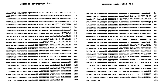
FIGURE 5: Nucleotide sequence of the TB-2 sub-
genomic DNA construct (single-stranded DNA in the
transcription ~] sense).
FI~URE 6: Schematic illustration of the
organisation of the TB-1 sub-genomic DNA construct. The
structure is represented as a double-stranded DNA
molecule which is suitable for cloning into the Bam Hl
site of a baculovirus expression vector. The R ribozyme
cleavage site are those which are active in the RNA
SUBSTITUTE SHEET
W093/OlX33 PCT/AU92/0036~
211 3572 24
transcript of this cloned DNA assuming transcription
occurs in the direction indicated.
FIGURE 7: Illustration of the sequence of the
transcript of the TB-l sub-genomic DNA construct
indicating the functional domains and the R ribozyme
cleavage site.
FIGURE 8: Nucleotide sequence of the TB-l sub-
genomic DNA construct (illustrated as a single-stranded
~NA in tha transcription [+] sense).
FIGURE 9: Nucleotide sequence of synthetic
oligonucleotides used for PCR in the construction of the
filler domain, the TB-2 sub-genomic DNA and the derived
TB-l sub-genomic DNA.
FI~URE 10: Autoradiograph of a 6% polyacrylamide-
urea ge- indica~ing the products of in vitro
transcription of uncut and Xba I-cut pTB2 DNA at 37C
using T3 RNA polymerase. Ribozyme cleavage products are
identified as bands A, B and C as described in the text
above.
FIGURE 11: Immunoblo~ of cell lysates and particle
preparations from mixed infections with recombinant
baculoviruses using polyclonal anti-rabies mouse serum as
described in the t~xt above. Lane ~: Lysate of cells
infected with wild type AcNPV; Lane 2: Lysate of cel~
in~ected with recombinant dual expression bac~loviruses
expressing rabie N/Ml proteins and M2/G proteins; Lane
3: Lysate of cells infected with recombinant single
expression baculoviruses expressing rabies N, M1, M2 and
G proteins~ Lane 4: Particles prepared from the culture
supernatant of cells infected with recombinant dual
expression baculoviruses expressing rabies N/Ml proteins
and M2/G proteins and recombinant baculovirus AcTB2
containing the TB-2 DNA construct; Lane 5: Par~icles
prepared from the culture supernatant of cells inferted
with recombinant dual expres~ion baculoviruses expressing
rabies N/M1 proteins and M2/G proteins.
FIGURE 12: Electron micrograph of rabies VLP
r~ ~_
~'
W093/01833 ~ 1 1 3 5 7 2 PCT/AU92/0036~
preparation. The sample was prepared as described in the
text above from the culture supernatant obtained from a
mixed infection of Spodoptera f~ugiperda cells with
recombinant baculovirus vectors expressing the rabies N,
Ml, M2 and G proteins and a recombinant baculovirus
expression vector Ac~TB2. As described in the text
above, the micrograph illustrates both rabies VLPs
(irregular par~icles 70-85 nm diameter, with surface
projections~ and particles of the recombinant
baculovirusas (rod-shaped particles 40 x 250 nm).
¦ SU~STITUTE SHEET