Note: Descriptions are shown in the official language in which they were submitted.
E69
45/10
2115171
DESCRIPTION
MONOCLONAL ANTIBODIES TO HUMAN PUT~MO~ARY SURFACTANT
-APOPROTEIN D AND USE THEREOF
Technical Field
The present invention relates to a monoclonal
antibody capable of specifically binding with human
pulmonary surfactant apoprotein D and also relates to use
thereof.
Background Art
The lung is a vascular cavity organ and has the
alveoli. The surfaces of the alveoli which take the
inhaled air directly are covered with the mantle called
the alveolar lining layer. The major components of the
alveolar lining layer consist of pulmonary surfactant
abundant in phospholipids.
The pulmonary surfactant is a lipoprotein
composed of about 10% proteins and phospholipids, the
components of which are mainly dipalmitoylphosphatidyl
choline and phosphatidyl glycerol (abbreviated as DPPC and
PG, respectively). The pulmonary surfactant exerts the
function in such a way that it reduces the surface tension
of the alveoli wherein phospholipids are aligned on the
surface at the air-fluid boundary. Therefore, for
2~1~171
example, the amount of the pulmonary surfactant in the
amniotic fluid is considered to reflect maturity of the
lung of the fetus.
The protein moiety in the pulmonary surfactant
is called pulmonary surfactant apoprotein, and there have
been hitherto known four (4) proteins of pulmonary
surfactant apoprotein family, i.e., A, B, C and D. Recent
studies have been clarifying that the protein moiety plays
an important role in exhibiting the function of pulmonary
surfactant, regulation of metabolism, defense mechanism in
the body, and the like.
As diseases in association with the pulmonary
surfactant, infant respiratory distress syndrome (IRDS),
adult respiratory distress syndrome (ARDS) and the like
have been reported. In the newborn infant with IRDS, the
pulmonary surfactant content in the alveoli decreases so
that the alveoli collapse. In such a case, the
respiratory function cannot be maintained to be stable.
Thus, by determining the pulmonary surfactant
content in the amniotic fluid prior to delivery, it may be
predicted whether the coming baby might suffer from IRDS.
When the fetus is suspected to suffer from IRDS,
immediately after birth, the newborn infant can be
medicated by liposome preparations of the surfactant.
In order to determine the pulmonary surfactant
content in the amniotic fluid, conventional methods have
been proposed wherein the proportion of lecithin to
211,~1 71
-- 3 --
sphingomyelin (L/S ratio) or the amount of DPPC has been
determined in consideration for phospholipids. However,
these methods encounter various problems and are still
unsatisfactory, due to low correlation to disease, lack of
quantitative determination, or difficulties in
operability.
To solve the problems in the prior arts which
have conventionally focused on phospholipids, it has been
attempted to detect or determine pulmonary surfactant by
taking notice of the protein moiety of pulmonary
surfactant, i.e., pulmonary surfactant apoprotein, and
using antibodies to the protein (cf., e.g., Japanese
Patent KOKAI (Laid-Open) Nos. 62-64956 and 4-9665, WO
89/02075, WO 89/01624).
As stated above, it is known that 4 kinds of
proteins A through D exist in human pulmonary surfactant
apoprotein. Human pulmonary surfactant apoprotein A is a
hydrophilic protein having a molecular weight of 28 to 38
kDa under a reduced condition, and participates
predo~in~ntly in regulation of pulmonary surfactant
metabolism. Human pulmonary surfactant apoproteins B and
C are hydrophobic proteins having molecular weights of 8
kDa under a reduced condition and 3 to 4 kDa under a
reduced condition, respectively. Both surfactant proteins
B and C play a role mainly in exhibiting the function of
pulmonary surfactant.
Human pulmonary surfactant apoprotein D is a
2~1~171
hydrophilic protein having a molecular weight of 43 kDa
under a reduced condition. While the function has not
been fully clarified, it has been reported that pulmonary
surfactant apoprotein D would have the action different
from those of pulmonary surfactant apoprotein A, B and C.
Furthermore, the change with time passage in pulmonary
surfactant apoprotein D content in the amniotic fluids is
also different from that of pulmonary surfactant
apoprotein A. In association with the function, research
interests have been directed to detection of pulmonary
surfactant apoprotein D in the lung tissues, and also
directed to quantitative determination of pulmonary
surfactant apoprotein D in blood, bronchoalveolar lavage
fluids and amniotic fluids, and accurate determination of
change with time passage in the pulmonary surfactant
apoprotein D content.
Therefore, an object of the present invention is
to provide a monoclonal antibody which makes it possible
to specifically detect or determine human pulmonary
surfactant apoprotein D.
Another object of the present invention is to
provide a method for specifically detecting or determining
human pulmonary surfactant apoprotein D and a kit for use
in the method, utilizing the monoclonal antibody thus
provided.
21 1~171
-- 5 --
Disclosure of the Invention
In order to achieve the foregoing objects, the
inventors have made extensive studies and succeeded in
efficiently obt~ining a monoclonal antibody for achieving
the above objects. Based on the finding that human
pulmonary surfactant apoprotein D can be specifically
detected or determined using the monoclonal antibody, the
present invention has thus been accomplished.
Therefore, as a first aspect, the present
invention is directed to a monoclonal antibody capable of
specifically binding with human pulmonary surfactant
apoprotein D.
As a second aspect, the present invention is
directed to a method for determination of human pulmonary
surfactant apoprotein D using the monoclonal antibodies as
reagents for the determination, and also directed to a kit
for use in the method.
As a third aspect, the present invention is
directed to a method for detecting the presence of human
pulmonary surfactant apoprotein D in human lung tissues
and a kit for use in the method, using the monoclonal
antibody as an antibody reagent.
Brief Description of the Drawings
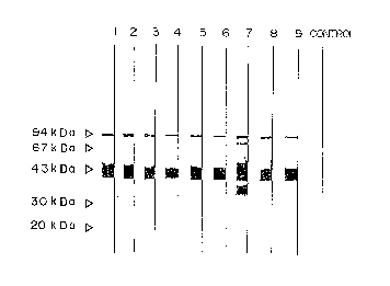
Fig. 1 shows specificity of the monoclonal
antibody in immunoblotting.
Fig. 2 shows reactivity of the monoclonal
211~171
-- 6
antibody, 6B2.
Fig. 3 shows reactivity of the monoclonal
antibody, 7C6.
Fig. 4 shows cross-reactivity of the monoclonal
antibodies, 6B2 and 7C62 in immunoblotting.
Fig. 5 shows cross-reactivity of the monoclonal
antibodies, 6B2 and 7C62 in the sandwich ELISA.
Fig. 6 shows a calibration curve in the sandwich
ELISA.
Fig. 7 shows the results of dilution test in the
sandwich ELISA.
Fig. 8 shows the results of pulmonary surfactant
apoprotein D (abbreviated as SP-D) levels in the amniotic
fluids from pregnant women which have been determined by
the sandwich ELISA.
Fig. 9 shows the results of SP-D levels in
bronchoalveolar lavage fluids from the patient with
pulmonary alveolar proteinosis and from healthy volunteers
which have been determined by the sandwich ELISA.
Fig. 10 shows the results of SP-D levels
determined by the sandwich ELISA in sera from patients
with various respiratory diseases including squamous cell
carcinoma, adenocarcinoma, sarcoidosis, tuberculosis,
pulmonary emphysema, bacterial pneumonia and small cell
carcinoma, and from healthy volunteers.
Fig. 11 shows the results of SP-D levels
determined by the sandwich ELISA in sera from patients
211~171
-- 7
with various respiratory diseases including idiopathic
interstitial pneumonia, interstitial pneumonia with
collagen disease, pulmonary alveolar proteinosis,
bronchial asthma, bronchiectasis, diffuse
panbronchiolitis, Hashimoto's disease and Basedow's
disease, and from healthy volunteers.
Fig. 12 shows the results obtained by
immunologically staining adenocarcinoma using the
monoclonal antibody, 6B2.
Fig. 13 shows the results obtained by
immunologically st~ining squamous cell carcinoma tissue
using the monoclonal antibody, 6B2.
Best Mode for Carrying Out the Invention
The present invention is described below in more
detail.
1. Monoclonal antibodies
(1) Properties
The monoclonal antibody of the present invention
is capable of specifically binding with human pulmonary
surfactant apoprotein D. The monoclonal antibody is not
limited by any other properties, but typically has the
properties as set forth hereinbelow. The use of such a
monoclonal antibody has enabled for the first time to
specifically detect or determine human pulmonary
surfactant apoprotein D in a sample solution.
(1)-1. Specificity
2115171
As shown in Examples herein, studies on the
specificity of the antibodies by immunoblotting reveal
that the monoclonal antibody reacts specifically with
human pulmonary surfactant apoprotein D.
(1)-2. Reactivity
As shown in Examples herein, studies on the
reactivity of the antibodies by ELISA reveal that the
monoclonal antibody reacts with human pulmonary surfactant
apoprotein D dependently on the concentration of the
antibody.
(1)-3. Cross-reactivity
As shown in Examples herein, studies on the
reactivity of the monoclonal antibody by immunoblotting
and sandwich ELISA reveal that the monoclonal antibody
does not substantially react with human-derived pulmonary
surfactant apoproteins A, B and C, or even if it reacts
with these apoproteins, there is no adverse influence on
measurement of human pulmonary surfactant apoprotein D.
(1)-4. Species specificity
As shown in Examples herein, studies on the
reactivity of the monoclonal antibody by immunoblotting
and sandwich ELISA reveal that the monoclonal antibody
does not substantially react with pulmonary surfactant
apoprotein D derived from other animal including rat, or
even if it reacts with these apoproteins, there is no
adverse influence on measurement of human pulmonary
surfactant apoprotein D.
211~171
(2) Production
The monoclonal antibody of the present invention
as described hereinabove can be produced in a conventional
manner. As an immunogen, human pulmonary surfactant
apoprotein D is used. The human pulmonary surfactant
apoprotein D can be prepared from, e.g., the
bronchoalveolar lavage fluids, preferably from the
bronchoalveolar lavage fluids of patients with pulmonary
alveolar proteinosis, according to the method of Persson
et al., J. Biol. Chem., 265, 5755, 1990 and the like.
There is no particular limitation to the purity degree of
the immunogen.
Alternatively, a peptide corresponding partially
to the amino acid sequence of human pulmonary surfactant
apoprotein D is chemically synthesized with a conventional
manner and the thus synthesized peptide may also be used
as the immunogen. Where the peptide synthesized has
merely a low antigenicity, the conjugate with a high
molecular carrier may be preferably used as the immunogen.
Such a carrier is conventionally used for preparing a
hapten antigen, and includes bovine serum albumin, Keyhole
limpet hemocyanin and the like. Furthermore, recombinant
human pulmonary surfactant apoprotein D prepared with a
recombinant DNA technology may also be used as an
immunogen.
An animal to which the immunogen is administered
may be any one of bovine, horse, sheep, goat, rat, mouse,
211~
-- 10 --
guinea pig, dog, swine, rabbit, monkey, pigeon, chicken,
and the like. Particularly, mouse, rat, guinea pig,
rabbit and goat are preferred.
The immunogen is administered to such an animal
in a conventional manner. For example, an emulsion is
prepared by mixing the immunogen with various adjuvants
such as complete Freund's adjuvant, incomplete Freund's
adjuvant, alum adjuvant, aluminum hydroxide adjuvant and
pertussis adjuvant. The emulsion is administered to the
animal intravenously, intraperitoneally, subcutaneously or
intracutaneously.
A preferred dose is in the range of from 0.01 to
10 mg protein/animal in the case of using rabbit and
guinea pig, and in the case of mouse and rat, in the range
of from 0.001 to 1 mg protein/animal.
After the first administration, the animal is
further boostered in the same dose as above approximately
1 to 5 times at the interval of 1 to 4 weeks to induce
production of an antibody to human pulmonary surfactant
apoprotein D. Then, antibody-producing cells such as
spleen cells, lymph node cells and peripheral blood
lymphocytes are collected from the antibody production-
induced animal in a conventional manner.
As myeloma cells used to fuse with the antibody-
producing cells, there may be used established cellsderived from various animals such as mouse, rat and human
which are readily accessible. Preferred cell lines used
21 1~171
-- 11
are those having such properties that they have a chemical
resistance, cannot survive in a selective medium in a non-
fused state but can survive only in the state fused with
the antibody-producing cells. In general, 8-azaguanine-
resistant cells are used. This cell line is deficient ofhypoxanthine guanine phosphoribosyl transferase and cannot
thus grow in hypoxanthine-aminopterin-thymidine (HAT)
medium. In terms of the cell properties, a cell line
which does not secrete immunoglobulin, so-called a non-
secretory cell line is preferred.
Specific examples of myeloma cell line includemyeloma cell line such as P3x63Ag8 (ATCC TIB-9) (Nature,
256, 495-497 (1975)), P3x63Ag8U.1 (P3Ul) (ATCC CRL-1597)
(Current Topics in Microbiology and Immunology, 81, 1-7
(1978)), P3x63Ag8.653 (ATCC CRL-1580) (J. Immunology, 123,
1548-1550 (1979)), P2/NSI/l-Ag4-1 (ATCC TIB-18) (European
J. Immunology, 6, 511-519 (1976)) and Sp2/0-Agl4 (ATCC
CRL-1581) (Nature, 276, 269-270 tl978)); rat myeloma cell
line such as 210.RCY.Agl.2.3 (Y3-Agl. 2. 3) (ATCC
CRL-1631) (Nature, 277, 131-133 (1979)); human myeloma
cell line such as U-266-ARl (Proc. Natl. Acad. Sci.
U.S.A., 77, 5429 (1980)), GM1500 (Nature, 288, 488 (1980))
and KR-4 (Proc. Natl. Acad. Sci. U.S.A., 79, 6651 (1982)).
For cell fusion, myeloma cells compatible with
the antibody-producing cells are chosen.
The cell fusion is carried out efficiently in a
conventional manner, e.g., by mixing 106 to 108 cells/ml of
211~ i 71
- 12 -
myeloma cells with the antibody-producing cells in a
mixing ratio of 1 : 4 to 10 and contacting the cells with
each other for 1 to 10 minutes at 37~C in animal cell
culture medium such as Eagle's minim~lm essential medium
(MEM), Dulbecco's modified Eagle~s medium (DMEM) and RPMI-
1640 medium. To accelerate the cell fusion, it is
advantageous to use a cell fusion accelerator such as
polyethylene glycol (PEG) having an average molecular
weight of 1,000 to 6,000, polyvinyl alcohol and Sendai
virus. The cell fusion between the antibody-producing
cells and myeloma cells may also be accelerated by means
of a commercially available cell fusion device utilizing
an electric pulse.
In order to select a desired hybridoma from the
cells after the cell fusion treatment, selective
proliferation of the cells in a selective medium can be
used. The selection is made, for example, by diluting the
cell suspension with 15% fetal calf serum (FCS)-cont~ining
RPMI-1640 medium and the like to an appropriate dilution
degree, inoculating the diluted suspension on a microplate
in approximately 103 to 106 cells/well, adding a selective
medium (e.g., HAT medium) to each well and then further
culturing by suitably exchanging the selective medium with
another. Where a 8-azaguanine-resistant strain is used as
the myeloma cell and HAT is used as the selective medium,
myeloma cells not fused are dead within ten (10) days from
the start of culture and the antibody-producing cells
211~171
- 13 -
which are normal cells cannot grow in vitro over long
periods of time. Thus, the cells which grow after 10 to
14 days can be acquired as the desired hybridoma cells.
- The hybridoma capable of producing the
monoclonal antibody which recognizes human pulmonary
surfactant apoprotein D can be surveyed by enzyme
immunoassay (EIA, ELISA), radioimmunoassay (RIA) and the
like. The survey can be made by, e.g., adding the culture
supernatants containing the monoclonal antibodies to 96
well microplates for ELISA, to which human pulmonary
surfactant apoprotein D has been previously adsorbed, to
react the monoclonal antibodies with human pulmonary
surfactant apoprotein D, then reacting the bound specific
antibodies either with enzyme-labeled anti-immunoglobulin
antibody or with biotin-labeled anti-immunoglobulin
antibody and then with enzyme-conjugated avidin D, and in
any case, then adding an enzyme substrate to each well to
form a color. By selecting the culture supernatant which
forms a color only in the human pulmonary surfactant
apoprotein D-adsorbed well, the desired hybridoma capable
of producing the desired antibody which specifically
reacts with human pulmonary surfactant apoprotein D can be
detected.
In the screening as described above, it is
preferred to use highly purified human pulmonary
surfactant apoprotein D. The monoclonal antibody of the
present invention can be efficiently screened by using
2115171
- 14 -
human pulmonary surfactant apoprotein D having a purity of
90% or more.
Cloning of the hybridoma can be performed by
limiting dilution method, soft agar method, fibrin gel
method, fluorescence-excited cell sorter method and the
like.
The monoclonal antibody can be produced from the
thus obtained hybridoma in a conventional manner such as
cell culture and ascites formation.
According to cell culture, the hybridoma is
cultured in a culture medium for animal cells, such as 10
to 15% FCS-containing RPMI-1640 medium and serum-free
medium in a conventional manner, and the monoclonal
antibodies can be collected from the culture supernatant.
In the method for recovering from the ascites,
mineral oil such as pristane (2,6,10,14-tetramethyl-
pentadecane) is intraperitoneally administered to animal
histocompatible with the hybridoma, and the hybridoma is
then intraperitoneally administered to the animal, e.g.,
in about 106 cells/animal in the case of mouse. The
hybridoma forms ascites tumor in about 10 to 18 days and
the antibodies are produced in a high level in serum and
the ascites.
Where it is required to purify the antibodies,
the monoclonal antibody may be purified by appropriately
choosing and combining therewith known methods such as
ammonium sulfate salting; ion exchange chromatography
2 ~ ~5 ~71 ~l
- 15 -
utilizing anionic exchangers such as DEAE cellulose;
affinity chromatography using protein A-Sepharose; and
molecular sieve chromatography.
2. Method for determination and a kit for use in the
method
The method for determination of human pulmonary
surfactant apoprotein D according to the present invention
is characterized by using the monoclonal antibody as
described above as a reagent. Principal, conditions and
the like of the deterrin~tion are not limited so long as
the monoclonal antibody according to the invention are
used as the reagent.
In terms of reaction mode for the determination,
a competitive reaction and a non-competitive reaction
(i.e., immunometric assay) are known. Any of those
reactions may be applied to the present invention.
In terms of detection, there are known a non-
labelling method (e.g., nephelometry) in which the result
of antigen-antibody reaction is directly detected, and a
labelling method in which the result may be detected using
any marker. Any of those methods may be adopted in the
present invention.
There are also known a heterogeneous method
which requires BF separation and a homogeneous method
which requires no BF separation. Any of those methods may
be adopted in the present invention.
In terms of reaction phase, there are known a
*Trademark
-
-
_ 16 -
liquid phase method in which an overall reaction proceeds
in the liquid phase and a solid phase method in which an
immune reaction is carried out after the one partner of
the immune reaction has been previously immobilized. Any
of those methods may be adopted in the present invention.
The determination method of the present
invention is performed by choosing from the various known
techniques as described above the one suitable method
depending on the purpose.
Details of these known techniques are described
in the following publications
(1) "ZOKU RADIOIMMUNOASSAY (Radioimmunoassay, Sequel)"
edited by Hiroshi Irie, published May 1, 1979 by
KODANSHA Publishing Co.
(2) "KOSO MENEKI SOKUTEI-HO (Enzyme immunoassay)" edited
by Eiji Ishikawa (second edition), published December
15, 1982 by IGARU SHOIN Publishing Co.
(3) "RINSHO BYORI (Clinical Pathology)'l, Extra Number,
Special Issue "RINSHO KENSA NO TAMENO IMMUNOASSAY -
GIJUTSU-TO-OYO (Immunoassay for Clinical Inspection -
Technique and Application)ll, published in 1983 by
RINSHO BYORI KANKOKAI
(4) "BIOTECHNOLOGY JITEN (Dictionary of Biotechnology)",
published October 9, 1986 by CMC
(5) Methods in ENZYMOLOGY Vol. 70 (Immunochemical
techniques (Part A))
,f. '~
211~171
- 17 -
(6) Methods in ENZYMOLOGY Vol. 73 (Immunochemical
techniques (Part B))
(7) Methods in ENZYMOLOGY Vol. 74 (Immunochemical
techniques (Part C))
(8) Methods in ENZYMOLOGY Vol. 84 (Immunochemical
techniques (Part D: Selected Immunoassay))
(9) Methods in ENZYMOLOGY Vol. 92 (Immunochemical
techniques (Part E: Monoclonal Antibodies and General
Immunoassay Methods))
(Articles (5)-(9) were published by Academic
Press)
The monoclonal antibody of the present invention
may be appropriately modified, if necessary and desired,
into a form suitable for practical use in the selected
method. Specific examples of such a form include a
labelled antibody, an immobilized antibody and the like.
The monoclonal antibody may be used without any
modifications, but in view of preventing non-specific
adsorption, it is desired to use an active fragment of the
monoclonal antibody.
The active fragment from the monoclonal antibody
may take any form so long as the fragment maintains the
characteristics of the monoclonal antibody. Such an
active fragment includes F(ab~ )2~ Fab~ and Fab. These
active fragments can be prepared by a known process such
as a restrictive digestion method wherein a purified
antibody is digested with a protease such as papain,
211~171
- 18 -
pepsin and trypsin (e.g., see "MENEKI SEIKAGAKU K~:~KYU-
HO - ZOKU SEIKAGAKU JIKKEN KOZA 5 (Immunobiological
Study - Sequel to Biochemical Experimental Lecture
Series), edited by Japanese Biochemical Association, 89
(1986)).
As a marker bound to the antibody, there may be
radioisotopes (e.g., 32p, 3H, 14C and l25I), enzymes (e.g.,
~-galactosidase, peroxidase, alkaline phosphatase,
glucose-6-phosphate dehydrogenase, catalase, glucose
oxidase, lactate oxidase, alcohol oxidase and monoamine
oxidase), coenzymes and prosthetic groups (e.g., FAD, FMN,
ATP, biotin and hem), fluorescein derivatives (e.g.,
fluorescein isothiocyanate and fluorescein thioflubamyl),
rhodamine derivatives (e.g., tetramethyl-rhodamine B
isothiocyanate), fluorescent dyes (e.g., umbelliferone and
l-anilino-8-naphthalenesulfonic acid), and luminol
derivatives (e.g., luminol, isoluminol and N-(6-
aminohexyl)-N-ethylisoluminol). These markers are used to
label the antibody or active fragment thereof, and the
labelling is performed in a conventional manner suitably
chosen from known techniques described in textbooks, e.g.,
~ZOKU SEIKAGAKU JIKKEN KOZA 5: MENEKI SEIKAGAKU K~KYu-HO
(Sequel to Biochemical Experimental Lecture Series -
Immunobiological Study), Tokyo Kagaku Dojin, 102-112,
(1986)).
For immobilizing the antibody, a carrier is
employed. Typical examples of the carrier include
21i~171
-- 19 --
synthetic organic high molecular compounds such as
polyvinyl chloride, polystyrene, styrene-divinylbenzene
copolymer, styrene-maleic anhydride copolymer, nylon,
polyvinyl alcohol, polyacrylamide, polyacrylonitrile,
polypropylene and polymethylene methacrylate;
polysaccharides such as agarose gel (e.g., Sepharose and
Biogel), cellulose (e.g., paper disc and filter paper);
and inorganic high molecular substances such as glass,
silica gel and silicone. These carriers may be introduced
with functional groups such as an amino, aminoalkyl,
carboxyl, acyl and hydroxy group, if desired. Substances
with a low ability of binding to a protein are preferred
as the carrier and in this regard, non-treated polystyrene
and polyvinyl chloride are advantageously used.
The carrier may take any shape selected from
plate-like (e.g., microtiter plate and disc), fibrous,
membrane-like, fine particulate (e.g., latex particles),
capsule-like, vesicular forms and the like. An
appropriate shape of the carrier can be chosen depending
upon assay mode to be practiced. Liposome (e.g., mono- or
multi-layer lipid membrane) may also be used as a carrier
for immobilizing the antibody.
The monoclonal antibody may be bound to the
carrier by conventional methods, e.g., through physical
adsorption, ionic bond, covalent bond, entrapping and the
like (see, e.g., KOTEIKA KOSO (Immobilized Enzyme), edited
by Ichiro Chihata, published March 20, 1975 by Kodansha
2115171
- 20 -
Publishing Co). Among them, physical adsorption is
preferred because of the convenience. The binding between
the monoclonal antibody and the carrier may be performed
either directly or indirectly through other substances.
There is no particular restriction to a sample
solution to be analyzed so long as the sample solution is
suspected to contain human pulmonary surfactant apoprotein
D therein. Typical examples of such a sample solution
include amniotic fluids, bronchoalveolar lavage fluids,
blood, serum and plasma.
The kit for use in the above assay method is
characterized in that the monoclonal antibody of the
present invention is contained as one of constituent
reagents for the kit. Other reagents for the kit may be
appropriately chosen depending upon assay method adopted.
Where a competitive reaction is adopted, the kit
comprises, for example:
(1) an immobilized antigen or antibody,
(2) a solution of a labeled antibody or antigen
and,
(3) a solution of an antigen having a known
concentration.
Where a sandwich method is adopted, the kit
comprises, for example:
tl) an immobilized first antibody,
(2) a solution of a second antibody,
(3) a solution of a labeled anti-immunoglobulin
2 ~ 7 1
- 21 -
antibody
and,
(4) a solution of an antigen having a known
concentration.
In the kits as described above, the terms
"antibody" and "antigen" are, of course, used to mean the
monoclonal antibody of the present invention and human
pulmonary surfactant apoprotein D, respectively. As is
recognized in the art, human pulmonary surfactant
apoprotein D is a polyvalent antigen, and in the kit for
use in the sandwich method, the "first antibody~ and the
"second antibody~ may be those that recognize the same or
different antigenic determinants on human pulmonary
surfactant apoprotein D.
A modified kit for use in the assay based on the
sandwich method comprises, for example:
(1) an immobilized first antibody,
(2) a solution of a labeled second antibody
and,
(3) a solution of an antigen having a known
concentration.
3. Method for detection and kit for use in the method
The characteristic feature for the method of the
present invention resides in using as an antibody reagent
the monoclonal antibody according to the present invention
when detecting pulmonary surfactant apoprotein D in the
211~
- 22 -
lung tissue. Therefore, any markers, labelling of
antibodies and methods for detection using labelled
antibodies which are conventionally employed in
immunological tissue diagnosis may also be applicable to
the present invention.
That is, the radioisotopes, enzymes, coenzymes
and prosthetic groups, fluorescent dyes, luminol
derivatives and the like as described hereinabove can be
used as markers. These markers may be bound either to the
monoclonal antibody itself or to the fragment. The
binding between the marker and the antibody or fragment
thereof is effected in a conventional manner. The
monoclonal antibody may also be indirectly labelled using
a labelled anti-immunoglobulin antibody and the like.
The labeled antibody thus prepared is reacted
with a lung tissue specimen in a conventional manner to
visualize the marker bound to the antibody, wherein
pulmonary surfactant apoprotein D in the lung tissue can
be detected.
In case that enzyme is used as a marker in the
assay method, the kit may comprise:
(1) an enzyme-labeled antibody
and
(2) a substrate solution.
When the biotin-avidin method is used, the kit
comprises:
(1) a biotinylated antibody,
211~171
- 23 -
(2) an enzyme-conjugated avidin
and,
(3~ a substrate solution.
In the kit as described above, the term
~antibody" means the monoclonal antibody of the present
invention.
The present invention is described below in more
detail by referring to examples.
ExamPle 1
Production of mouse monoclonal antibody to human pulmonary
surfactant apoprotein D (SP-D)
(1) Production of monoclonal antibody-producinq hybridoma
cells
Human SP-D prepared by known method (Persson, A.
et al., J. Biol. Chem., 265, 5755 (1990)) was dissolved in
physiological saline (0.4 mg/ml). The solution was mixed
with complete Freund's adjuvant in 1:1 proportion. The
resulting emulsion was intraperitoneally (i.p.)
administered to BALB/c mouse (female, age of 6 weeks) in a
dose of 20 ~g/100 ~l for initial immunization. After the
initial immunization, the animal was boostered (i.p.)
several times every other weeks in a similar manner. For
the final booster, a physiological saline solution of
human SP-D was intravenously (i.v.) administered to the
animal at the tail vein in a dose of 5 ~g/200 ~l.
Three days after the final booster, mouse spleen
211~171
- 24 -
was withdrawn and washed with RPMI-1640 medium to prepare
spleen cells suspension. At the same time, mouse myeloma
cells P3x63Ag8Ul (P3Ul) (ATCC CRL-1597~ were washed with
RPMI-1640 medium. After the spleen cells were mixed with
P3Ul in 10 : 1, the mixture was centrifuged and the
resulting pellets were gradually added with 1 ml of RPMI-
1640 medium cont~ining polyethylene glycol (PEG) 1000 to
perform cell fusion. RPMI-1640 medium was further added
to the system to make the volume 10 ml. The mixture was
then centrifuged and the resulting pellets were suspended
in RPMI-1640 medium containing 1% fetal calf serum (FCS)
in a concentration of 3 x 10⁴ cells/0.1 ml when counted as
P3Ul. The suspension was separately charged by 0.1 ml in
each well of 96-well microtiter plates. One day after,
0.1 ml each of hypoxanthine-thymidine-aminopterin-
cont~ining RPMI-1640 medium (HAT medium) was further added
to each well. Then the half volume of the medium was
replenished with fresh HAT medium every 3 or 4 other days.
Fourteen (14) days after the fusion, hybridoma
cells were screened. That is, human SP-D (10 ~g/ml) was
previously coated and 50 ~1 of the culture supernatant was
supplemented to each well of 96-well microtiter plates
blocked with PBS cont~ining 25% BLOCK ACE (manufactured by
Dainippon Pharmaceutical Co., Ltd.), followed by reacting
them at room temperature for an hour. After washing three
(3) times with 200 ~1 of PBS, 50 ~1 of biotinylated anti-
mouse IgG (manufactured by Vector Laboratories Inc.)
211~171
- 25 -
solution was added to the system followed by reacting at
room temperature for further an hour. After the reaction,
the system was washed three (3) times with PBS, 50 ~1 of
peroxidase-conjugated avidin D (manufactured by Vector
Laboratories Inc.) solution was added to react them at
room temperature for 30 minutes, the system was likewise
washed with PBS, and then 200 ~1 of substrate solution
(containing 0.25 mg/ml of 4-aminoantipyrine, 0.25 mg/ml of
phenol and 0.425 M hydrogen peroxide) was added to react
them at room temperature. By measuring absorbance at 550
nm, antibodies which specifically reacted with human SP-D
were detected and specific antibody-producing hybridoma
cells were selected as shown in Table 1.
Table 1
Number of
Specific Number of
Antigen Antibody- Wells where Number of
Positive Cells Grew Total Wells
Wells
Human SP-D 94 660 940
The thus selected hybridoma cells were cloned by
the limiting dilution method and nine (9) hybridoma cell
lines were established (lG11, 3E4, 3H4, 5A4, 6s2, 7A10,
7C6, 9E1 and lOH11). These hybridoma cells produced
antibodies to human SP-D which showed extremely high
specificity.
1 7 1
- 26 -
(2) Production and purification of monoclonal antibodies
These nine (9) hybridoma cells established were
intraperitoneally administered in 3 x 10⁶, respectively, to
mouse previously treated with 0.5 ml of pristane. About
two (2) weeks after, the ascites was collected. Then the
ascites was subjected to affinity chromatography on
Protein A-Sepharose CL4B column to purify IgG from the
fluid. First, 20 ml of Protein A-Sepharose CL4B
(manufactured by Pharmacia Biotech AB) was packed in a
glass column of 1.5 x 20 cm and then equilibrated with 1.5
M glycine buffer solution (pH 8.9) containing 3M sodium
chloride. Then, the ascites was diluted to 2-fold with
the equal volume of the glycine buffer solution. The
diluted ascites was passed through the column. After
washing and removing non-adsorbed protein with a
sufficient volume of the glycine buffer solution, the
adsorbed IgG was eluted with 0.1 M citrate buffer solution
(pH 3.0). The thus obtained IgG fraction was immediately
dialyzed against PBS overnight to avoid denaturation.
Example 2
Analyses (1) of monoclonal antibodies on properties
(survey of class and type):
After human SP-D (10 ~g/ml) was coated, the
culture supernatants of each hybridoma cell or each
solution of the purified monoclonal antibodies was added
to 96-well microtiter plates blocked with PBS containing
25% BLOCK ACE (manufactured by Dainippon Pharmaceutical
Co., Ltd.). Class and type of the antibodies were
identified using MonoAb-ID EIA ~it (manufactured by Zymed
Laboratories Inc.). The results are shown in Table 2.
Table 2
Clone IgG Class/Type
IGll IgGl /K
3E4 IgGl/K
3H4 IgGl/~
5A4 IgG2a/K
6B2 IgGl/K
7A10 IgGl /K
7C6 IgG1/K
9E1 IgG1/K
lOH11 IgG1 /K
Example 3
Analyses (2) of monoclonal antibodies on properties
(antigenic specificity of monoclonal anti-human SP-D
antibody):
The antigenic specificity of the monoclonal
antibodies produced by the hybridoma cells (i.e., lGll,
3E4, 3H4, 5A4, 6B2, 7A10, 7C6,-9El and lOHll) were
verified by immunoblotting using sodium dodecyl sulfate-
polyacrylamide gel electrophoresis (SDS-PAGE) and by
enzyme immunoassay (ELISA). The name of each monoclonal
antibody is the same as the hybridoma producing the
*TMC1~m~rk
7 ~
- 28 -
antibody.
(A) Analyses of specificity by immunoblottinq
SDS-PAGE was performed by the method of Laemmli
(Nature, 227, 680, 1972). The basic procedures of
immunoblotting are as follows. That is, the separated gel
obtained by SDS-PAGE was laid on nitrocellulose membrane.
By applying 60V for 12 hours thereto, protein was
transferred onto the nitrocellulose membrane. The thus
obtained nitrocellulose membrane was cut into strips along
the moving line of a sample solution. A part of the
membrane strips was used to stain proteins with Amide
Black. The other membrane strips were immersed at 37~C
for an hour in PBS containing 0.5% Triton X-100 and 2%
skimmed milk for blocking and reacted at room temperature
for an hour with the monoclonal antibody solution
appropriately diluted with PBS. After washing with PBS,
the nitrocellulose membrane was reacted with peroxidase-
labelled anti-mouse IgG antibody at room temperature for
an hour. The nitrocellulose membrane was washed likewise
and reacted with a substrate solution (con~ining 30 mg of
COLOR DEVELOPER manufactured by Bio-Rad Laboratories Inc.,
10 ml of methanol, 50 ml of PBS and 30 ~l of 30% hydrogen
peroxide). At the time when a color was appropriately
,
formed, the system was washed with water to tërmin~te the
reaction.
Human SP-D shows a molecular weight of 43 kDa
under a reduced condition. After the human SP-D fraction
*Tr~ m~rk
211~171
- 29 -
was electrophoresed under a reduced condition, the
reaction specificity of each monoclonal antibody was
ex~mined by immunoblotting. As shown in Fig. l, the nine
(9) monoclonal antibodies (i.e., Lane 1: lG11, Lane 2:
3E4, Lane 3: 3H4, Lane 4: 5A4, Lane 5: 6B2, Lane 6: 7A10,
Lane 7: 7C6, Lane 8: 9El and Lane 9: lOH11) all showed
extremely strong reactivity with human SP-D having a
molecular weight of 43 kDa. Furthermore, the monoclonal
antibody 7C6 showed extremely strong reactivity also with
the degradation product of human SP-D having a molecular
weight of about 38 kDa which was considered to be by-
produced at the preparation process.
(B) AnalYses of sPecificity by ELISA
A solution of purified human SP-D in 5 mM Tris
(1.0 ~g/ml, pH 7.4) was added by 50 ~l to each well of 96
well microtiter plates. After allowing to stand at 4~C
overnight, the plates were washed three (3) times with
PBS. Each well was added with 200 ~l of PBS cont~ining
0.5~ Triton X-100 and 2~ skimmed milk. The plates were
allowed to react at room temperature for an hour to effect
blocking. After washing three (3) times with PBS, 50 ~l
each of the monoclonal antibody solution was added to
react at room temperature for an hour. After washing
three (3) times with 200 ~l of PBS, 50 ~l of biotinylated
anti-mouse IgG (manufactured by Vector Laboratories Inc.)
solution was added to the system followed by reacting at
room temperature for further an hour. After the reaction,
211~171
- 30 -
the system was washed three (3) times with PBS and 50 ~l
of peroxidase-conjugated avidin D (manufactured by Vector
Laboratories Inc.) solution was added to react them at
room temperature for 30 minutes. The reaction mixture was
likewise washed with PBS and 100 ~l of substrate solution
(0.1 M citrate buffer solution, pH 5.9, cont~ini~g 0.2
mg/ml of o-phenylenediamine and 0.425 M hydrogen peroxide)
was added to react them at room temperature. After adding
100 ~l of 2 N sulfuric acid to terminate the reaction, the
absorbance was measured at 492 nm. As shown in Figs. 2
and 3, the reactivity of the monoclonal antibodies 6B2 and
7C6 with human SP-D (1.0 ~g/ml) was dependent on the
concentration of the antibodies. While the results are
not shown, similar phenomena were confirmed with other
monoclonal antibodies lG11, 3E4, 3H4, 5A4, 7A10, 9E1 and
lOH11.
Example 4
Analyses (3) of monoclonal antibodies on properties
(cross-reactivity of monoclonal anti-human SP-D antibody)
The cross-reactivity of the monoclonal
antibodies 6B2 and 7C6 produced by the hybridoma cells
with human SP-A and rat SP-D was examined by
immunoblotting using SDS-PAGE and sandwich ELISA.
Human and rat SP-D were prepared by the Persson
et al. method suPra and human SP-A was prepared by the
method of Kuroki et al., Proc. Natl. Acad. Sci. U.S.A.,
85, 5566, 1988.
2115171
(A) Analyses of cross-reactivity by immunoblottinq
Human SP-D fraction, human SP-A fraction, rat
SP-D fraction, and human amniotic fluids (38 weeks of
pregnancy) were subjected to electrophoresis under a
reduced condition and the cross-reactivity of the
monoclonal antibodies was examined by immunoblotting.
Based on the results of blotting (Lane 1: human
SP-D, Lane 2: human SP-A, Lane 3: rat SP-D, Lane 4: human
amniotic fluids), the respective monoclonal antibodies 6B2
and 7C6 showed extremely strong reactivity with human SP-D
having a molecular weight of 43 kDa, protein of about 90
kDa which was considered to be a dimer of human SP-D, and
SP-D in human amniotic fluids. Furthermore, the
monoclonal antibody 7C6 showed extremely strong reactivity
also with the degradation product of human SP-D having a
molecular weight of about 38 kDa which was considered to
be by-produced at the preparation process. On the other
hand, the both monoclonal antibodies did not show any
reactivity at all with human SP-A having a molecular
weight of approximately 26 to 38 kDa under a reduced
condition. The monoclonal antibodies only slightly
reacted with human SP-D having a molecular weight of 43
kDa which was considered to be intermingled in the human
SP-A fraction in a trace amount. The both monoclonal
antibodies did not show significant reactivity with rat
SP-D (having a molecular weight of about 43 kDa under a
reduced condition, as shown in Fig. 4.
211~i171
- 32 -
(B) Analyses of cross-reactivity by sandwich ELISA
The cross-reactivity of the monoclonal
antibodies 6B2 and 7C6 of the invention to human SP-A and
rat SP-D was examined by sandwich ELISA using the
monoclonal antibodies. The basic procedures of sandwich
ELISA are as follows.
A PBS solution (10 ~g/ml) of each monoclonal
antibody was added by 50 ~l to each well of 96 well
microtiter plates. After allowing to stand at 4~C
overnight, the plates were washed three ( 3) times with
PBS. Each well was added with 200 ~1 of PBS containing
0.5% Triton X-100 and 2% skimmed milk. The plates were
allowed to stand for an hour at room temperature for
blocking. After washing three ( 3) times with PBS, 50 ~l
of a solution of human SP-D in PBS was added to each well.
The plates were allowed to stand at 4~C overnight. After
washing three (3) times with 200 ~1 of PBS, 50 ~l of 10
~g/ml PBS solution (containing 0.5% Triton X-100 and 0.1%
skimmed milk) of biotinylated monoclonal antibody 7C6 was
added to the system followed by reacting at room
temperature for 4 hours. After the reaction, the system
was washed three (3) times with PBS and 50 ~l of
peroxidase conjugated avidin D (manufactured by Vector
Laboratories Inc.) solution was added to react them at
room temperature for 30 minutes. The reaction mixture was
likewise washed with PBS and 100 ~l of substrate solution
(0.2 M citrate buffer solution, pH 3.8, cont~ining 3 mM
1 7 1
- 33 -
3,3',5,5'-tetramethylbenzidine and 0.005% hydrogen
peroxide) was added to react them at room temperature.
After adding 100 ~1 of 2 N sulfuric acid to terminate the
reaction, the absorbance was measured at 450 nm.
The cross-reactivity (%) was calculated
according to the following equation.
Cross-reactivity (%)
human SP-D level for achieving 50% binding
property (corresponding to absorbance of 0.6)
(50 ng/ml)
x 100
level of analogous substance for achieving 50%
binding property (corresponding to absorbance of
0.6) (ng/ml)
The results indicate that the cross-reactivity
to human SP-A was less than 0.2% and that to rat SP-D was
less than 0.25% as shown in Fig. 5 and Table 3.
According to analyses of the cross-reactivity by
the aforesaid immunoblotting using the same samples, the
monoclonal antibodies 6B2 and 7C6 did not show any cross-
reactivity at all with human SP-A of 26 to 38 kDa, but
reacted with SP-D which was considered to be present in
the SP-A fraction in a trace amount. It is thus
considered that the actual cross-reactivity to human SP-A
would be much less.
2115171
- 34 _
Table 3
AnalogousConcentration for
SubstanceAchieving 50% Cross-reactivity
binding (ng/ml) (%)
Human SP-D 50 100
Human SP-A >25,000 <0.2
Rat SP-D >20,000 <0.25
(C) As stated above, the monoclonal antibodies
of the present invention react specifically with human SP-
D. By using immobilized monoclonal antibody 7C6 in
combination with horseradish peroxidase-labelled
monoclonal antibody 6B2 prepared by the method of Nakane
et al., Immunoassays in the clinical laboratory, Alan R.
Liss Inc., New York, 81, 1979, high sensitive sandwich
ELISA specific to human SP-D was established. The basic
procedure of sandwich ELISA are as follows.
A PBS solution (10 ~g/ml) of the monoclonal
antibody 7C6 was added by 100 ~l to each well of 96 well
microtiter plates. After allowing to stand at 4~C
overnight, the plates were washed three (3) times with
PBS. Each well was added with 200 ~l of PBS containing 1%
bovine serum albumin (BSA). The plates were allowed to
stand for an hour at room temperature for blocking. After
washing three (3) times with PBS, each well was added with
100 ~l of a solution of human SP-D in PBS containing 0.5%
- 211~171
- 35 -
Triton X-100. The plates were allowed to react at room
temperature overnight. After washing three (3) times with
200 ~l of PBS, 100 ~l of 2 ~g/ml PBS solution (containing
0.5% Triton X-100 and 1% BSA) of horseradish peroxidase-
labelled monoclonal antibody 6B2 was added to each well toreact at room temperature for 2 hours. After the
reaction, the plates were washed three (3) times with PBS
and, 100 ~l of a substrate solution (0.2 M citrate buffer
solution, pH 3.8, containing 0.3 mM 3,3',5,5'-
tetramethylbenzidine and 0.005% hydrogen peroxide) wasadded to react them at room temperature for 20 minutes.
Then 100 ~l of 2 N sulfuric acid was added to terminate
the reaction and, the absorbance was measured at 450 nm.
(1) Calibration curve
Sandwich ELISA was performed using as a standard
substance 0.5% Triton X-lO0-containing PBS solution of
purified human SP-D prepared by the method of Persson et
al. supra. As the result, an excellent calibration curve
dependent on concentration of human SP-D was obtained in
the range of 3.13 ng/ml to 200 ng/ml, as shown in Fig. 6.
(2) RecoverY test and dilution test
For the purpose of evaluating the basic
efficiency of sandwich ELISA according to the present
invention, a test of adding purified human SP-D to human
amniotic fluids and recovering the added human SP-D from
the fluids was carried out. A dilution test of human
amniotic fluids was also carried out. In the recovery
211~171
test, 0.5% Triton X-100-containing PBS solution of
purified human SP-D (0, 12.5, 25, 50 ng/ml) was added to
human amniotic fluid samples and the human SP-D was
assayed by sandwich ELISA. The results reveal that the
purified human SP-D was recovered with a good recovery
rate (i.e., 94.4 to 111.2%) as shown in Table 4. In the
dilution test using human amniotic fluid samples, an
excellent linear relationship was noted between the
dilution magnification of the sample and the concentration
of SP-D determined by ELISA, as shown in Fig. 7.
Table 4
Sample Amount of
Amount of Data SP-D Recovery
SP-D Added Observed Recovered Rate (%)
(ng/ml) (ng/ml) (ng/ml)
1 0 8.3
12.5 20.4 12.1 96.8
25.0 31.9 23.6 94.4
50.0 56.0 47.7 95.4
2 0 14.0
12.5 27.4 13.4 107.2
25.0 38.4 24.4 97.6
50.0 62.4 48.4 96.8
3 0 35.1
12.5 49.0 13.9 111.2
25.0 59.8 24.7 98.8
50.0 82.4 47.3 94.6
211~171
- 37 -
(3) Cross-reactivity
In order to explore the cross-reactivity of
sandwich ELISA to a substance analogous to human SP-D,
serial dilutions of human SP-A, rat SP-D and human SP-D
for control were prepared, and the reactivity was
examined. The cross-reactivity was also calculated in
accordance with the following equation.
Cross-reactivity (%)
human SP-D level for achieving 50% binding
property (60 ng/ml)
x 100
level of analogous substance for achieving 50%
binding property (ng/ml)
The results indicate that the cross-reactivity
to human SP-A was less than 0.3% and that to rat SP-D was
0.6%, as shown in Table 5.
According to analyses of the cross-reactivity by
the aforesaid immunoblotting using the same samples, the
monoclonal antibodies 6B2 and 7C6 did not show any cross-
reactivity at all with human SP-A of 26 to 38 kDa but
reacted with human SP-D which was considered to be present
in the SP-A fraction in a trace amount. It is thus
considered that the actual cross-reactivity to human SP-A
would be much less.
211~171
- 38 -
Table 5
Concentration for
Analogous achieving 50% Cross-reactivity
Substance binding (ng/ml) (%)
Human SP-D 60 100
Human SP-A >20480 <0.3
Rat SP-D 10240 0.6
Example 5
Determination of SP-D levels in vital samples
Following the sandwich ELISA in Example 4 (c),
the SP-D levels were determined in the amniotic fluids
from healthy normal pregnant women, in the bronchoalveolar
lavage fluids obtained from the patients with pulmonary
alveolar proteinosis, and in sera collected from the
patients with interstitial pneumonia, pulmonary alveolar
proteinosis and other respiratory diseases.
(A) SP-D levels in the amniotic fluids from healthy normal
preqnant women
The concentrations of SP-D in 21 amniotic fluid
specimens obtained from healthy normal pregnant women of
26 to 40 weeks were determined. The results indicate that
the SP-D concentrations in 13 amniotic fluids from mid-
trimester pregnancies (over 30 weeks of gestation) were
significantly higher than those of 8 amniotic fluids from
late pregnancies (less than 30 weeks of gestation), as
21 1
- 39 -
shown in Fig. 8. The results indicate that SP-D levels in
amniotic fluids will reflect maturity of fetal lung and
also suggest the possibility that determination of SP-D
levels in amniotic fluids would be useful for diagnosis of
IRDS in fetus.
(B) SP-D levels in bronchoalveolar lavaqe fluids
With respect to bronchoalveolar lavage fluids of
the patients with pulmonary alveolar proteinosis (13
cases), idiopathic pulmonary fibrosis (IPF), and
sarcoidosis (Sar), or that of healthy volunteers (13
cases), the SP-D levels were assayed. The results
indicate that the SP-D levels were obviously higher in the
patients with pulmonary alveolar proteinosis as compared
to the healthy volunteers, as shown in Fig. 9. This
suggests that determination of the SP-D levels in
bronchoalveolar lavage fluids would be effective for
diagnosis of patient with pulmonary alveolar proteinosis.
In the patients with IPF and Sar, the SP-D levels were
almost the same as that of the healthy volunteers.
(C) SP-D levels in sera obtained from Patients with
various resPiratorY diseases
The SP-D levels were assayed in sera obtained
from patients with various respiratory diseases such as
idiopathic interstitial pneumonia, interstitial pneumonia
with collagen disease, pulmonary alveolar proteinosis,
squamous cell carcinoma, adenocarcinoma, small cell
carcinoma, sarcoidosis, tuberculosis, pulmonary emphysema,
2 1 ~
- 40 -
bacterial pneumonia, bronchial asthma, bronchiectasis and
diffuse panbronchiolitis, and for control, sera from
healthy volunteers. As the result, the SP-D levels were
obviously higher in the patients with interstitial
pneumonia such as idiopathic interstitial pneumonia and
interstitial pneumonia with collagen disease, and with
pulmonary alveolar proteinosis, as compared to that of
healthy volunteers, as shown in Figs. 10 and 11. It is
thus demonstrated that it would be effective for judgment
and diagnosis of the various diseases described above to
determine the SP-D levels in sera.
Example 6
Immunologically st~ining of tissues
Lung cancer tissues (i.e., adenocarcinoma,
squamous cell carcinoma, large cell carcinoma, small cell
carcinoma) obtained by surgical operation and autopsy and
cancer tissues of other organs were fixed with formalin
and embedded in paraffin. Then immunohistological
examination was performed according to ABC method.
That is, after sufficiently removing the
paraffin with xylene, tissue slices were hydrated by
changing an ethanol concentration stepwise and then washed
with water. Next, the slices were immersed at room
temperature for 30 minutes in methanol containing 0.3%
hydrogen peroxide thereby to remove the endogenous
peroxidase activity, and then in PBS for 5 minutes for
rinsing. This lavage procedure was repeated three (3)
211~171
- 41 -
times. Next, the slices were immersed in 10% horse serum-
containing PBS at room temperature for 30 minutes to
perform blocking. Thereafter, the monoclonal anti-human
SP-D antibody or monoclonal anti-human SP-A antibody
appropriately diluted with PBS was dropped onto the slices
to react them at room temperature for 30 minutes. The
slices were then washed with PBS in a similar manner.
Next, biotinylated anti-mouse IgG antibody (manufactured
by Vector Laboratories Inc.) was dropped on the slices to
react at room temperature for 30 minutes. The slices were
then washed with PBS likewise. Subsequently, ABC Reagent
(manufactured by Vector Laboratories Inc.) was dropped
onto the slices to cause reaction at room temperature for
30 minutes. After washing three (3) times with PBS,
peroxidase substrate solution (manufactured by Vector
Laboratories Inc.) was dropped on the slices to cause
reaction at room temperature. At the time when a color
was appropriately formed, the slices were washed to
terminate the color-forming reaction. The slices were
then sealed.
As the result, SP-D was positive in 25 out of 36
cases with adenocarcinoma and 4 out of 5 cases with
squamous cell carcinoma, respectively, whereas SP-D was
negative in all other cases of tissue type lung cancer and
cancers of other organs. With regard to SP-A levels
tested simply for reference, SP-A was positive in 18 out
of 36 cases with adenocarcinoma and, at least one of SP-D
21 1 ~
- 42 -
and SP-A was positive in 31 out of 36 cases. From the
foregoing results it was confirmed that use of the
antibodies to SP-D and SP-A in combination improved a
positive rate and was thus more useful for diagnosis of
lung-primary adenocarcinoma. Typical examples of the
immunologically stained lung tissue of adenocarcinoma and
squamous cell carcinoma using the monoclonal anti-human
SP-D antibody 6B2 are shown in Figs. 12 and 13.
Industrial Applicability
The monoclonal antibody of the present invention
is capable of specifically binding with human pulmonary
surfactant apoprotein D. By using such a monoclonal
antibody as an antibody reagent, human pulmonary
surfactant apoprotein D can be specifically detected or
determined for the first time. The monoclonal antibody of
the present invention is therefore useful as a tool for
clarifying the function of human pulmonary surfactant
apoprotein D. In addition, the method for detection or
determination of human pulmonary surfactant apoprotein D
and the kit for use in the method are useful for diagnosis
of respiratory diseases such as IRDS, ARDS, pulmonary
alveolar proteinosis, interstitial pneumonia,
adenocarcinoma and squamous cell carcinoma.