Note: Descriptions are shown in the official language in which they were submitted.
i'"v WO 93/10814 PCT/US92/10309
CA2111303
1
Anti-cancer immunotherapeutic vector constructs.
~ 5
~'echnical Field
The present invention relates generally to the field of anti-cancer
immunotherapy, and more specifically, to methods of killing selected tumor
cells
by generating an immune response agains~ °he tumor cells.
ground of the Invention
Cancer accounts for one-fifth of the total mortality in the United
States, and is the second leading cause of death. Cancer is typically
character-
ized by the uncontrolled division of a population of cells. This uncontrolled
division typically leads to the formation of a tumor, which may subsequently
metastasize to other sites.
Primary solid tumors can generally be treated adequately by
surgical resection. However, the majority of patients which present with solid
tumors also possess micrometastases beyond the primary tumor site. If treated
with surgery alc , approximately 70% of these patients will experience recur-
rence of the cancer. In addition to surgery, many cancers are now also treated
with a combination of therapies involving cytotoxic chemotherapeutic drugs
(e.g., vincristine, vinblastine, cisplatin, etc.) and/or radiation therapy.
One
difficulty with this approach, however, is that radiotherapeutic and
chemothera-
peutic agents are toxic to normal tissues, and often create life-threatening
side
effects. In addition, these approaches often have extremely high
failure/remission rates (up tc )% depending upon the type of cancer).
In addition to chemo- and radiation therapies, many have
attempted to bolster or augment an individual's own immune system in order ~o
eliminate the cancer cells. Several immunotherapies have utilized bacterial or
viral components in order to stimulate the immune system to destroy the tumor
cells. Examples of such components include immunomodulatory agents (such as
BCG, endotoxin, and mixed bacterial vaccines), interferons (a, ~, and 7),
inter-
feron inducers (e.~ , Brucella abortus, and various viruses), and thymic
factors
. 35 (eg., thymosin fraction ~, and thymosin alpha-1) (see generally
"Principles of
Cancer Biotherapy," Oldham (ed.), Raven Press, New York, 1987). Such agents
WO 93/ 10814 0 A 21 1 l 3 0 3
PCT/US92/1
2
have generally been useful as adjuvants and as nonspecific stimulants in
animal
tumor models, but have not yet proved generally effective in humans.
Lymphokines have also been utilized in the treatment of cancer.
Briefly, lymphokines are secreted by a variety of cells, and generally have an
effect on specific cells in the generation of an immune response. Examples of
lymphokines include Interleukins (IL)-1, -2, -3, and -4, as well as colony
stimu
lating factors such as G-CSF, GM-CSF, and M-CSF. Recently, one group has
utilized IL-2 to stimulate peripheral blood cells in order to expand and
produce
large quantities of cells which are cytotoxic to tumor cells (Rosenberg et
al., N.
Eng~ .l. Med 313:1485-1492, 1985).
Others have suggested the use of antibody-mediated anti-cancer
therapies. Briefly, antibodies may be developed which recognize certain cell
surface antigens that are either unique, or more prevalent on cancer cells
compared to normal cells. These antibodies, or "magic bullets," may be
utilized
either alone or conjugated with a toxin in order to specifically target and
kill
tumor cells (Dillman, "Antibody Therapy," Principles of Cancer Biotherapy,
Oldham (ed.), Raven Press, Ltd., New York, 1987). For example, Ball et al.
(Blood 62:1203-1210, 1983) treated several patients with acute myelogenous
leukemia with one or more of several monoclonal antibodies specific for the
leukemia, resulting in a marked decrease in circulating leukemia cells during
treatment. Similarly, others have used toxin-conjugated antibodies therapeuti-
cally to treat a variety of tumors, including, for example, melanomas,
colorectal
carcinomas, prostate carcinomas, breast carcinomas, and lung carcinomas (see
Dillman, supra). One difficulty however, is that most monoclonal antibodies
are
of murine origin, and thus hypersensitivity against the murine antibody may
limit its efficacy, particularly after repeated therapies. Common side effects
include fever, sweats and chills, skin rashes, arthritis, and nerve palsies.
Therefore, agents which can augment natural host defences
against tumor induction or progression may increase remission rates and
enhance survival of patients, without the cytotoxic side effects of prior
methods.
The present invention provides such agents, and further provides other related
advantages.
Summary of the Invention
The present invention provides methods for destroying selected
tumor cells with an altered cellular component which is normally associated
with the selected tumor cells. Within one aspect, a method is provided for
,~" WO 93/10814 C A 21 17 3 0 3 PGT/US92/10309
3
destroying selected tumor cells comprising the step of administering to a warm-
blooded animal a vector construct which directs the expression of at least one
' immunogenic, non-tumorigenic form of an altered cellular component normally
associated with the selected tumor cells. Within another aspect of the
invention,
' S a method is provided for destroying selected tumor cells in a warm-blooded
animal comprising the steps of (a) removing cells from a warm-blooded animal,
(b) administering to the removed cells a vector construct which directs the
expression of at least one immunogenic, non-tumorigenic form of an altered
cellular c; nponent normally associate : with the selected tumor cells, and
(c) returning the cells to a warm-blooded animal, such that the selected tumor
cells are destroyed. As will be evident to one of ordinary skill in the art,
the
animal from which the cells are removed need not be the same animal to which
they are returned, although preferably, they should be histocompatible. In
addi-
tion, it should be understoo- ° that within the context of the present
invention
when reference is made to a a:ral construct which "expresses" any substance in
a
cell, that this in fact refers to protein production of the resulting provirus
following reverse transcription of the viral RNA into the cell. Within various
embodiments of the invention, the vector construct may be carried by a recom-
binant retrovirus, or by other recombinant viruses such as those selected from
the group consisting of adeno-associated virus, canary pox virus, adenovirus,
and
pox virus. Alternatively, an immunogenic form of an altered cellular component
may be manufactured in vitro, and given to patients with an appropriate
adjuvant, preferably one which leads to induction of cellular immunity.
Within another aspect of the present invention, a vector construct
is provided which directs the expres~on of at least one immunogenic, non
tumorigenic form of an altered cellular component. Within various embodi
ments, the cellular component may be altered by a point mutation, by a
deletion, or by a chromosomal translocation. Within other embodiments, the
altered cellular components include, ras*, p53*, Rb*, altered protein encoded
by the Wilms' tug ~v.r gene, ubiquitir~ *, DCC, APC, MCC, neu, an altered
~ receptor, or polyt~ides resulting from chromosomal translocations such as
bcr/abl. Withir. mother embodiment, non-tumorigenic altered cellular
~ components are provided, including for example, D ras'12, D ras'~, and D
ras'61.
Within other aspects of the invention, the altered cellular component is
mucin*.
Also. provided are vector constructs which direct the expression of several
altered cellular components, including, for example, a vector construct which
WO 93/ 10814
PCT/US92/lf
4
directs the expression of both ras' and pS3*, or a vector construct which
directs
the expression or ras*, mucin*, and DCC.
Within another aspect of the invention, recombinant reuoviruses
as well as other recombinant viruses, such as polioviruses, rhinoviruses, pox
viruses, adenoviruses, parvoviruses, herpes viruses and sindbis viruses, are
provided for carrying the above-described vector constructs. Target cells
infected with these recombinant viruses are also provided, including, for
example, embodiments wherein the target cells are selected from the group
consisting of human, macaque, dog, rat, and mouse cells.
Also provided are pharmaceutical compositions comprising the
above-described recombinant viruses, in combination with a pharmaceutically
acceptable carrier or diluent.
These and other aspects of the present invention will become
evident upon reference to the following detailed description and attached
drawings.
Brief Description of the Drawines
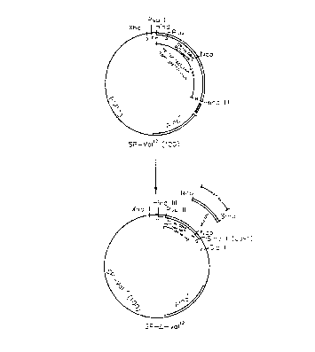
Figure 1 is a schematic illustration which outlines the construction
of the plasmid SP-Val~(100).
Figure 2 is a schematic illustration which outlines the construction
of the plasmid SP-D-Va112.
Figure 3 is a schematic illustration which outlines the construction
of the plasmid N2-ras-Va112.
Figure 4 is a schematic illustration which outlines the construction
of the plasmid N2-A-ras-Val~.
Figure ~ is a schematic illustration of mucin cDNA cloned into the
KT-3 retroviral backbone.
Figure 6 is a Western Blot which illustrates the expression of
mucin from various cell types.
Figure 7 is a graph which illustrates the CTL response for several
BC10ME clones.
Figure 8 is a FACS analysis of mucin expression on several
different BC10ME clones.
Figure 9 is a bar graph which illustrates the tumorigenicity of two
B16F10 clones.
Figure 10 is a bar graph which illustrates protection in mice
injected with the B16F10-Mucin clone.
CA 02117303 2000-07-14
5
Detailed Description of the Invention
Prior to setting forth the invention, it may be helpful to an understanding
thereof
to first set forth definitions of certain terms that will be used hereinafter.
"Altered Cellular Component" refers to proteins and other cellular
constituents
which are either associated with rendering a cell tumorigenic, or are
associated with
tumorigenic cells in general but are not required or essential for rendering
the cell tumorigenic.
Within one aspect of the invention, before alteration of the cellular
component, the cellular
component may be essential to normal cell growth and regulation. Examples
include proteins
which regulate intracellular protein degradation, transcriptional regulation,
cell-cycle control,
and cell-cell interaction. After alteration, the cellular components no longer
perform their
regulatory functions, and hence the cell may experience uncontrolled growth.
Representative
examples of such altered cellular components include ras*, p53*, Rb*, altered
protein encoded
by the Wilms' tumor gene, ubiquitin*, protein encoded by the DCC, APC, and MCC
genes, as
well as receptors or receptor-like structures such as neu, and altered or
mutated forms of the
thyroid hormone receptor, platelet derived growth factor (PDGF) receptor,
insulin receptor,
epidermal growth factor (EGF) receptor, and the colony stimulating factor
(CSF) receptor.
Within other aspects of the present invention, the cellular component may
become altered by
expression in a cell type that does not normally express the cellular
component. For example,
mucin will experience abberant glycosylation upon expression in cell types
other than normal
breast or pancreatic epithelium, and hence become "altered". These as well as
other cellular
components are described in more detail below, as well as discussed in cited
references.
"Non-tumorigenic" refers to altered cellular components which will not cause
cellular transformation or induce tumor formation in nude mice. Representative
assays which
distinguish tumorigenic cellular components from non-tumorigenic cellular
components are
described in more detail below and in Example 4.
"Immuno~enic" as utilized within the present invention refers to altered
cellular
components which are capable, under the appropriate conditions, of causing an
immune
response. This response must be cell-mediated and may also include a humoral
response.
Representative assays which may be
WO 93/10814 PCT/US92/1''~"~9
.-.
C~ 21 17303
utilized to determine immunogenicity are described in more detail below and in
Example 5.
"Vector construct" refers to an assembly which is capable of '
expressing the sequences) or genes) of interest. The vector construct must
S include promoter elements and preferably includes a signal that directs poly-
'
adenylation. In addition, the vector construct must include a sequence which,
when transcribed, is operably linked to the sequences) or genes) of interest
and acts as a translation initiation sequence. Preferably, the vector
construct
may also include a selectable marker such as Neo, SV2 Neo, TK, hygromycin,
phleomycin, histidinol, or DHFR, as well as one or more restriction sites and
a
translation termination sequence. In addition, if the vector construct is
placed
into a retrovirus, the vector construct must include a packaging signal and
long
terminal repeats (LTRs) appropriate to the retrovirus (if these are not
already
present).
As noted above, the present invention provides methods and
compositions suitable for destroying selected tumor cells. Within one aspect
of
the present invention, a method is provided which comprises the step of admin-
istering to a warm-blooded animal a vector construct which directs the expres-
sion of at least one immunogenic, non-tumorigenic form of an altered cellular
component normally associated with the selected tumor cells. Within another
aspect of the present invention, a method is provided for destroying selected
tumor cells in a warm-blooded animal comprising the steps of (a) removing
cells
from a warm-blooded animal, (b) administering to the removed cells a vector
construct which directs the expression of at least one immunogenic, non-
tumorigenic form of an altered cellular component normally associated with the
selected tumor cells, and (c) returning the cells to a warm-blooded animal,
such
that the selected tumor cells are destroyed. Within a third aspect of the
present
invention, a method is provided for introducing proteins manufactured
elsewhere which correspond to altered cellular components with a suitable
adjuvant into a warm-blooded animal such that an immune response is
generated and selected tumor cells are destroyed. Utilizing these methods, an
immune response may be generated which destroys tumor cells that are
associated with the altered cellular component.
Briefly, the ability to recognize and defend against foreign
pathogens is central to the function of the immune system. 'This system,
through
immune recognition, is capable of distinguishing "self' from "nonself'
(foreign),
which is essential to ensure that defensive mechanisms are directed towards
~~~ WO 93/10814 C A 21 17 3 0 3 pCT/US92/10309
7
invading entities rather than against host tissues. The fundamental features
of
the immune system are the presence of highly polymorphic cell surface
recognition structures (receptors) and effector mechanisms (antibodies and
cytolytic cells) for the destruction of invading pathogens.
~ 5 Cyt~: rtic T lymphocytes (CTL) are normally induced by the
display of processed pathogen-specific peptides in conjunction with MHC
molecules along with accessory molecules such as CD3, ICAM-1, ICAM-2,
LFA-1, or analogs thereof (eg., Altmann et al., Nature 338:512, 1989). Other
genes coding for proteins that enhance the stimulation or recognition of cell
mediated responses may also be used in this context. Antigenic peptide
presentation in association with MHC (major histocompatibility) Class I
molecules leads to CD8+ CI"L production. Peptides presented in association
with MHC Class II molecules leads to production of antibodies, helper cells
and
B-cell memory and may induce CD4+ CTIs. The methods which are described
in greater detail below provide an effective means of inducing potent class I-
restricted protective and therapeutic CTL responses, as well as humc~.-al
responses.
As noted above, altered cellular components refers to proteins
and other cellular constituents which are either associated with rendering the
cell tumorigenic, or are associated with tumorigenic cells in general, but are
not
required or essential for rendering the cell tumorigenic. Representative
examples of alterations which occur in cellular components include point
mutations, deletions, and chromosomal translocations. These alterations serve
to generate an altered cellular component which the host immune system may
not recognize as "self," and thereby eliminate the neoplastic or pre-
neoplastic
cells containing the altered cellular component.
Within one embodiment of the present invention, a vector
construct is provided which directs the expression of a non-tumorigenic,
altered
ras (ras~) gene. Briefly, the ras' gene is an attractive target because it is
causally
linked to the neoplastic phenotype, and indeed may be necessary for the
induction and maintenance of tumorigenesis in a wide variety of distinct
cancers, such as pancreatic carcinoma, colon carcinoma and lung
adenocarcinoma. In addition, ras' genes are found in pre-neoplastic tumors,
and therefore immune intervention therapy may be applied prior to detection of
a malignant tumor.
Normal ras genes are non-tumorigenic and ubiquitous in all
mammals. They are highly conserved in evolution and appear to play an
WO 93/10814 ~ ~ ~ ~ ~ ~ ~ PCT/US92/?' ~ 9
8
important role in maintenance of the cell cycle and normal growth properties.
The normal ras protein is a G-protein which binds GTP and has GTPase
activity, and is involved in transmitting signals from the external milieu to
the
inside of the cell, thereby allowing a cell to respond to its environment.
Ras'
genes, on the other hand, alter the normal growth regulation of neoplastic
cells
by uncoupling cellular behavior from the environment, thus leading to the
uncontrolled proliferation of neoplastic cells. Mutation of the ras gene is
believed to be an early event in carcinogenesis (Kumar et al., "Activation of
ras
Oncogenes Preceding the Onset of Neoplasia," Science 248:1101-1104, 1990),
which, if treated early, may prevent tumorigenesis.
Ras' genes occur in a wide variety of cancers, including for
example, pancreatic, colon, and lung adenocarcinomas (see Table 1 below).
. TABLE 1
Tumor tune Incidence of ras mutations
Pancreatic Adenocarcinoma 90%
Colon Adenoma 50%
Colon Adenocarcinoma 50%
Seminoma 40%
Lung Adenocarcinoma 30%
Myelodisplatic Syndrome 30%
Acute Myelogenous leukemia 30%
Keratinoacanthoma 30%
Thyroid carcinoma 25%
Melanomas 20%
Bladder carcinoma 6%
The spectrum of mutations occurring in the ras' genes found in a
variety of cancers is quite limited. These mutations alter the GTPase activity
of
the ras protein by converting the normal on/off switch to a constitutive ON
position. Tumorigenic mutations in ras* occur primarily (in vivo) in only 3
codons: 12, 13 and 61; with mutations at codon 12 being the most prevalent in
both human and animal tumors. Table 2 below sets forth the incidence of
mutations at codons 12 and 13 for carious human tumors. (The normal codons
for positions 12 and 13 are GGT and GGC, respectively, both of which code for
'
the amino acid glycine.)
CA 02117303 2000-07-14
9
TABLE 2
Approximate percentage of specific mutations at codons 12 and 13 of ras'
Tumor typelMutation GAT AGT CGT TGT GTT GCT GAC
Asp(12) Ser(12) Arg(12) Cys(12) Val(12) Ala(12) Asp(13)
Pancreatic Carcinoma47% 2% 10% 12% 27% <1% 2%
Colorectal Adenoma39% 3% <1% 9% 23% 2% 23%
or Carcinoma
Lung Carcinoma 17% 4% 4% 40% 30% <1% 4%
Table 3 summarizes known in vivo mutation (codons 12,13 and 61 ) which
activate
human ras, as well as potential mutations which have in vitro transforming
activity. Briefly,
potential mutations with in vitro transforming activity may be produced by the
systematic
substitution of one nucleic acid of a normal codon, in order to produce upon
expression another
amino acid (e.g., in this manner other amino acids were substituted for normal
glycine a
position 12). Such mutations, while not presently known to occur in humans or
animals, may
serve as the basis for an anti-cancer immunotherapeutic if they are eventually
found to arise
in vivo.
WO 93/10814 PCT/US92/1' ~'~
Cp2111303 to
Table 3
Amino acid substitutions that activate human ras proteins
$ Amino AcidGly Gly Ala Gln Glu Asn Lys Asp
Mutant 12 13 59 61 63 116 117 119
Codon
In vivo Val Asp Arg
Arg Val His
Asp Arg Leu
Cys
Ala
Ser
Phe
1$ In vitro Ala Ser Thr Val Lys His Glu His
Asn Ala Ile Arg Glu
Gla Cys Ala
Glu Asn Asn
His Ile
Ile Met
Leu Thr
Lys Tyr
Met Trp
Phe Phe
2$ Ser Gly
Thr
Trp
Tyr
Alterations as described above result in the production of proteins
containing novel coding sequence(s). The novel proteins encoded by these
sequences) may be used as a marker of tumorigenic cells, and an immune
response directed against these novel coding regions may be utilized to
destroy
tumorigenic cells containing the altered sequences (ras~).
3$ Within another embodiment of the present invention, a vector
construct is provided which directs the expression of an altered p$3 (p$3 )
gene.
Briefly, p$3 is a nuclear phosphoprotein which was originally discovered in
extracts of transformed cells, and thus was initially classified as an
oncogene
(Linzer and Levine, Cell 17:43-$2, 1979; Lane and Crawford, Nature 278:261-
263, 1979). It was later discovered that the original p$3 cDNA clones were
mutant forms of p$3 (Hinds et al., I. Virot; 63:739-746, 1989). It now appears
that p$3 is a tumor suppressor gene, which negatively regulates the cell
cycle,
and that mutation of this gene may lead to tumor formation. Of colon
carcinomas that have been studied, 7$%-80% show a loss of both p$3 alleles,
~"' WO 93/10814 C A 21 17 3 0 3 pCT/US92/10309
11
one through deletion, and the other through point mutation. Similar mutations
are found in lung cancer, and in brain and breast tumors.
- The majority of p53 mutations (e.g., p53* 1, p53*2, etc.) are
clustered between amino-acid residues 130 to 290 (see Levine et al., Nature
351:453-456, L991; see also the following references which describe specific
mutations in more detail: Baker et al., Science 244:217-221, 1989; Nigro et
al.,
Nature 342:705-708, 1989 (p53 mutations cluster at four "hot spots" which
coincide with the four highly conserved regions of the genes and these
mutations
are observed in human brain, breast, lung and colon tumors); Vogelstein,
Nature
348:681-682. :990; Takahashi et al., Science 246:491-494, 1989; Iggo et al.,
Lancet 335 ~~ ~~ °.-679, 1990; James et al., Proc. Nato Acad. Sci. USA
86:2858-2862,
198; Mac: et al., Lancet 11:1384-1385,1988; Kelman et al., Blood 74:2318-
2324, 1989; ~falkin et al., Science 250:1233-1238, 1990; Baker et al., Cancer
Res.
50:7717-7722, 1991; Chiba et al rncogene 5:1603-1610, 1990 (pathogenesis of
early stage non-small cell lung C.~t~eT is associated with somatic mutations
in the
p53 gene between codons 132 . ~ 283); Prosser et al., Oncogene 5:1573-1579,
1990 (mutations in the p53 gene coding for amino acids 126 through 224 were
identified in primary breast cancer); Cheng and Hass, Mod Cel~ Biol. 10:5502-
5509, 1990; Bartek et al., Oncogene 5:893-899, 1990; Rodrigues et al., Proc.
NatL
Acad. Sci. USA 87: 7 X55-7559, 1990; Menon et al., Proc. NarL Acad Sci. USA
87:5435-5439, 1990; Mulligan et al., Proc. Natl. Acad Sci. USA 87:5863-X867,
1990; and Romano et al., Oncogene 4:1483-1488, 1990 (identification of a p53
mutation at codon 156 in human osteosarcoma derived cell line HOS-SL)).
Certain alterations of the p53 gene may be due to certain specific
toxins. For example, Bressac et al. (Nature 350:429-431, 1991 ) describes
specific
G to T mutations in codon 249, in patients affected with hepatocellular
carcinoma. One suggested causative agent of this mutation is aflatoxin B1, a
liver carcinogen which is known to be a food contaminant in Africa.
WO 93/10814 C A 21 17 3 0 ~ PCT/US92/1 ~9
12
Four regions of the gene that are particularly affected occur at
residues 132-145, 171-179, 2i9-248, and 272-286:
NN NN
~ ~' .i v
00 44
tp ~ ~ N N ~ ~ ~ NNNN
~~
N4iP ~ U fJ~ p - V p m
o~C 0N4a
> li li
~ >>>n
~
~
~
s 'f '0
~ C ~Clu
~0~ ~ ~
'
o
v
1 1 l ~ o
l t .:',S vo
~ 1 1 1 1 ~ ~
15
c~«~ ~: so too Q tso p zoo p zso p 300 3so
A B C 0
t32-ta5 t71-179 239-248 272-286
Three "hot spots" of particular interest occur at residues 175, 248
and 273 (Levine et al., Nature 351:453-456, 1991). These alterations as well
as
others which are described above result in the production of proteins) which
contain novel coding sequence(s). The novel proteins encoded by these
sequences may be used as a marker of tumorigenic cells, and an immune
response directed against these novel coding regions may be utilized to
destroy
ttimorigenic cells containing the altered sequence (p53 ~).
Within another embodiment of the present invention, a
vector construct is provided which directs the expression of an altered Rb (Rb
)
gene. Briefly, retinoblastoma is a childhood eye cancer associated with the
loss
of a gene locus designated Rb, which is located in chromosome band 13q14. A
gene from this region has been cloned which produces a nuclear phosphoprotein
of about 110kd (Friend et al., Nature 323:643, 1986; Lee et al., Science
235:1394,
1987; and Fung et al.. Science ?36:1657, 1987).
Rb is believed to be a negative regulator of cellular proliferation,
and has a role in transcriptional control and cell-cycle regulation. Rb binds
to at
least seven proteins found in the nucleus, and in particular, appears to be
involved with a cellular transcription factor which has been designated both
E'_F
CA 02117303 2000-07-14
13
(Bagchi et al., Cell 62:659-669, 1990) and DRTF (Shivji and La Thangue, Mol.
Cell Biol.
11:1686-1695, 1991). Rb is believed to restrict cellular growth by
sequestering a variety of
nuclear proteins involved in cellular proliferation.
Deletions within the Rb gene have been detected which evidence that the Rb
gene
may be responsible for tumorigenicity. These deletions include, for example, a
deletion in exon
21 in a prostrate cancer and bladder cancer cell line (Bookstein et al.,
Science 247:712-715,
1990; Horowitz et al., Science 243:937, 1989), a deletion of exon 16 in a
small-cell carcinoma
of the lung {Shew et al., Cell Growth and Diff 1:17, 1990), and a deletion
between exons 21
and 27 (Shew et al., Proc. Natl. Acad. Sci USA 87:6, 1990), Deletion of these
exons results in
the production of a protein containing a novel coding sequence at the junction
of the deleted
exons. This novel protein coding sequence may be used as a marker of
tumorigenic cells, and
an immune response directed against this novel coding region may eliminate
tumorigenic cells
containing the Rb exon deletion.
Within another embodiment of the present invention, a vector construct is
provided which directs the expression of an altered gene which causes Wilms'
tumor. Briefly,
Wilms' tumor is typically found in children younger than 16 years of age. One
child in 10,000
will develop this tumor, which comprises approximately 5% of childhood
cancers. The tumor
usually presents itself as a large abdominal mass which is surrounded by a
fibrous
pseudocapsule. Approximately 7°/. of the tumors are multifocal in one
kidney, and 5.4% are
involved with both kidneys. The Wilms' tumor gene has been localized to
chromosome 11 p 13,
and a cDNA clone (wtl) has been isolated that is characteristic of a tumor
suppressor gene
(Call et al., Cell 60:509, 1990; Gessler et al., Nature 343:744, 1990; Rose et
al., Cell 60:495,
1990; and Haber et al., Cell 61:1257, 1990). The wtl gene encodes a protein
which contains
four zinc forgers and a glutamine and proline rich amino terminus. Such
structures are believed
to be associated with transcriptional and regulatory functions.
Mutations of the Wilms' tumor gene include the insertion of lysine, threonine,
and
serine between the third and forth zinc fingers. A wtl protein which contains
such insertions
does not bind to the EGR-1 site. A second alternative mutation results in the
insertion of about
17 amino acids in the region immediately NHZ terminal to the zinc finger
domain (Madden et
al., Science 253:1550-1553, 1991; Call et al.,Cell 60:509, 1990; Gessler et
al.,Nature 343:744,
1990; Rose et al., Cell 60:495, 1990; Haber et al., Cell 61:1257, 1990; and
Buckler et al.,Mol.
Cell. Biol. 11:1707, 1991).
WO 93/10814 C ~ ~ ~ ~ PCT/US92/1
14
Alterations as described above result in the production of
proteins) containing novel coding sequence(s). The novel proteins) encoded
by these sequences) may be used as a marker of tumorigenic cells, and an
immune response directed against these novel coding regions) may be utilized
to destroy tumorigenic cells containing the altered sequences) or gene(s),
which
cause Wilms' tumor.
Within another embodiment of the present invention, a vector
construct is provided which directs the expression of an altered DCC (deleted
in
colorectal carcinomas) gene. Briefly, a very common region of allelic loss in
colorectal tumors is chromosome 18q, which is lost in more than 70% of
carcinomas, and in almost SO% of late adenomas. A presumptive tumor
suppressor gene (DCC) from this region has been identified (Fearon et al.,
1990), which encodes a protein with significant homology to cell-surface
adhesion molecules, such as neural cell-adhesion molecule (NCAM) and
contactin (reviewed by Edelma.n in Biocf:em 27:3533-3543, 1988). This protein
is believed to play a role in the development of colorectal tumors, perhaps
through alterations in normal cell-cell and/or cell-extracellular matrix
interactions.
The DCC gene is expressed in normal colonic mucosa, but its
expression is reduced or absent in the majority of colorectal carcinomas
(Solomon, Nature 343:412-414, 1990). This loss of expression has been
associated in some cases with somatic mutations of the DCC gene. A
contiguous stretch of DNA comprising 370kb has been cloned which encodes an
approximately 750 amino acid protein (Fearon et al., "Identification of a
Chromosome 18q Gene That Is Altered in Colorectal Cancers," Science 247:49-
56, 1990).
Within another embodiment of the present invention, a vector
construct is provided which directs the expression of MCC or APC. Both MCC
(mutated in colorectal cancer) and APC have been identified as tumor
suppressor genes (Kinzler et al., Science 251:1366-1370, 1991) which undergo
mutation in familial adenomatous polyposis (FAP). FAP is believed to be the
most common autosomal dominant disease which leads to cancer, and it affects
at least 1 in 5,000 individuals in the United States (Nishiho et al., Science
253:665-669, 1991 ). Affected individuals usually develop hundreds to
thousands
of adenomatous polyps of the colon and rectum, which may progress to
carcinoma. Gardner's syndrome ("GS," a variant of FAP) presents desmoid
tumors, osteomas, and other neoplasms together with multiple adenomas of the
CA 02117303 2000-07-14
IS
colon and rectum. This proliferation is believed to be induced by loss or
inactivation of the
familial adenomatous polyposis gene (and in particular, MCC and APC) which is
found on
chromosome Sq.
For example, in Nishiho et al. (supra), the following germ line mutations of
the
APC gene were found in FAP and GS patients: (1) codon 280, a serine to stop
mutation (in a
patient with mandibular osteoma), (2) codon 302, an arginine to stop mutation
in two separate
patients, one with a desmoid tumor, (3) codon 414, an arginine to cysteine
mutation in a patient
with mandibular osteoma, and (S) codon 713, a serine to stop mutation in
another patient with
mandibular osteoma (Nishiho et al., Science 253:665-669, 1991). In addition,
six point
mutations were identified in MCC codon numbers 12, 145, 267, 490, 506, and
698, as well as
an additional 4 somatic mutations in APC (codons number 289, 332, 438, and
1338).
Alterations as described above result in the production of proteins)
containing
novel coding sequence(s). The novel proteins) encoded by these sequences) may
be used as
a marker of tumorigenic cells, and an immune response directed against these
novel coding
regions) may be utilized to destroy tumorigenic cells containing the altered
sequences) or
genes) which cause DCC, APC, or MCC.
Within another embodiment of the present invention, a vector construct is
provided which directs the expression of altered ubiquitin. Briefly, ubiquitin
is a cellular protein
which is involved in cell-cycle control and DNA replication. Other functions
of ubiquitin
include intracellular protein degradation, heat-shock response,
transcriptional regulation, cell-
cycle control, and cell-cell interaction. Ubiquitin is believed to be a marker
molecule that
targets proteins for a variety of metabolic fates, and a cDNA sequence which
encodes this
protein has been identified (Lund et al., "Nucleotide sequence analysis of a
cDNA encoding
human ubiquitin reveals that ubiquitin is synthesized as a precursor,"
J. Biol. Chem. 263:4926-4931, 1985).
A mutant ubiquitin (ubiquitin') has recently been identified in a human colon
carcinoma cell line (Mafune et al.,Arch. - Surg. 126:462-466, 1991). This
tumor cell contains
a novel fusion protein consisting of a hybrid ubiquitin-ribosomal protein
S27a. The fusion
junction of this protein results in a novel nonself protein sequence which may
be immunogenic,
and therefore used to eliminate tumor cells carrying this fusion protein.
Within another embodiment of the present invention, a vector construct is
provided which directs the expression of altered ber/abl. Briefly, in
WO 93/10814 PCT/US92/a
.I~ :,~,,:;
16
tumor cells from almost all patients with chronic myelogenous leukemia, the
Philadelphia chromosome, a fusion of chromosomes 9 and 22, directs the
synthesis of the fused PZlObcr/abl protein. This hybrid gene encodes a 210kD
phosphoprotein with disregulated protein-kinase activity which leads to the
chronic myelogenous leukemia (Daley et al., Science 247:824-829, 1990;
Shtivelman et al., Nature 315:550-554, 1985; Ben-Neriah et al., Science
233:212
214, 1986; and Shtivelman et al., Cell 47:277-284, 1986). The fusion junction
of
these two chromosomes results in a novel nonself protein sequence which may
be immunogenic, and thus used to eliminate tumor cells carrying this fusion
protein.
Within other embodiments of the invention, a vector construct is
provided which directs the expression of an altered receptor which is
functionally locked or stuck in an "ON" or "OFF" mode. Briefly, many cellular
receptors are involved in cell growth by monitoring the external environment
and signaling the cell to respond appropriately. If either the monitoring or
signalling mechanisms fail, the cell will no longer respond to the external
environment and may exhibit uncontrolled growth. Many different receptors or
receptor-like structures may function as altered cellular components,
including,
for example, neu and mutated or altered forms of the thyroid hormone receptor,
the PDGF receptor, the insulin receptor, the Interleukin receptors (e.g., IL-
1, -2,
-3, etc. receptors), or the CSF receptors, such as the G-CSF, GM-CSF, or M-
CSF receptors.
For example, neu (also referred to as the Human Epidermal
Growth Factor Receptor "HER" or the Epidermal Growth Factor "EGF"'
receptor) is an altered receptor which is found in at least 28% of women with
breast cancer. A cDNA clone which encodes this protein has been isolated
(Slamon et al., Science 244:707-712, 1989; Slamon et al., Cancer Cells 7:371-
380,
1989; Shih et al., Nature 290:261, 1981 ). This clone encodes a protein that
has
extracellular, transmembrane, and intracellular domains (Schechter, Nature
312:513, 1984; Coussens et al., Science 230:1132, 1985) and thus is believed
to
encode the neu receptor.
Studies of the rat neu gene isolated from chemically induced
neuroglioblastoma cells indicate that it contains a single mutation at
position
664 from valine to glutamic acid (Bargmann et al., EMBO J. 7:2043, 1988). In
other studies, baby rats which ~ were treated with N-ethyl-N-nitrosourea
developed malignant tumors of the nervous system. All 47 trigeminal
schwannomas and 12 neurinomas which developed carried a T to A transversion
! ~"' WO 93/10814 C A 21 17 3 0 3 pL'1'/LJS92/10309
17
at position 664 of the neu gene (Nikitin et al., Proc. Nail. Acad Sci USA
88:9939-
9943, 1991 ).
Other altered receptors may also be expressed by vector
constructs in order to destroy selected tumor cells. For example, a deletion
in
S chromosome 3p21-p25 has been associated with small-cell lung carcinomas
(Leduc et al., Am. J. Hum. Genet. 44:282-287, 19$9). A deletion is believed to
occur in the ERBA~ gene which otherwise codes for a DNA-binding thyroid
hormone receptor (THR).
Alterations in receptors as described above result in the
production of proteins) (or receptors) containing novel coding sequence(s).
The novel proteins) encoded b~~ these sequences) may be used as a marker of
tumorigenic cells, and an immune response directed against these novel coding
regions) may be utilized to destroy tumorigenic cells containing the altered
sequences) or gene(s).
As noted above, within other aspects of the present invention, a
vector construct is provided which, when expressed in cell types other than
normal breast or pancreatic epithelium, directs the expression of mucin which
is
abberantlyglycosylated (mucin~). Briefly, mucins are large molecular weight
glycoproteins which contain ap!~roximately 50% carbohydrate. Polymorphic
epithelial mucin (PEM) is a ~;:mor-associated mucin (Girling et al., Int. J.
Cancer 43: i~472-1076, 1989) which is found in the serum of cancer patients.
The
full-length cDNA sequence has been identified (Gendler et al., J. Bio~ Chem.
265(25):15286-15293, 199Q; Lan et al., J. BioL Chem. 265(25):15294-15299,
1990;
and Ligtenberg et al., I BioL Chem. 265:5573-5578, 1990). Breast tumors and
pancreatic tumors both express a mucin with an identical core sequence,
containing a 20 amino-acid tandem repeat (Jerome et al., Cancer Res. 51:2908-
2916, 1991). C'TL lines which have been developed to breast tumors cross-react
with pancreatic tumor targets, and further, appear to spccifically recognize
the
specific 20 amino-acid tandem repeat (Jerome et al., supra). A sequence
encoding one or more of the 20 amino-acid tandem repeats, or even the entire
mucin cDNA, may be expressed by a vector construct of the present invention,
in order to develop an immune response against tumor cells which express
abberantly glycosylated mucin. A particularly preferred embodiment of the
invention is set forth in more detail below in Examples 6 to 10.
Sequences which encode the above-described altered cellular
component:: may be obtained from a variety of sources. For example, piasmids
which contain sequences that encode altered cellular products may be obtained
WO 93/10814 PCT/US92/1C
18
from a depository such as the American Type Culture Collection (ATCC,
Rockville, Maryland), or from commercial sources such as Advanced
Biotechnologies (Columbia, Maryland). Representative examples of plasmids
containing some of the above-described sequences include ATCC No. 41000
(containing a G to T mutation in the 12th codon of ras), and ATCC No. 41049
(containing a G to A mutation in the 12th codon).
Alternatively, plasmids which encode normal cellular components
may also be obtained from depositories such as the ATCC (see, for example,
ATCC No. 41001 which contains a sequence which encodes the normal ras
protein, ATCC No. 57103 which encodes abl, and ATCC Nos. 59120 or 59121
which encode the bcr locus) and mutated to form the altered cellular
component. Methods for mutagenizing particular sites may readily be
accomplished using methods known in the art (see Sambrook et al., supra., 15.3
et seq. ). In particular, point mutations of normal cellular components such
as
ras may readily be accomplished by site-directed mutagenesis of the particular
codon, for example, codons 12, 13 or 61.
In like manner, sequences which encode normal cellular
components may be obtained from cells, and mutated by site-directed
mutagenesis in order to obtain sequences which encode the altered cellular
component. Such sequences may be readily obtained by, for example, preparing
primers on either side of the sequence, and amplifying the sequence by PCR
(see U.S. Patent Nos. 4,683,202; 4,683,195; and 4,800,159) (see also PCR
Technology: Principles and Applications for DNA Amplification, Erlich (ed.),
Stockton Press, 1989). Briefly, double-stranded DNA is denatured by heating in
the presence of heat stable Taq polymerase, specific DNA primers, ATP, CTP,
GTP and TTP. Double-stranded DNA is produced when synthesis is complete.
This cycle may be repeated many times, resulting in a factorial amplification
of
the desired DNA.
Sequences which encode altered cellular components may also be
synthesized, for example, on an Applied Biosystems Inc. DNA synthesizer (e.g.,
ABI DNA synthesizer model 392 (Foster City, California). Such sequences may
be ligated to form a long single-stranded DNA molecule. Briefly, short,
overlapping antisense linkers are mixed with the primary sequences, after
which
the primary sequences may be ligated to form a long, single-stranded DNA
molecule. '
Once a sequence encoding the altered cellular component has
been obtained, it is necessary to ensure that the sequence encodes a non-
CA 02117303 2000-07-14
19
tumorigenic protein. Various assays are known and may easily be accomplished
which assess
the tumorigenicity of a particular cellular component. Representative assays
include a rat
fibroblast assay (which is described in more detail below in Example 4), tumor
formation in
nude mice or rats, colony formation in soft agar, and preparation of
transgenic animals, such
as transgenic mice.
Tumor formation in nude mice or rats is a particularly important and sensitive
method for determining the tumorigenicity of a particular cellular component.
Nude mice lack
a functional cellular immune system(i.e., do not possess CTLs), and therefore
provide a useful
in vivo model in which to test the tumorigenic potential of cells. Normal non-
tumorigenic cells
do not display uncontrolled growth properties if infected into nude mice.
However, transformed
cells will rapidly proliferate and generate tumors in nude mice. Briefly, in
one embodiment the
vector construct is administered to syngeneic murine cells, followed by
injection into nude
mice. The mice are visually examined for a period of 2 to 8 weeks after
injection in order to
determine tumor growth. The mice may also be sacrificed and autopsied in order
to determine
whether tumors are present. (Giovanella et al., J. Natl. Cancer Inst. 48:1531-
1533, 1972;
Furesz et al., "Tumorigenicity testing of cell lines considered for production
of biological
drugs," Abnormal Cells, New Products and R. Hopps and Petricciani (eds.),
Tissue Culture
Association, 1985; and Levenbook et al.,
J. Biol. Std. 13:135-141, 1985).
Tumorigenicity may also be assessed by visualizing colony formation in soft
agar
(Macpherson and Montagnier, Vir. 23:291-294, 1964). Briefly, one property of
normal non-
tumorigenic cells is "contact inhibition" (i.e., cells will stop proliferating
when they touch
neighboring cells). If cells are plated in a semi-solid agar support medium,
normal cell rapidly
become contact inhibited and stop proliferating, whereas tumorigenic cells
will continue to
proliferate and form colonies in soft agar.
Transgenic animals, such as transgenic mice, may also be utilized to assess
the
tumorigenicity of an altered cellular component. (Stewart et al.,Cell 38: 627-
637, 1984; Qulife
et al., Cell 48:1023-1034, 1987; and Koike et al., Proc. Natl. Acad. Sci. USA
86:5615-5619,
1989). In transgenic animals, the gene of interest may be expressed in all
tissues of the animal.
This dysregulated expression of the transgene may serve as a model for the
tumorigenic
potential of the newly introduced gene.
WO 93/10814 PCT/US92/1~ ~ '~
'~; A 21 I ~~0~ 20
If the altered cellular component is associated with making the
cell tumorigenic, then, it is necessary to make the altered cellular component
non-tumorigenic. For example, within one embodiment, the sequence or gene
of interest which encodes the altered cellular component is truncated in order
to
render the gene product non-tumorigenic. The gene encoding the altered
cellular component may be truncated to a variety of sizes, although it is
preferable to retain as much as possible of the altered cellular component. In
addition, it is necessary that any truncation leave intact at least some of
the
immunogenic sequences of the altered cellular component. Alternatively,
multiple translational termination codons may be introduced into the gene
which encodes the altered cellular component, downstream of the immunogenic
region. Insertion of termination codons will prematurely terminate protein
expression, thus preventing expression of the transforming portion of the
protein.
Within one embodiment, the ras* gene is truncated in order to
render the ras protein non-tumorigenic. Briefly, the carboxy-terminal amino
acids of ras* functionally allow the protein to attach to the cell membrane.
Truncation of these sequences renders the altered cellular component non-
tumorigenic. Preferably, the ras* gene is truncated in the purine ring
formation,
for example around the sequence which encodes amino acid number 110. The
ras gene sequence may be truncated such that as little as about 20 amino acids
(including the altered amino acids) are encoded by the vector construct,
although preferably, as many amino acids as possible should be expressed
(while
maintaining non-tumorigenicity).
Within another embodiment, the p53 protein is modified by
truncation in order to render the cellular component non-tumorigenic. As
noted above, not all mutations of the p53 protein are tumorigenic, and
therefore, not all mutations would have to be truncated. Nevertheless, within
a
preferred embodiment, p53* is truncated to a sequence which encodes amino
acids 100 to 300, thereby including all four major "hot spots."
Other altered cellular components which are oncogenic may also
be truncated in order to render them non-tumorigenic. For example, both neu
and bcr/abl may be truncated in order to render them non-tumorigenic. Non-
tumorigenicity may be confirmed by assaying the truncated altered cellular
component as described above, or as described in Facample 4.
It should be noted, however, that if the altered cellular component
is only associated with non-tumorigenic cells in general, and is not required
or
,~"'~. W"O 93/10814
PCT/US92/10309
~A 21 17303
21
essential for making the cell tumorigenic, then it is not necessary to render
the
cellular component non-tumorigenic. Representative examples of such altered
cellular components which are not tumorigenic include Rb , ubiquitin , and
mucin .
As noted above, in order to generate an appropriate immune
response, the altered cellular component must also be immunogenic.
Immunogenicity of a particular sequence is often difficult to predict,
although T
cell epitopes often possess an immunogenic amphipathic alpha-helix
componen~. In general, however, it is preferable to determine immunogenicity
in an assay. Representative assays include an ELISA which detects the presence
of antibodies against the newly introduced vector, as well as assays which
test for
T helper cells such as gamma-interferon assays, IL-2 production assays, and
proliferation assays. A particularly preferred method for determining
immunogenicity is the CTL assay which is described in detail below in Example
S.
As noted above, within another aspect of the present invention,
several different altered cellular components may be co-expressed in order to
form a general anti-cancer therapeutic. Generally, it will be evident to one
of
ordinary skill in the art that a variety of combinations can be made. Within
preferred embodiments, this therapeutic may be targeted to a particular type
of
c:: w per. For example, nearly all colon cancers possess mutations in ras,
p53,
I .C APC or MCC genes. A vector construct which co-expresses a number of
these altered cellular components may be administered to a patient with colon
cancer in order to treat all possible mutations. This methodology may also be
utilized to treat other cancers. Thus, a vector construct which co-expresses
mucin*, ras', neu, and p53' may be utilized to treat breast cancer.
In addition, the altered cellular components of the present
invention may also be co-expressed with a lymphokine and/or immune
modulator. Representative examples of lymphokines include tumor necrosis
factor, IL-1, IL-2, IL-3, IL-4, IL-5, IL-6, IL-7, IL-8, IL-9, IL-10, IL-11, GM-
CSF,
CSF-1, and G-CSF. Representative examples of immune modulators include
CD3, ICAM-1, ICAM-2, LFA-1, LFA-3, ~-2-microglobulin, chaperones, alpha
interferon and gamma interferon, and major histocompatibiIity complex
(MHC).
Once a particular altered cellular component has been selected, it
is placed into a vector construct which directs its expression. Vector
constructs
of the present invention may he used as an alternative to surgery, or may be
CA 02117303 2000-07-14
22
used in combination with surgical or adjuvant modalities, and may prove more
effective post-
surgically then chemotherapy or radiotherapy since a specific cytotoxicity
against remaining
tumor cells is elicited. Construction of retroviral vector constructs is
described in greater detail
below in Example 2. In addition, construction of additional vector constructs
as well as
administration of retroviral constructs by direct injection is described in
greater detail in an
application entitled "Recombinant Retroviruses" (published International
patent application
WO 91/02305).
Other viruses may also be utilized to administer vector constructs, including,
for
example, poliovirus (Evans et al., Nature 339:385-388, 1989, and Sabin, J. of
Biol.
Standardization 1:115-118,1973); rhinovirus (Arnold,J Cell. Biochem. L401-
405,1990); pox
viruses, such as canary pox virus or vaccinia virus (Fisher-Hoch et aI.,PNAS
86:317-321,1989;
Flexner et al., Ann. N. Y. Acad. Sci. 569:86-103, 1989; Flexner et al.,
Vaccine 8:17-21, 1990;
U.S. Patent Nos. 4,603,112 and 4,769,330; WO 89/01973); SV40 (Mulligan et
al.,Nature
277:108-114, 1979); influenza virus (Luytjes et al., Cell 59:1107-1113, 1989;
McMicheal et
al., The New England Journal ofMedicine 309:13-17, 1983; and Yap et al.,
Nature 273:238-
239, 1978); adenovirus (Berkner, Bitechniques 6:616-627, 1988, and Rosenfeld
et al., Science
252:431-434, 1991); parvovirus such as adeno-associated virus (Samulski et
al.,Journal of
Virology 63:3822-3828,1989, and Mendelson et al., Virology 166:154-165,1988);
herpes (Kit.
Adv. Exp. Med. Biol. 215:219-236, 1989); SV40; HIV; measles (EP 0 440.219);
and Sindbis
virus (Xiong et al., Science 234:1188-1191, 1989). Furthermore, viral carriers
may be
homologous, non-pathogenic (defective), replication component virus (e.g.,
Overbaugh et al.,
Science 239:906-910, 1988), and yet induce cellular immune responses,
including CTL.
Various methods may be utilized to administer the vector construct, or nucleic
acids which encode the altered cellular component to patients directly,
including, for example,
transfection by methods utilizing various physical methods, such as
lipofection (Felgner et al.,
Proc. Natl. Acad. Sci. USA 84:7413-7417, 1989), direct DNA injection (Acsadi
et al.,Nature
352:815-818, 1991); microprojectile bombardment (Williams et al., PNAS 88:2726-
2730,
1991); liposomes (Wang et al., PNAS 84:7851-7855, 1987); CaP04
(Dubensky et al., PNAS 81:7529-7533, 1984); or DNA ligand (Wu et al., J of
Biol Chem.
264:16985-16987, 1989).
~'''- WO 93/10814 ~ A 21 17 3 0 ~ PGT/US92/10309
23
In addition, a CTL response may also be generated by
administration of a bacteria which expresses the altered cellular components)
on its cell surface. Representative examples include BCG (Stover, Nature
351:456-458, 1991) and salmonella (Newton et al., Science 244:70-72, 1989).
Cell mediated and humoral responses may also be induced against
tumors by parenteral administration of the altered cellular components
themselves. Briefly, altered cellular components (ras*, p53*, etc.) or
peptides
carrying relevant epitopes can be produced in a number of known ways (Ellis
and Gerety,1. Mec~ G irol 31:54-58, 1990), including chemical synthesis
(Bergot
et al., Applied Biosystems Peptide Synthesizer User Bulletin No. 16, 1986,
Applied
Biosystems, Foster City California) and DNA expression in recombinant
systems, such as the insect-derived baculovirus system (Doerfler, Current
Topics
in Immunology 1,31:51-68, 1986), mammalian-derived systems (such as CHO
cells) (Berman et al., J. IrroL 63:3489-3498, 1989), yeast-derived systems
(McAleer et al., Nature 307:178-180), and prokaryotic systems (Burrel et al.,
Nature 279:43-47, 1979).
The proteins or peptides can be purified by conventional means
and delivered by a number of methods to induce cell-mediated responses,
including class I and class II responses. These methods include use of
adjuvants
of various types, such as ISCOMS (Morein, Immunology Letters 25:281-284,
1990; Takahashi et al., Nature 344:873-875m, 1990), liposomes (Gergoriadis
et al., Vaccine 5:145-151, 1987), lipid conjugation (Deres et al., Nature
342:561-
564, 1989), coating of the peptide on autologous cells (Staerz et al., Nature
329:449-451, 1987), pinosomes (Moore et al., Cell 54:777-785, 1988), alum,
complete or incomplete Freund's adjuvants (Hart et al., Proc. Nati: Acad Sci.
USA 88:9448-9452, 1991), or various other useful adjuvants (e.g., Allison and
Byars, Vaccines 87:56-59, Cold Spring Harbor Laboratory, 1987) that allow
effective parenteral administration.
Alternatively, the proteins or peptides corresponding to altered
cellular components can be encapsidated for oral administration to elicit
immune response in enteric capsules (Channock et al., J. Amer. Med Assoc.
195:445-452, 1966) or other suitable carriers, such as poly (DL-lactide-co
glycolate) spheres (Eldridge et al. in Proceedings of the International
Conference
on Advances in AIDS Vaccine Development, DAIDS, NIAID, U.S. Dept of
Health & Human Services, 1991 ), fc5r gastrointestinal release.
In addition, the proteins or peptides can be manipulated to render
them more immunogenic (e.~., by adding amino acid sequences that correspond
WO 93/10814 PCT/US92/lf
~~ ~~y 2117 ~~ 24
to T helper epitopes), to promote cellular uptake by adding hydrophobic
residues, to particulate structures, or any combination of these (Hart et al.,
PNAS 88:9448-9452, 1991; Milich et al., Prop NatL Acad. Sci. USA 85:1610-1614,
1988; Willis, Nature 340:323-324, 1989; Griffiths et al., J. Irro~ 65:450-456,
1991).
Within one aspect of the invention, a method is provided for
destroying selected tumor cells in a warm-blooded animal comprising the steps
of (a) removing cells from a warm-blooded animal, (b) administering to the
removed cells a vector construct which directs the expression of at least one
immunogenic, non-tumorigenic form of an altered cellular component normally
associated with the selected tumor cells, and (c) returning the cells to a
warm-
blooded animal, such that said selected tumor cells are destroyed. Within the
context of the present invention it should be understood that the removed
cells
need not necessarily be returned to the same animal, but may be utilized to
destroy selected tumor cells in another animal. In such a case it is generally
preferable to have histocompatibility matched animals (although not always,
see,
Wig., Yamamoto et al., "Efficacy of Experimental FIV Vaccines," 1st
International Conference of FIV Researchers, University of California at
Davis,
September 1991). In addition, it should be understood that a variety of cells
(target cells) may be utilized within the context of the present invention,
including for example, human, macaque, dog, rat, and mouse cells.
Cells may be removed from a variety of locations, including for
example from the skin (dermal fibroblasts) and the blood (peripheral blood
leukocytes). If desired, particular fractions of cells such as a T cell subset
or
stem cells may also be removed from the blood for administration of the vector
construct (eg., PCT WO 91/16116, an application entitled "Immunoselection
Device and Method"). Vector constructs may then be administered to the
removed cells utilizing any of the above-described techniques, followed by the
return of the cells to the warm-blooded animal.
Within another aspect of the present invention, a vector construct
is provided which directs the expression of a tumorigenic cellular component
and a prodrug activator. For example, within one embodiment, genes for an
altered cellular component and a prodrug activator, such as Herpes Simplex
Virus Thymidine Kinase (HSVTK), are incorporated into the vector construct.
This vector construct is then administered to cells in the presence of an
exogenous substance, such as acyclovir, which kills cells that express the
HSVTK. As one of ordinary skill in the art will readily appreciate, this
vector
CA 02117303 2000-07-14
25
construct may also be utilized to ensure that even if the delivered genes
contribute to a
tumorigenic event in cells which have taken up the vector, these cells can be
skilled by, for
example, acyclovir.
Prior to administering the vector constructs, it may first be desirable to
determine
what altered cellular components) are associated with the tumor cells. This
may be determined
in a number of ways. For example, ELISA-based assays may be utilized to detect
specific
tumor markers or altered cellular components.
Alternatively, presence of an altered cellular component may also be
determined
on a genetic level. For example, DNA or cDNA may be obtained directly from a
tumor and
subjected under hybridizing conditions with a labeled probe specific for the
altered cellular
component. If the number of tumor cells is small, PCR (as described above) may
be utilized
to amplify selected nucleic acid regions, which may then similarly be
subjected to hybridization
with the labeled probe. The hybridization probe should be selected and
utilized under
conditions which allow it to specifically bind to the sequence which encodes
the altered cellular
component (see Orkin et al., J. Clin. Invest. 71:775-779, 1983). In addition,
it should be
recognized that one of ordinary skill in the art could readily apply other
detection methods to
the native or amplified nucleic acids, including, for example, use of the
RNase A mismatch
cleavage method (Lobez-Galindez et al., Proc. Natl. Acad. Sci. USA 85: 3522-35-
26, 1988).
Within preferred embodiments of the present invention, pharmaceutical
compositions are provided comprising one of the above described recombinant
viruses, such
as a recombinant retrovirus or recombinant virus selected from the group
consisting of adeno-
associated virus, canary pox virus, adenovirus, and pox virus, or a
recombinant DNA vector
with or without attached ligands, in combination with a pharmaceutically
acceptable carrier or
diluent. The composition may be prepared either as liquid solution, or as a
solid form (e.g.
lyophilized) which is suspended in a solution prior to administration. In
addition, the
composition may be prepared with suitable carriers or diluents for either
injection, oral, or
rectal administration. Within certain embodiments of the invention,
compositions may be
prepared such that they may be directly injected into a selected tumor.
Pharmaceutically acceptable carriers or diluents are nontoxic to recipients at
the
dosages and concentrations employed. Representative examples of carriers or
diluents for
injectable solutions include water, isotonic
WO 93/10814 ., . PCT/US92/1~~ ~ ~
.'! ? 1 17 3 0 ~
26
saline solutions which are preferably buffered at a physiological pH (such as
phosphate-buffered saline or Tris-buffered saline), mannitol, dextrose,
gl!cerol,
and ethanol, as well as polypeptides or proteins such as human serum albumin.
A particularly preferred composition comprises a vector or recombinant virus
in
10 mg/ml mannitol, 1 mg/ml HSA, 20mM Tris pH = 7.2 and 150 mM NaCl. In
this case, since the recombinant vector represents approximately 1 ~cg of
material, it may be less than 1% of high molecular weight material and less
than
1/100,000 of the total material (including water). This composition is stable
at -
70°C for at least six months. The composition may be injected
intravenously
(i.v.) or subcutaneously (s.c.), although it is generally preferable to inject
it
intramuscularly (i.m.). The individual doses normally used are 10~ to 10g
c.~u. ~.
(colony forming units of neomycin resistance titered on HT1080 cells). These
are administered at one to two week intervals for three or four doses
initially.
Subsequent booster shots may be given as one or two doses after 6-12 months,
and thereafter annually.
Oral formulations may also be employed with carriers or diluents
such as cellulose, lactose, mannitol, poly (DL-lactide-co-glycolate) spheres,
and/or carbohydrates such as starch. The composition may take the form of, for
example, a tablet, gel capsule, pill, solution, or suspension, and
additionally may
be formulated for sustained release. For rectal administration, preparation of
a
suppository may be accomplished with traditional carriers such as polyalkalene
glucose, or a triglyceride.
The following examples are offered by way of illustration, and not
by way of limitation.
PGT/US92/10309
r~ WO 93/10814
27
EXAMPLES
Isolation of ras'~~
A 700 base pair Hind III fragment containing the entire T24
ras~'~ cod~ng region is obtained from ply smid HRAS1 (ATCC No. 41000) and
ligated into the Hind III site of pSP73 CPromega, Madison, Wisconsin). This
plasmid is designated SP-Va112(100) (see Figure 1). Plasmids containing rasxl2
may also be obtained from other sources, such as the Advanced Biotechnologies
(Columbia, Maryland).
In order to determine proper orientation of ras~~ in pSP73,
clones are subjected to Pvu II digestion, and a clone containing a 100 by
digest
is selected. This clone is designated SP-Va112(100).
E. coli (DHS alpha) (Bethesda Research Labs, Gaithersburg,
Maryland) is transformed with the SP-Va112 vector construct, and propagated to
generate a quantity of plasmid DNA. The plasmid is then isolated and purified,
essentially as described by Birnboim et al. (Nuc. Acid Res. 7:1513, 1979; see
also,
ZO "Molecular Cloning: A Laboratory Manual," Sambrook et al. (eds.), Cold
Spring x Iarbor Press, p. 1.25 et seq., 1989).
E~cample 2
Preparation of a vector construct containing a rasxl2
A. PREPARATION OF a RAS~~
A Nco I-Sma I fragment from SP-Va112( 100) is removed by
restriction endonuclease cleavage (see Figure 2). A Xba I linker (New England
Biolabs, Beverly, Massachusetts) containing a universal stop codon in all
three
reading frames is inserted 3' to the ras coding sequence. This process forms a
poly Xba I region which can be removed by restriction endonuclease cleavage at
Xba I sites followed by ligation. This mutant is designated SP-e-Valh and
x
expresses non-active truncated ras (ras ) protein.
B. INSERTI(7NnF a RAS;1~ INTnTItERETROViRALBACKBONE
.- 21 1 73 03
28
N2-ras and N2-ras*-neo retroviral vectors are constructed
essentially as described in published International patent application WO
91 /02305. Briefly, this engineered N2 murine recombinant retrovirus
contains the SV40 early promoter and the neomycin phosphotransferase
gene to facilitate isolation of the infected and transfected call lines. The
N2 Mo MLV gag ATG initiator codon is also altered t ATT by in vitro
site-directed mutagenesis in order to increase retroviral titer and enhance
the level of expression of tranduced genes.
A 350 by Xho I-Cla I fragment from SP- 4Val'Z( 100) is then
ligated into the retroviral vector. This construct was designated N2- -
ras*-Val'2 (see Figure 4).
The full length Sp-Val'Z(100) cDNA is similarly ligated into
the retroviral vector to be used as a positive control for transformation.
This construct is designated N2-ras-Val'2 (see Figure 3).
Example 3
Transfection of Mammalian Cells
The murine fibroblast cell lines BC10ME (BC with MHC I
type H-2d1 and L33 (also H-2d) (obtained from Gunther Dennert,
University of Souther California), and human fibroblast cell line HT1080
(HT) (ATCC No CCL121 ), are grown in DMEM (Irvine Scientific, Santa
Ana, California), containing 10% fetal bovine serum (Gemini, Calabasas,
California). BC or HT cells are transduced or transfected with the vector
constructs described above. BC-ras' cells are used for immunization of
mice.
Recombinant retrovirus is transfected by the CaP04 method
in CA cells (an amphotropic packaging line) made from the dog cell line
CF2; see published International patent application WO 91 /02305. Cells
are 6418 selected, cloned, and expanded in DMEM supplemented with
_t,
. 21 17303
28a
10% fetal bovine serum. Viral supernatant from the highest titer clone is
filtered with a 0.45 um filter and stored at -70°C.
Alternatively, higher titers may be obtained when retroviral
vectors are introduced into packaging cell lines (PCLs) by infection (Miller
et al., Somat., Cell Mol. Genet. 12:175-183, 1986). Briefly, although
amphotropic MLV vectors are known to infect PCLs, they may be blocked
from infecting such cell lines due to expression of ampho env("viral
interference"). In order to overcome this problem, vectors containing
other viral envelopes (such as xenotropic env or VSG G protein, which
bind to cell receptors other than the
'~~ 3:.,
CA 02117303 2000-07-14
29
ampho receptor) may be generated. Briefly, 10 ug of the vector DNA of interest
is co-
transfected with 10 ug ofDNA which expresses either xeno env (pCMVxeno,
above), or a VSV
G protein expression vector, MLP G, onto a cell line which expresses high
levels of MoMLV
gag/pol such as 2-3 cells. The resultant vector containing xenotropic env or
VSV G protein,
respectively, may then be produced transiently in the co-transfected cells,
and after 2 days cell-
free supernatants may be added to the potential PCLs. Vector-infected cells
may be identified
by selection in 6418.
The mouse fibroblast cell lines BClOM and L33 may be transfected with the
retroviral vector DNA using the CaP04 technique, and clones selected using 800
ug/ml 6418
for 8 days. Cells may then be lysed and assayed for ras' protein expression
using western blots
(see generally Sambrook et al., 18.60 et seq. ).
Example 4
Transformation (Tumorigenicity) Assay
Rat 2 cells (ATCC No. CRL 1764) are grown in Dulbecco-Vogt modified Eagle
medium supplemented with 10% fetal bovine serum. Rat 2 cells are plated at 106
cells per
5 cm dish 1 day before transfection. The cells are transfected with 0.1-1.0 ug
of construct DNA
as previously described (Graham and Van Der Eb, 1973; Corsaro and Pearson,
1981 ). The next
day the cells are trypsinized and seeded into three 5 cm dishes and fed every
three days
thereafter with medium containing 5% fetal bovine serum plus 2 x 10⁻⁶ M
dexamethasone (this
enhances the contrast between transformed and non-transformed rat 2 cell
morphology).
Transformed foci are visible after about 1 week. The plates are stained and
foci counted after
about three weeks (Miller et al., Cell 36:51, 1984).
Cells transfected with ras' recombinant retroviruses formed transformed foci,
whereas those transfected with o ras* recombinant retroviruses did not.
Example 5
Cytotoxicity Assay
WO 93/10814 '~~ ~ '~ ~~ PGT/US92/1~ 9
Six- to eight-week old female BALB/c mice (Harlan Sprague-
Dawley, Indianapolis, Indiana) are injected once intraperitoneally (i.p.) with
S x
106 irradiated ( 10,000 rads, 60°C) vector transfected cells (eg., BC-
ras ~ ).
Animals are sacrificed 7 days later and the splenocytes (3 x 10⁶/ml) cultured
in
5 vitro with irradiated syngeneic uansduced cells (6 x 10⁴/ml) in flasks (T-
25,
Corning, Corning, New York). Culture medium consists of RPMI 1640, heat-
inactivated fetal bovine serum (5%, Hyclone, Logan, Utah), sodium pyruvate
(1 mM), gentamicin (SO ug/ml) and 2-mercaptoethanol (10-5 M, Sigma
Chemical, St. Louis, Missouri.). Effector cells are harvested 4-7 days later
and
10 tested using various Effector:Target cell ratios in 96 well microtiter
plates
(Corning, Corning, New York) in a standard 4-6 hour assay. The assay employs
Na251Cr04-labeled (Amersham, Arlington Heights, Illinois) (100 uCi, 1 hr at
37°C) target cells ( 1 x 10⁴ cells/well) in a final volume of 200 ul.
Following
incubation, 100 ul of culture medium is removed and analyzed in a Beckman
15 gamma spectrometer. Spontaneous release (SR) is determined as CPM from
targets plus medium and maximum release (MR) is determined as CPM from
targets plus 1M HCI. Percent target cell lysis is calculated as: [(Effector
cell +
target CPM) - (SR)/(MR) - (SR)] x 100. Spontaneous release values of targets
are typically 10%-20% of the MR.
x 1
Preparation of Murine Provector DNA
1. Cloning of Mucin Gene into 1CT-3
In order to develop immunotherapeutics based upon the altered
form of mucin, the following experiments may be undertaken to show that the
gene for the cDNA of polymorphic epithelial mucin ("PEM") can be packaged,
delivered, and expressed in various cell types. Briefly, mucin cDNA is
obtained
from Dr. Joyce Taylor-Papadimitriou at the Imperial Cancer Research Fund
(ICRF) in the SK+ vector (Stratagene, San Diego, Ca.). The mucin cDNA
fragment containing the full coding sequence but lacking the polyadenylation
site is isolated by digestion with Xmn I (nucleotide 38) and Bam HI
(nucleotide
1766) (EC 3.1.21.4, Boehringer Mannheim, Indianapolis, Indiana). This
numbering corresponds to the published sequence having a single hypothetical
tandem repeat while the actual cDNA clone contains approximately 32 tandem
repeats. The 4.0 kilohase (Kb) Xmn I-Bam HI mucin cDNA fragment is blunt
,~~ WO 93/10814 C A 21 17 3 0 3 pCT/US92/10309
31
ended using Klenow (EC 2.7.7.7, Boehringer Mannheim, Indianapolis, Indiana),
and cloned into a replication defective MoMuLV KT-3 retroviral backbone
containing a neomycin resistance gene (see Figure S). The retroviral backbone
is digested with Xho I and Cla I, blunt ended using Klenow and the ends.
dephosphorylated with calf intestinal phosphatase (CIP EC 3.1.3.1, Boehringer
Mannheim, Indianapolis, Indiana).
The ligated vector is transformed into bacterial cells and the
orientation of the cDNA is determined using restriction enzyme as well as
sequencing the 5' and 3' junctions. Two clones are selected, one with PEM in
the sense orientation and the other with PEM in the antisense orientation.
2. Transduction of Packaging Cell Line CA
A cell line (CA) for the packaging of replication defective vectors
based on the CF-2 dog cell line ' ATCC CRL 6574) may be prepared essentially
as described in patent application WO 92/05266. Briefly, the CA packaging cell
line expresses MoMuLV amphotrophic envelope and gag-pol proteins encoded
by two different plasmids which possess non-LTR promoters. In addition, a
MoMuLV gag-pol expressing human cell line 293 (derived from ATCC No.
CRL 1573) may also be established as described in patent application WO
92/05266. This is a versatile partial packaging cell line in which different
envelope specificities could be expressed by cotransfection of an envelope
expression vector, in this case the VSVG protein described in the above
application, with the replication defective retroviral vector containing the
mucin
cDNA.
The recombinant retroviral vector (designated "PEM vector")
which is prepared as described above, may then be utilized to transduce cell
lines such as L33, BC10ME and CA.
Example 7
Murine Tumor Line Transduction
L33, BC10ME, and CA cell lines are transduced with the PEM
vector containing the mucin cDNA at a multiplicity of infection ("M.O.I.") of
between 1-10 in the presence of polybrene (4 mg/ml,
1,5-dimethyl-1,5-diazaundeca-methylene, polymethobromide, Sigma, St.
Louis, Missouri). 6418 selection is initiated 24 hours after infection. Cell
types
WO 93/10814 ~ ~ ~ ;~ ~ ~ PCT/US92/lf
32
which should express PEM are lysed and analyzed by Western blot using
HMFG-1 (a human milk-fat globule monoclonal antibody) which recognizes the
epitope expressed by both AGM and normal mucin. The HMFG-1, HMFG-2,
and the SM3 antibodies were obtained from Dr. Joyce Taylor-Papadimitriou at
the ICRF. Mucin expression is demonstrated in all three cell lines with a
predominance toward lower molecular weight forms as compared to the human
breast cancer cell line MCF-7 (ATCC HTB-22) (see Figure 6). The
electrophoretic pattern resulted in a smear of various molecular weight forms
presumably due to the heterogeneity of glycosylation. The data suggests that
the
mucin protein is predominantly underglycosylated in these cell types, and
therefore most likely expresses the AGM epitope which is recognized by the
SM3 antibody.
xam 1
BC10ME Assays
In order to determine whether retroviral vectors which direct the
expression of the altered forms of mucin could generate a CTL response,
Balb/C mice are injected with either BC10ME cells, or one of two other
BC10ME clones that express AGM. These mice were injected once
intraperitoneally (i.p.) with 1.0 x 10~ irradiated ( 10,000 Rads) vector-
transduced
cells. Animals are sacrificed 7 to 14 days later and the harvested splenocytes
(3,000,000 cells/ml) are cultured with irradiated mucin-vector transduced
cells
(either BC10ME, BC10ME AGM #6 or BC10ME AGM #10, respectively, at
60,000 cells/ml). Culture medium consisted of RPMI 1640 (Irvine Scientific,
Calif.) supplemented with 5% heat-inactivated fetal calf serum (FBS, Hyclone
Logan, Utah) 1mM sodium pyruvate, 10 mM of HEPES, pH=7.4, SO ug/mL
gentimicin (Sigma, St. Louis Missouri) and 1.0 x 10-~ 2-mercaptoethanol. After
4-7 days, these in vitro restimulated splenocytes are harvested and tested
using
various effector to target cell ratios in 96-well microtiter plates in a
standard 4-6
hour assay. The assay employed radioactive chromium-labeled (CR51 target
cells. (10,000 cells/well) in a final volume of 200 ul. Following incubation,
100 ul of supernatant is removed from the various wells and analyzed in a
Beckman Gamma Counter (Beckman, Calif.). Percent target cell lysis is
calculated as ((Effector cell + target CPM)-(SR)/(MR)-SR] x 100. Only one of
r~ WO 93/ 10814
~ A 2' > > 3 0 3 PCT/US92/10309
,-
33
the AGM expressing clones (BC10ME #6) generated a CTL response (see
Figure 7).
In order to understand why the CTL response is different for the
two AGM clones, it is necessary to determine the level of expression of AGM
on the surface of these respective clones. Single cell suspensions of AGM #6
and AGM # 10 are incubated with mouse HMFG-2 or SM3 monoclonal
antibodies, washed, and incubated with a FITC-conjugated rabbit anti-mouse
antibody and then analyzed in a Fluorescence Activated Cell Sorter (FACS).
The FACS analysis of AGM #10 confirmed the expression of AGM on the cell
surface. Clone AGM #6 which generated the CTL response demonstrated that
there was no surface expression of AGM (see Figure 8). However, previous
Western blot analysis data confirmed that clone AGM #6 did express AGM
ir_~racellularly. Thus, the induction of the CTL response suggests the
possibility
that mouse T cells may recognize AGM in the context of self MHC molecules,
and that large amounts of AGM expressed on the surface could block the
presentation of peptide by MHC.
:Effect of PEM on Tumorigenesis
The following experiment may be conducted in order to test
whether mucin can inhibit tumor formation. Briefly, the B16F10 cell line was
transduced with replication defective virus particles containing murin cDNA at
a M.O.I of 1-10 in the presence of polybrene (4 mg/ml). 6418 selection is
started 24 hours later. Mice are then injected with 400,000 cells
intravenously of
either parent B16F10, or one of two B16F10 clones expressing mucin. One of
the clones was shown to be slightly less tumorigenic than either parent 816F10
or the other mucin expressing clone. This is presumably due to the increased
immunogenicity created by the surface expression of one of the forms of mucin
(Figure 9).
Vaccination of Mice for PEM-Expressing Tumors by
Injection of PEM Vector
WO 93/10814 ~~ ~ ~ ~ ~ ~ ~ ~ PCT/US92/1( ~
34
In order to test whether retroviral vectors which direct the
expression of altered forms of mucin could function as a vaccine, mice were
injected with mucin encoding retroviral vectors, and then challenged with
B16F10 or B16F10 cells expressing mucin. Briefly, pretreated C571B1/6 mice
S are injected intraperitoneally with 1.8 x 10⁶ virus particles in a 2 ml
volume in a
two dose regimen with 1 week between each dose. Two weeks after the second
dose of vector, mice are challenged with 400,000 cells intravenously with
either
B16F10 or B16F10 expressing mucin. As the data in Figure 10 demonstrates,
the immunization with the mucin vector protected against subsequent growth of
the mucin expressing tumors.
From the foregoing, it will be appreciated that, although specific
embodiments of the invention have been described herein for purposes of
illustration, various modifications may be made without deviating from the
spirit
and scope of the invention. Accordingly, the invention is not limited except
as
by the appended claims.