Note: Descriptions are shown in the official language in which they were submitted.
- 1 - 2119772
DETACHABLE EMBOLIC COIL ASSEMBLY
FIELD OF THE INVENTION
This invention is a surgical instrument and
specifically is a device for delivering embolic coils to
a selected site within the vasculature or other lumen of
a human body via use of a catheter. The invention
further includes the coils. In particular, the device
(typically a "pusher assembly" in conjunction with a
catheter) uses embolic coils having interlocking ends,
e.g., clasps or hooks, on at least one end of the coils.
The coils may further be secured to each other by a
control wire within the catheter. The coils are pushed
out the end of the catheter for placement and retraction
of the optional control wire into the catheter body
uncouples the distal coil. If no control wire is used,
the coil is self-disengaging.
BACKGROUND OF THE INVENTION
The endovascular treatment of a variety of
vascular maladies throughout the body is an increasingly
- 2 - 2119772
more important form of therapy. Catheters have been used
to place various treatment materials, devices, and drugs
within arteries and veins in the human body. Examples of
these devices and their use in such treatments are shown
in U.S. Patent Application No. 07/806,898, issued August
10, 1993, as U.S. Patent No. 5,234,437 ("Detachable
Pusher-Vasoocclusive Coil Assembly with Threaded
Coupling").
These show methods and devices for delivery of coils or
wires within the human body to sites such as aneurysms,
to occlude those sites. Coils such as are discussed in
U.S. Patent No. 4,994,069, may be of a regular or helical
configuration or assume a random convoluted configuration
at the selected site. The coils normally are made of a
radiopaque, biocompatible metal such as platinum, gold,
tungsten, or alloys of these and other metals.
In treating aneurysms it is common to place a
number of coils within the aneurysm. The coils occlude
the site by posing a physical barrier to blood flow and
by promoting thrombus formation at the site.
Coils have typically been placed at the desired
site within the vasculature using a catheter and a
pusher. The site is first accessed by the distal end of
a catheter. In treating peripheral or neural conditions
requiring occlusion, the sites are accessed with
flexible, small diameter catheters such as those shown in
U.S. Patent Nos. 4,739,768 and 4,813,934. The catheter
may be guided to the site through the use of guidewires
(see U.S. Patent No. 4,884,579) or by flow-directed means
such as balloons placed at the distal end of the
catheter. Use of guidewires involves the placement of
relatively long, torqueable proximal wire sections within
the catheter, which proximal sections are attached to
more flexible distal end wire section assigned to be
advanced across sharp bends at vessel junctions. The
guidewire is visible
W094/06503 PCT/US93/08581
-3- 2ll 9 772
using x-ray and allows a catheter to be manipulated-through
extremely tortuous vessels, even when such vessels are
surrounded by soft tissue such as the brain.
Once the selected site has been reached, the
catheter lumen is cleared by removing the guidewire (if a
guidewire has been used), and the coil is placed into the
pro~lmAl open end of the catheter and advanced through the
catheter with a pusher. Pushers are wires having a distal
end that is adapted to engage and push the coil through the
catheter lumen as the pusher is advanced through the
catheter. When the coil reaches the distal end of the
catheter, it is discharged from the catheter by the pusher
into the vascular site. This technique of discharging the
coil from the distal end of the catheter has a number of
undesirable limitations. First, because of the plunging
action of the pusher and the coil, the positioning of the
coil at the site cannot be controlled to a fine degree of
accuracy. Second, once the coil has left the catheter, it
is difficult to reposition or retrieve the coil if such is
desired. Nevertheless, the technique has the benefit of
delivering multiple coils at low cost with a short delivery
time.
Several techniques have been developed to en~hle
more accurate placement of coils within a vessel. In one
technique (U.S. Patent No. 5,122,136, issued June 16, 1992)
the coil is hQn~e~ via a metal-to-metal joint to the distal
end of the pusher. The pusher and coil are made of
dissimilar metals. The coil-carrying pusher i9 advanced
through the catheter to the site and a low electrical
current is passed through the pusher-coil assembly. The
current causes the joint between the pusher and the coil to
be severed via electrolysis. The F~lshPr may then be
retracted leaving the detached coil at an exact position
within the vessel. In addition to enabling more accurate
coil placement, the electric current may facilitate
-
.
_ 4 _ 2ll 9 7 72
thrombus formation at the coil site. The only perceived
disadvantage of this method is that the electrolytic
release of the coil requires a period of time so that
rapid detachment of the coil from the pusher does not
occur.
Another technique for detaching an embolic coil
is also possible. In that technique, a coil having an
enlarged portion is mated with a pusher having a keyway
adapted to receive the enlarged portion of the coil in an
interlocking relationship is covered by a coaxial member
about the pusher and the coil. The coaxial member is
movable by sliding the member axially. As the coaxial
member is moved away from the junction where the coil's
member engages the member of the keyway of the pusher,
the coil disengages and the pusher is removed.
Another device for placement of coils is shown
in U.S. Patent Application 07/806,898, issued on August
10, 1993, as U.S. Patent No. 5,234,437. This device
includes a coil having a helical portion at one end and a
pusher which is threaded to the inside of the helical
coil by the use of a threaded section on the outside of
the pusher. The device operates to release the coil by
engaging the proximal end of the coil with a sleeve while
the pusher is unthreaded. Once the pusher is free, the
sleeve may be used to push the coil out into the
treatment area.
Another method of placing an embolic coil is
shown in U.S. Patent No. 5,108,407. This patent shows
the use of a device in which embolic coils are separated
from the distal end of a catheter by the use of heat-
releasable adhesive bonds. The coil adheres to the
therapeutic device via a mounting connection using a heat
sensitive adhesive. Laser energy is transferred through
a fiber optic cable, which cable terminates at the
connector. The connector becomes warm and releases the
adhesive bond between the connector and the coil.
..,
W094/06503 PCT/US93/08581
2t t~772
None of these disclosed devices suggest coils
having interlocking ends which allow an embolic coil to be
positioned within a vessel and then released upon ejection
of the coil from the catheter distal end or, optionally,
upon retraction of a control wire positioned within that
interlocking end.
SUMMARY OF THE lNv~NllON
This invention is a device for placing detachable
coils within the vasculature of the human body so to
occlude that site with the coils. The device includes a
coil that carries an interlocking connector or end such as
a slot, hook, or clasp at at least one end of the coil,
preferably at its proximal end. The device includes a
pusher (positioned within the catheter) which has a
cooperating connector at its distal end w~.ich interlocks
with the connector or end situated on the coil. The coils
may have interlocking clasps at each end thereby allowing
a mlmher of coils to be strung together and yet
individually released. The coil may alternatively carry a
receiving slot on at least one end of the coil adapted to
cooperatively receive a hook which is shaped to engage the
receiving slot. The coil having such a receiving slot may
also have such a hook at the end of the coil distant from
the receiving slot. An optional control wire passing
through the catheter, the pll~h~r assembly, the pusher
clasp, and the coil ends releases the coil as the control
wire is retracted through axial passageways or openings
provided in the two clasps.
Another portion of the invention is a method for
occluding a selected vascular site comprising the steps of:
(a) accessing the site with a distal end of a catheter; (b)
advancing the assembly described above through the catheter
with the coil interlocked with the pusher to a position out
the end of the distal end of the catheter; (c) disengaging
W094/06503 PCT/US93/08581
` 2t t9772
the coil optionally by withdrawing the control wire from
the coil; and (d) withdrawing the catheter and pusher from
the vessel.
BRIEF DES~TPTION OF THE DRAWINGS
Figures lA and lB show, respectively, a partial
sectional view of a pusher assembly and an engaged coil
assembly having an interlocking clasp at only one end and
a front three-quarters view of one variation of the
interlocking clamp.
Figure 2 shows a series of coil assemblies having
either one or two interlocking clasps at their ends.
Figure 3 shows deployment of the interlocking
coil within a catheter.
lS Figures 4 and 5 show the operation of the
assembly as it places a coil within a target site.
Figures 6A and 6B show, respectively, a partial
sectional view of a pusher assembly and an engaged coil
assembly having a variation of an interlocking clasp and a
front three-quarter view of that variation of the
interlocking clasp.
Figures 7A, 7B, and 7C show a method of attaching
coils having the interlocking clasp shown in Figures 6A and
6B to a pusher body within the catheter lumen.
Figure 8 shows a variation of the invention in
which both the coils and the pusher body have simple loops
as interlocking clasps.
Figure 9A shows a side view of a clasp, joinable
to a coil or p~1~h~r, s;milAr in shape to the Figure lA to
5 clasp but, in design without, without the control wire.
Figure 9~ shows a front three-quarter view of the
Figure 9A clasp.
Figure l0A shows a side view of a clasp similar
to the clasp shown in Figures 9A and 9B but with a ramped
end.
W094/06503 PCT/US93/08581
-7-
2 1 1 9772
Figure lOB shows a front three-quarter view of
the Figure lOA cla~p.
Figure 11 shows the Figure 9A clasp mounted on a
pusher and on a coil as would be qeen in a catheter.
Figure 12 shows in partial cross-sectional view,
a guidewire pusher assembly having a W-shaped hook at the
distal end for engaging coils having a cooperatively shaped
slot.
Figure 13 shows the distal tip of a variation of
the guidewire pusher shown in Figure 12 but having a simply
V-shaped hook positioned at the distal end.
Figure 14 shows the distal tip of a variation of
the guidewire pusher shown in Figure 12 but having a solid
hook positioned at the distal end.
Figure 15 shows an embolic coil having the
desired engaging slot at one end.
Figure 16 shows a coil similar in construction to
that found in Figure 15, but also having a hook at its
other end.
Figure 17 shows an embolic coil si m; 1 A~ to that
in Figure 15 and a pusher 8;m;l~r to Figure 14 using a
control wire to allow disengagement of the coil.
Figure 18 depicts the mAnnPr in which the
invention operates.
DESCRIPTION OF l~E lNv~NllON
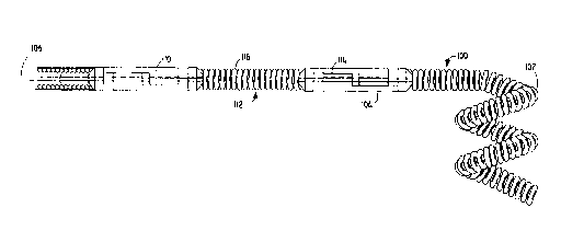
m e coil assembly (100) is shown in Figure 1.
The coil (102) is shown as helical in form, although it m.~ay
be any other suitable form. The coil should be of a size
sufficiently small that it may be advanced through a
catheter that is appropriately sized for accessing the
- targeted va~cular site. For instance, when accessing a
brain aneurysm in a 9 s ll vessel, an appropriately sized
catheter is quite small and very flexible. The coil in
W O 94/06503 8- PC~r/US93/08581
21 19772
such a situation must be small enough to fit through the
catheter and out its distal end at the treatment site.
The coil is desirably made up of a radiopaque,
physiologically compatible material. For instance, the
material may be platinum, gold, tungsten, or alloys of
these. Certain polymers are also suitable as coil material
either alone or in conjunction with metallic markers
pro~iding radiopacity. These materials are chosen 90 that
the procedure of locating coils within the vessel may be
viewed using radiography. However, it i9 also contemplated
that these coils may be made of various other biologically
inert polymers or of carbon fiber.
The size of the coil and its constituent winding
will depend upon the use to which the coil will be placed.
For occluding peripheral or neural sites, the coils will
typically be made of 0.05 to 0.15 mm diameter wire
(platinum or platinum/tungsten alloy) that may be wound to
have an inner diameter of 0.15 to 1.5 mm with a m;n~lm
pitch -- that is to say that the pitch is equal to the
diameter of the wire used in the coil. The outer diameter
is then typically between 0.25 mm to 1.8 mm. The length of
the coil will normally be in the range of 0.5 to 60 cm,
preferably 0.5 to 40 cm.
If desired, the coil may be formed in such a way
that the coil is essentially linear as it passes through
the catheter and yet assumes a r~n~omly oriented relaxed
condition after it is released from the distal end of the
catheter. A discussion of this ~ariation may be found in
U.S. Patent No. 4,994,069.
Fixedly attached to coil (102), as is shown in
Figure lA, is interlocking clasp (104). Interlo~king clasp
(104) as is depicted in the front three-guarter view in
Figure 13, has an interior passageway allowing the control
wire (106) to pass completely therethrough. As is shown in
Figure lA, the male portion of the next adjacent
WO 94/06503 PCr/US93/08581
g
21 l9772
interlocking clasp (110) fits into the area (108) left
within clasp (104) so to allow the interlocking to take
place. Said another way, the distal portion of
interlocking clasp (104) is generally cylindrical in shape
5 but has a surface (107), which may be cut or milled away,
allowing the portion to mesh within the middle area t108)
of an adjacent clasp. The pro~tim~l section is adapted for
attaching to a coil or to a pusher assembly. The
attachment may be by welding, soldering, gluing, or the
10 like. With a control wire (106) passing through the axis
of both interlocking clasps (104) and (110), the two are
locked together. As is shown in Figure lA, the control
wire rnay extend through the length of coil (102).
Figure 2 shows an interm~l;Ate coil assembly
15 (110) comprising coil (102) and interlocking clasp (104)
(joined with coil assembly (112)) which has interlocking
clasp (114) fixedly attached at both ends of the
intervening coil (116). As was the situation in Figure lA,
the proY;mAl interlocking clasp (114) is joined by control
20 wire (106) with interlocking clasp (110). In this way, a
significant number of coils (112) may be loaded onto a
control wire (106) and delivered to the treatment site
without L ~.o~/~l of the control wire from the catheter.
Figure 3 shows the relationship of coil assembly
25 (100) and the pusher assembly (118) with its distal
interlocking clasp (110) as it fits within catheter sheath
(120). Also shown is movable inner core member (122) and
the sheath (124) which fits within catheter sheath (120)
and supports interlocking clasp (110). Shown in Figure 3
30 is the stiffener spring (126) which pro~rides form and
support for the distal end of the pusher assembly (118) and
in particular rigidly adheres to interlsrking clasp (110).
Inner core member (122) allows the control wire (106) to be
moved axially along the interior of the catheter sheath
(120) and the pusher assembly (118). Mo~rement of the inner
W094/06503 PCT/US93/08581
- 10-
2~ ~ 977~
core member (122) in a proYi m~ 1 direction permits
uncoupling of the coil as will be discussed in more detail
below. -
The length of pusher assembly (118) will be such
as to be capable of being ad~anced entirely through thecatheter to place coil (102) at the target site but yet
with a 3ufficient portion of the proY;mAl end of the pusher
assembly (118) protruding from the pro~;mAl end of the
catheter to enable the control wire (106) to be
manipulated. For use in peripheral or neural surgeries,
the pusher will normally about 100-200 cm in length, more
normally 130-180 cm in length. The diameter of the pusher
assembly (118) is usually in the range of 0.25 to about
o.g0 mm.
As indicated pre~iously, conventional catheter
insertion and na~igational techniques in~ol~ing guidewires
or flow-directed devices may be used to access the site
with a catheter. Once the distal end of the catheter is
positioned at the site, often by locating its distal end
through the use of radiopaque marker material and
radiography, the catheter i9 cleared. For instance, if a
guidewire has been used to position the catheter, it is
withdrawn from the catheter and then the pusher assembly
(118) ha~ing coil assembly (100) at the distal end is
ad~anced through the catheter. The pusher assembly (118)
is advanced past the distal end of the catheter 90 that the
coil is free of the catheter and with the coil positioned
precisely at the desired treatment site.
As is shown in Figures 4 and 5, control wire
(106) is withdrawn from the junction between coil
interlocking cla~p (104) and the other interlocking clasp
(110). Coil assembly (100) is then free. The entire
catheter may then be removed or the p~he~ assembly ~118)
may be withdrawn from the catheter lumen to pro~ide for
installation of other coils. If additional coils are to be
W O 94/06503 PC~r/US93/08581
2 1 1 9772
placed at the target site, the procedure is repeated.
After the desired number of coils have been placed at the
site, the catheter is withdrawn from the vessel.
Figure 6A shows a variation in which coil
assembly (128) is interlocked with pusher assembly (130) by
control wire (106). The depicted coil assembly (128) and
pusher as~embly (130) are different in that they
incorporate the interlocking clasp (132) design shown more
clearly in Figure 6B. The interlocking clasp (132), as
with the clasp depicted in Figure lB, utilizes an open area
(134) within the clasp (132) to accept the mating ramp
latch (136) from another similar clasp. The ramp latch
(136) typically has a slot (138) and a passageway (140) to
permit passage of the control wire through the clasp (132)
from end to end without obstruction.
The ramp latch (136) allows easy assembly of a
string of coils within the catheter for subsequent
placement using the device.
Such an assembly process is shown in Figures 7A,
7B, and 7C.
Figure 7A shows a pusher assembly (130)
approaching a coil assembly (128) which has been previously
placed within a catheter sheath (120). The distal
interlocking clasp (132) on the ~qher assembly (130) is
positioned to interlock with the proY;~-l interlocking
clasp (134) on the coil assembly (128).
Figure 7B shows the two interlockng clasps (132
and 134) as they approach their respective ramps contacting
and causing the two clasps to displace ~Y;~lly within the
catheter sheath.
Figure 7C ~hows the location of the coil aRsembly
(128) and the pusher assembly (132) after the respective
clasps are interlocked and the control wire (106) has been
placed through the passageways within the clasps.
W094/06503 PCT/US93/08581
-12-
2 ~ ~ 97 1 ~
Figure 8 shows an elegantly simple variation of
the invention in which the pusher (138) i9 a tubing member
having a control wire (106) within its core. The clasp
portion (140) is a simple loop comprising, e.g., wire or
small rod. The correspon~;ng interlocking loop ~142) on
the coil (144) forms the junction with the clasp on the
pusher.
The variation of the invention shown in Figures
6A, 6B, 7A, 7B, 7C, and 8 m.ay be placed within the
vasculature in the same m~nnPr as shown for the variation
shown in Figs. 4 and 5.********~****~
Figure 9A shows a side view of a clasp (160)
s;m;l~r in design to the clasp discus~ed in conjunction
with Figures lA, lB, 2, 3, 4, and 5. In contrast to the
clasp found on those vasooclusive devices, the Figure 9A
clasp (160) contains no lumen therethrough for a control
wire. As was the case with the clasp above, the clasp is
generally cylindrical in shape but has a surface (162)
which may be cut or milled away to allow the to mesh with
a receiver area (164) in the next adjacent clasp as is
shown in Figure 11. The other end of the clasp is adapted
to allow joining with the end of a coil or pusher.
Although that other end is shown with a reduced diameter
(166), to allow insertion of the end (166) into the coil or
pusher, other end shapes are certainly appropriate, e.g.,
helical to accept the coil, square, bulbed, etc., the
reduced diameter cylinder is very desirable. The vertical
or mating surface (168) pulls the next de~ice in the chain
along when it contacts the s;mllAr surface in that next
de~ice. m e end surface (170) pushes against the next
device when in a chain.
Figure 9B shows a front, ele~ated, three-quarter
view of the clasp found in Figure 9A. Figure 9B shows the
end surface (170), the receiver area (164), and the reduced
diameter shaft (166) for mating with the coil or pusher.
WO94/06503 PCT/US93/08581
-13-
2 1 1 97 7 2
Figure lOA shows a clasp (172) much like that
shown in Figure 9A except that the end surface is a ramp
(174) to penmit assembly ease in placing the coils in the
catheter or introducer.
Figure 10~ shows a front, elevated, three-quarter
view of the clasp found in Figure lOA. Figure lOB shows
the end ramp (174), the receiver area (164), and the
reduced diameter shaft (166) for mating with the coil or
pusher.
Figure 11 shows how the clasps found in Figures
9A and 9~ mesh when installed on a pusher (178) or a coil
(1~0). It is within the scope of this variation of the
invention that the clasp be mounted on the ends of pushers,
coils (one or both ends), and that multiple coils or other
vasooclusive devices be joined in multiple end-to-end
trains for introduction into the vasculature.
An additional variation of the pusher assembly
(200) is shown in Figure 12. The configuration of the body
of the pusher assembly (200) is not particularly critical,
and many variations known in the art would likely be
suitable. The variation shown here entails, at the distal
end, a stainless steel core ~202) having a small diameter
section (204) covered by a desired polymeric material (206)
such as tetrafluoroethylene, or other suitable fluorinated
hydroc~rhon polymers; hydrophilic polymers such as
polyvinylpyrrolidone, polyethyleneoxide, or
polyhydroxyethylmethacrylate, or copolymers, or mixtures,
or blends thereof; or various ~ilicone-based polymeric
materials; or polyolefins such as polyethylene,
pol~ ylene, or their copolymers, mixtures, or blends; or
appropriate polyurethane polymers. This coating provides
a slippery surface allowing ease of insertion and traverse
through the catheter body.
It is desirable to include a radiopaque marker
(208). Such markers are common in this art and sy be made
W O 94/06503 -14- PC~r/US93/08581
2 1 1 9772
of known radiopaque materials such as platinum, palladium,
or other such materials. Cnmmonly, the radiopaque marker
(208) is a coil which i5 brazed or soldered to the
guidewire and may be coated with the polymeric materials
(206). This marker allows the tending physician to monitor
the progress of the guidewire tip via fluoroscopy and,
obviously, allow proper placement of the coil which is
attached to the end of the pusher guidewire (200).
More distal of the radiopaque marker (208) may be
found a flexible coil (210). This coil covers a tapered
section of the core wire (202). Tapering the inner wire
and enclosing it in a wire coil increases the column
strength of the tapered wire section without significant
loss of flexibility and increases the radial capacity of
the guidewire to allow fine manipulation of the guidewire
through various tortuous portions of the va~culature. The
tip of the core wire (202) and the distal portion of the
wire coil (210) are typically joined by uQe of a solder
joint (212). To this point, the guidewire is of a typical
guidewire respected in this art. See, for instance, those
guidewires shown in U.S. Patent Nos. 3,789,841; 4,545,390;
and 4,619,274.
Unique to this variation of the invention i9 the
hook (214) placed at the most distal end of the guidewire
assembly (200) which transforms it into a p~sher.
Engaging hook (214) has two legs (216) which are
based in solder joint (212). The outer hook portion (218)
is configured 90 that it slides into the conform;ng slot in
the coils as discussed below. The diameter (220) of the
hook (214) is typically no larger than the inside diameter
of the catheter as~embly into which it i5 placed.
Ob~iously, if the diameter is larger, it will bind in the
catheter and be of little use. The most distal portion of
the hook (218) is configured in such a way that the "W"
portion is in a plane which is generally perpendicular to
W094/06503 -15- PCT/US93/08581
21 19772
the longit~ nAl axis of the guidewire pusher assembly
(200). The engaging hook (214) need be made only of a
material which i9 adequate under the circumstances of use.
For instance, the hook may be of a stainless steel wire
which may be soldered onto the end of the guidewire
assembly (200) and bent into desirable shape. In this way,
the hook may be used to push the attached coil through the
catheter without bending. The length of guidewire pusher
assembly (200) should be such as to be capable of being
advanced entirely through a catheter to place a coil such
as discussed below at the target site, but yet retain a
sufficient portion of the proY; m~ 1 end of the guidewire
pusher assembly (200) protruding from the proY;mal end of
the catheter to enable the pusher to be manipulated. For
use in peripheral and neural surgeries, the pusher will
normally be about 100-200 cm in length, more normally 130-
180 cm in length. The diameter of the guidewire pusher
assembly (100) is usually in the range of 0.25 to about
0.90 mm.
Figure 13 shows a ~ariation of the distal tip of
guidewire pusher assembly (200) having a slightly different
configuration than that shown in Figure 12. In this
instance, the hook is a simple "vn- or "Un-~h~pP~ hook
which also will engage with the slotted coils described
below. The materials of construction and other such
variables are similar to those for the hook of Figure 12.
Figure 14 shows an additional variation of the pusher
assembly (200) having a hooked distal tip. m is hook (223)
is of a bent ribbon rather than the wire configuration
shown in Figures 12 and 13. The materials of construction
and method of attachment are 9im;1~ to those used in the
Figure 12 and 13 variations. The ribbon is bent in such a
way as to allow insertion of the hook's bent lip into the
slot found in the coils discussed below.
W094/06503 PCT/US93/08581
-16-
2 1 1 9772
The coil typical of that which might be used with
this invention, is shown in Figure 15. The coil (300) i9
shown as helical in form, although it may be any other
suitable form. The coil shown is one having a primary and
a secondary diameter. The primary diameter (302) is
sufficiently small that the coil (300), when straightened,
would fit inside the lumen of the catheter assembly. The
coil assembly shown assumes a second diameter t304) when
ejected from the tip of the catheter using the pusher
guidewire (200) shown in Figure 12.
Coil (300) may be made up of the same or similar
radiopaque, physiologically compatible materials discussed
in relation to coil (100) applicable to Figures 1-5 above.
The size, length, diameter (inner and outer), pitch, and
configuration all may be as discussed above.
Whatever the configuration may be, the coil
typically has caps at each end. Specifically, the distal
end of the coil (300) will have a distal cap to (306) which
may be solder or epoxy or other filling adhesive or fused
from the coil metal, preferably fonming a rounded form to
prevent the coil from hanging up within the catheter or an
inappropriate place within the patient's vasculature. The
unique aspect of this invention i9 found at the proYim~l
end of the coil (308). The proY;m~l end typically will be
soldered or glued, much in the way that the distal end has
been, but is configured in such a way that a slot (310) is
opened during the soldering or gluing process and will
accept the hook, variously (214) in Figure 12 or (222) in
Figure 13 or (223) in Figure 14 into the 910t . Obviously,
the receiving slot (310) may be generally substantially
perpendicular to the local axi~ of the coil.
Figure 16 depicts a variation of the coil shown
in Figure 15. This variation, however, includes, at the
distal end of the coil (316), a hook (318) of configuration
similar to that found in discussing the guidewire pusher
W O 94/06503 -17- PC~r/US93/08581
2~ 19772
assemblies in Figure9 12 and 13. This configuration allows
the introduction of discrete segments of coils into the
catheter and ~eparate placement of them should such a
situation be desirable. In such an instance, the hook
(318) would be introduced into the receiver slot (310) in
the 8~m; 1 ~r coil next in line. The most proY;~-l of the
coils would, in turn, be engaged with a hook on a pusher
assembly such as (200) shown in Figure 12.
Figure 17 shows a variation of the invention
shown in Figures 12-16 in which a control wire (240) is
placed generally through an axial pas~ageway between the
distal (or prox;m~l) end (242) of a coil or pusher having
a hook (244) and the corresponding end (246) of a coil
(243) having a slot (248) cooperating with the hook (244).
As with the variation shown in Figures 1-8, the control
wire (240) may be withdrawn when the coil (243) is situated
at the selected ~ascular site to disengage the coil (243).
Use of the control wire permits more precise placement of
coil (243) and its gentle disengagement.
Figure 18 is a side view depicting how the hook
(122), as depicted here, is placed in slot (310) of the
coil assembly (300). The tip of a typical catheter (400)
is shown in the Figure. Again, the o~erall diameter of the
various assemblies as put together for introduction into or
out of catheter must be of a diameter smaller than the
diameter lumen in catheter (400). Ob~iously, too large a
coil/pl~her combination will not be particularly ~aluable
in a situation where such is needed.
A~ indicated previously, conventional catheter
insertion and na~igational techniques involving guidewires
or even flow-directed de~ices may be used to access a
chosen ~ascular site with a catheter. Once the distal end
of the catheter is positioned at that chosen site, often by
locating its distal end through the use of a radiopaque
marker material and radiography, the catheter is cleared.
W O 94/06503 PC~r/US93/08581
-18- 2 1 1 9772
For instance, if a guidewire has been used to position a
catheter, it is withdrawn from the catheter and then the
guidewire pusher assembly such as (200) shown in Figure 12
having coil assembly such as (300) in Figure 15 is
S assembled and introduced into the pro~mAl end of the
catheter. The guidewire pusher assembly is then advanced
so that its distal end is free of the distal end of the
catheter and the coil positioned precisely at the desired
site. The pusher assembly (200) may require a twisting
movement to free the distal hook from the receiving slot in
the coil.
Modifications of the device described above and
methods of using it in keeping with this invention that are
apparent to those having skill in this mechanical and
surgical instrument design art and related fields are
intended to be within the scope of the claims which follow.