Note: Descriptions are shown in the official language in which they were submitted.
W O 93/09222 2 1 2 2 3 ~ 1 PC~r/US92/09627
~uuNsFEx~rIoN OF iEI~lEsRATE OE LLS EG. sY HK~L~XX~US RExx~MsINATloN
Description
~ackround of the InventiQn
Efforts to develop human gene therapies have their
roots in the 1950s, when early successes with kidney
transplantation led to speculation that it might be
possible to transplant cells from a normal individual into
a patient suffering from a genetio disease. Soon after
the discovery of the enzymatic defects in Gaucher's and
Niemann-Pick disease, scientists considered organ and bone
marrow transplantation and enzyme supplementation to treat
rare genetic disorders (Brady, R., NEJM 27S:312 (1966)).
By the late 1960s and early 1970s, several investigators
speculated that it also might be possible to introduce
~enes into a patient's own cells, and the cloning of the
first human genes only a few years later intensified work
in the field.
Until recently, almost all of the theoretical and
experimental work on h~man gene therapy was centered on
extremely rare genetic diseases, and gene therapy has come
to mean, to many in the field, the modification of a
patient's genes to treat a genetic disease. However, gene
therapy has far wider applications than simply treatment
of a genetic disease. Gene therapy is perhaps more
appropriately descri~ed as medical intervention in which
cells, either from the individual to be treated or another
appropriate source, are modified genetically to treat or
cure any condition, regardless of etiology, that will be
ameliorated by the long-term delivery of a therapeutic
protein. Gene therapy can ther~fore be thought of as an
in vivo protein production and elivery system, and almost
.. all diseases that are currently treated by the
W093/09222 PCT/US92/09l
2~229~1 -2-
administration of proteins are candidates for treatment
using gene therapy.
Gene therapy can be divided i~to two areas: germ cell
~nd somatic cell gene therapy. Germ cell gene therapy
S refers to the modification of sperm cells, egg cells,
zygotes, or early stage embryos. On the basis of both
ethical and practical criteria, germ cell gene therapy is
inappropriate for human use. In contrast to germ cell
gene therapy, somatic cell gene therapy would affect only
the per~on under treatment ~somatic cells are cells that
are not capable of developing into whole individuals and
include all of the body's cells with the exception of the
germ cells). As such, somatic cell gene therapy is a
reasonable approach to the treatment and cure of certain
disorders in human beings.
In a^somatic cell gene therapy system, somatic cells
(e.g., fibroblasts, hepatocytes, or endothelial cells) are
removed from the patient, cultured i~ vitro, transfected
with the gene~s) of therapeutic interest, characterized,
and reintroduced into the patient. The means by which
these five steps are carried out are the distinguishing
features of a given gene therapy system.
Presently-available approaches to gene therapy make
use of infectious vectors, such as retroviral vectors,
which include the genetic material to be expressed. Such
approaches have limitations, such as the potential of
generating replication-competent virus during vector
production; recombination between the therapeutic virus
and endogenous retroviral genomes, potentially generating
infectious agents with novel cell specificities, host
ranges, or increased virulence and cytotoxicity;
independent integration into large numbers of cells,
increasing the risk of a tumorigenic insertional event;
limited cloning capacity in the retrovirus (which
- wo 93/og222 2 1 2 2 9 9 1 PCT/US92/09627
restricts therapeutic applicability) and short-lived n
vivo expression of the product of interest. A better
approach to providing gene products, particularly one
w~ich avoids the risks associated with presently available
S methods and provides long-term production, would be
valuable. Proteins of therapeutic interest are generally
produced by introducing exogenous DNA encoding the protein
of therapeutic interest into appropriate cells.
Sum~ary of the Invention
The present invention relates to transfected primary
~nd secondary somatic cells of vertebrate origin,
particularly of mammalian origin, transfected with
exogenous DNA which encodes a therapeutic product,
exogenous DNA which is itself a therapeutic product and/or
exogenoûs DNA which causes the transfected cells to
express a gene at a higher level than occurs in the
corresponding nontransfected cell. Further, the present
invention relates to transfected primary and secondary
somatic cells of vertebrate origin, particularly mammalian
origin, transfected with exogenous genetic material (DNA)
which encodes a ~esired (e.g., a therapeutic) product or
is itself a desired (e.g., therapeutic) product, methods
by which primary and secondary cells are transfected to
include exogenous genetic material, methods of producing
clonal cell strains or heterogenous cell strains, methods
of gene therapy in w~ich the transfected primary or
secondary cells are used, methods of producing a
therapeutic protein through the use of transfected primary
or secondary cells made by the present method and methods
of producing antibodies using the transfected primary or
secondary cells.
In one embodiment, the present invention relates to
transfected primary and secondary somatic cells of
W093/09222 PCT/US92/09~
~12~991
vertebrate origin, particularly mammalian origin,
transfected with exogenous genetic material (DNA or RNA)
which encodes a clinically useful product, such ~s human
growth hormone (hGH), erythropoietin (EPO) or
S insulinotropin, methods by which primary and secondary
cells are transfected to include exogenous genetic
material encoding hGH, EPO or insulinotropin, methods of
producing clonal cell strains or heterogenous cell strains
which express exogenous genetic material encoding hGH, EPO
or insulinotropin, a method of providing hGH, EPO or
insulinotropin in physiologically useful quantities to an
individual in need thereof, through the use of transfected
cells of the present invention or by direct injection of
DNA encoding hGH, EPO insulinotropin into an individual;
and methods of producinq antibodies against the encoded
product ûsing the transfected primary or secondary cells.
In another embodiment, the present invention relates
to a method of gene or DNA targeting in cells of
vertebrate, particularly mammalian, origin. That is, it
relates to a method of introducing DNA into primary or
secondary cells of vertebrate origin through homologous
recombination or targeting of the DNA, which is introduced
into genomic DNA of the primary or secondary cells at a
preselected site. The preselected site determines the
2~ targeting sequences used. The present invention further
relates to homoloqously recombinant primary or secondary
cells t referred to as homologously recombinant (HR)
primary or secondary cells, produced by the present method
and to uses of the HR primary or secondary cells.
The present invention also relates to a method of
turning on or activating a gene present in primary cells,
secondary cells or immortalized cells of vertebrate
origin, which is normally not expressed in the cells or is
not expressed at significant levels in the cells.
W093/09222 2 1 2 2 9 .9 1 PCT/US92/~9627
Homologous recombination or t~rgeting is used to replace
or disable the regulatory rec on normally associated with
the gene with a regulatory sequence which causes the gene
to be expressed at levels higher than evident in the
corresponding nontransfected cell. The present invention,
therefore, relates to a method of making proteins by
turning on or activating an endogenous gene which encodes
the desired product in transfected primary, secondary or
immortalized cells.
As used herein, the term primary cell includes cells
present in a suspension of cells isolated from a verte-
brate tissue source ~prior to their being plated i.e.,
attached to a tissue culture substrate such as a dish or
flask), cells present in an explant derived from tissue,
both of the previous types of cells plated for the first
time, and cell suspensions derived from these plated
cells. The term secondary cell or cell strain refers to
cells at all subsequent steps i~ culturing. That is, the
first time a plated primary cell is removed from the
culture substrate and replated (passaged), it is referred
to herein as a secondary cell, as are all cells in
subsequent passages. Secondary clells are cell strains
whi~h consist of secondary cells ~which have been passaged
one or more times. A cell strain consists of secondary
cells that: 1) have been passaged one or more times; 2)
exhibit a finite num~er of mean population doublings in
culture; 3) exhibit the properties of contact-inhibited,
anchorage dependent growth (anchorage-dependence does not
apply to cells that are propagated in suspension culture);
and 4) are not immortalized. A "clonal cell strair is
defined as a cell strain that is derived from a si~ ~
founder cell. A "heterogenous cell strain`' is def~ l as
a cell strain that is derived from two or more fou;~ r
cells.
W093/09222 PCT/US92/09
~ 1 ,? 2 .q .~ 1
The present invention includes primary and secondary
somatic cells, such as fibroblasts, keratinocytes,
epithelial cells, endothelial cells, glial cells, neural
cells, formed elements of the blood, muscle cells, other
somatic cells which can be cultured and somatic cell
precursors, which have been transfected with exogenous DNA
wbich is stably integrated into their genomes or is
expressed in tbe cells episomally. The resultinq cells
are referred to, respectively, as transfected primary
cells and transfected secondary cells. The exogenous DNA:
1) encodes a product, such as a translational product
(e.g., a protein) or a transcriptional product (e.g., a
ribozyme or an anti-sense nucleic acid seguence) which is
a therapeutic product; 2) is itself a therapeutic product
(e.g., DNA which binds to a cellular regulatory protein or
alters gêne expression) or 3) is DNA which undergoes
homologous recombination with genomic DNA of recipient
cells and results in alteration of (increase or decrease
in) expression of an endogenous gene.
In the embodiment in which the exogenous DNA encodes
a translational or transcriptional product to be expressed
by tbe recipient cells, the resulting product is retained
within the cell, incorporated into the cell membrane or
secreted from the cell. In this embodiment, tbe exogenous
DNA encoding the therapeutic product is introduced into
cells along with additional DNA sequences sufficient for
expression of the exogenous DNA in transfected cells and
is operatively linked to those sequences.
In the embodiment in which the exogenous DNA is not
expressed, there is no gene product and the DNA itself is
the therapeutic product. In this embodiment, exogenous
DNA is, for example, DNA sequences which ~ind to a
cellular regulatory protein, DNA sequences sufficient for
sequestration of a protein or nucleic acid present in the
wo 93/0g222 2 1 2 ~ ~ ~ 1 PCr/US92/~g6i7
transfected primary or secondary cell, DNA sequences which
aiter secondary or tertiary chromosomal structure or DNA
sequences which are transcriptional regulatory elements.
Such primary cells modified to express or render available
exogenous DNA are referred to herein as transfected
primary cells, which include cells removed from tissue and
placed on culture medium for the first time. S~condary
cells modified to express or render available exogenous
DNA are referred to herein as transfected secondary cells.
In the embodiment in which exogenous DNA undergoes
homologous recombination with genomic DNA of transfected
(recipient) cells, introduction of the exogenous DNA
results in disablement of the endogenous sequences which
control expression of the endogenous gene, either by
replacing all or a portion of the endogenous (genomic)
~;~ sequence or disrupting the endogenous sequence.
Primary ~nd secondary cells transfected by the
subject method fall into three types or categori~s: 1)
cells which do not, as obtained, make or contain the
therapeutic product, 2) cells which make or contain the
therapeutic product but in lower quantities than normal
~ (in quantities less than the physiologically normal lower
; level) or in defective form, and 3) cells which make the
therapeutic product at physioloqically normal levels, but
are to be augmented or enhanced in their content or
production.
Exogenous DNA is introduced into primary or secondary
cells by a variety of techniques. For example, a
construct which includes exogenous DNA encoding a thera-
peutic protein and additional DNA sequences necessary forexpression in recipient cells is introduced into primary
or secondary cells by electroporation, microinjection, or
other means (e.g., calcium phosphate precipitation,
modified calcium phosphate precipitation, polybrene
W093/09222 PCT/US92/09~
2122991 -8-
precipitation, liposome fusion, receptor-mediated DNA
delivery). Alternatively, a vector, such as a retroviral
vector, which includes exogenous DNA can be used and cells
can be genetically modified as a result of infection with
the vector.
In addition to the exogenous DNA, transfected primary
and secondary cells may optionally contain DNA encoding a
selectable marker, which is expressed and confers upon
recipients a selectable phenotype, such as antibiotic
resistance, resistance to a cytotoxic-agent, nutritional
prototrophy or expression of a surface protein. Its
presence makes it possible to identify and select cells
containing the exogenous DNA. A variety of selectable
, ~arker genes can be used, such as neo, gpt, dhfr, ada,
pac, hyg, mdr and hisD.
Transfected cells of the present invention are
useful, as populations of transfected primary cells,
transfected clonal cell strains, transfected heterogenous
cell strains, and as cell mixtures in which at least one
representative cell of one of the three preceding
categories of transfected cells is present, as a delivery
system for treating an individual with an abnormal or
undesirable condition which responds to delivery of a
therapeutic product, which is either: 1) a therapeutic
protein (e.g., a protein which is absent, underproduced
relative to the individual's physiologic needs, defective
or inefficiently or inappropriately utilized in the
individual; a protein with novel functions, such as
enzymatic or transport functions) or 2) a therapeutic
~0 nucleic acid (e.g., DNA which binds to or sequesters a
regulatory protein, RNA which inhibits gene expression or
has intrinsic enzymatic activity). In the method of the
present invention of providing a therapeutic protein or
nucleic acid, transfected primary cells, clonal cell
2122~91
W093/0~222 PCT/US92/09627
strains or heterogenous cell strains are administered to
an individual in whom the abnormal or undesirable condi-
tion is to be treated or prevented, in sufficient quantity
~nd by an ~ppropriate route, to express or make available
the exogenous DNA at physiologically relevant levels. A
- physiologically relevant level is one which either
approximates the level at which the product is produced in
the body or results in improvement of the abnormal or
undesirable condition. Cells administered in the present
method are cells transfected with exogenous DNA which
encodes a therapeutic product, exogenous DNA which is
itself a therapeutic product or exoqenous DNA, such as a
regulatory sequence, which is introduced into a
preselected site in genomic DNA through homologous
recombination and functions to cause recipient cells to
produce~a product which is normally not expressed in the
cells or to produce the product of a higher level than
occurs in the corresponding nontransfected cell. In the
embodiment in which a regulatory sequence (e.g., a
promoter~ is introduced, it replaces or disables a
regulatory sequence normally associated with a gene, and
results in expression of the gene at a higher level than
occurs in the corresponding nontransfected cell.
Brief Descri~tion of the Drawinas
Figure 1 is a schematic representation of plasmid
pXGH5, which includes the human growth hormone (hGH) gene
under the control of the mouse metallothionein promoter.
Figure 2 is a schematic representation of plasmid
pcDNE0, which includes the neo coding region (BamHI-BglII
- 30 fragment) from plasmid pSV2neo inserted into the BamHI
site of plasmid pcD; the Amp-R and pBR3220ri sequences
from pBR322; and the polyA, 16S splice junctions and early
promoter regions from SV40.
W093/09222 PCT/US9210~
21229~1
--10--
Figure 3 is a schematic representation of plasmid
pXGH301 which includes the human growth hormone gene and
the neo gene.
Figure 4 is a flow chart of the ~ethod of the present
invention.
Figure 5 is a schematic representation of plasmid
pXEP01. The solid black arc represents the pUCl2 backbone
and the arrow denotes the diraction of transcription of
the ampicillin resistance gene. The stippled arc
represents the mouse metallothionein promoter (pmMTl).
The unfilled arc interrupted by black boxes represents the
human erythropoietin EP0 gene (the black boxes denote
exons and the arrow indicates the direction hEP0
transcription). The relative positions of restriction
endonuclease recognition sites are indicated.
Figure 6 is a schematic representation of plasmid
pE3neoEP0. The positions of the human erythropoietin gene
and the neo and amp resistance genes are indicated.
Arrows indicate the directions of transcription of the
various genes. pmMTl`denotes the mouse metallothionein
promoter (driving hEP0 expression) and pTK denotes the
Herpes Simplex Virus thymidine kinase promoter (driving
neo expression). The dotted regions of the map mark the
positions of human HGPRT sequences. The relative
positions of restriction endonuclease recognition sites
are indi~ated.
Figure 7 is a schematic diagram of a strategy for
transcriptionally activating the hEP0 gene.
Figure 8 is a schematic diagram of a strategy for
transcriptionally activating the hEP0 gene,
Figure 9 shows the results of an assessment of long-
term in vitro hGH production by transfected primary human
skin fibroblasts (two strains, HF96-11 and HF96-23).
2122~.9~
W093/09222 PCT/US92/09~27
--11--
Figure lo is a graphic representation of human growth
hormone (hGH) expression by transfected primary rabbit
skin fibroblasts Ln vitro.
Figure 11 s~ows the results of an assay to detect
serum levels of hGH over time in mice implanted with
transfected r~bbit fibroblasts expressing hGH.
Figure 12 is ~ graphic representation of ~uman growth
hormone (hGH) expression in cells recovered from subrenal
oapsule implants.
Figure 13a shows hematocrit (HCT~ levels in control
~ice and mice implanted with transfected rabbit
fibroblasts expressing hEPO.
Figure 13b shows results of an assay to detect hEPO
in t~e serum of mice implanted with transfected rabbit
fibroblasts expressing hEPO.
Detailed Descri~tion of the Invention
The present invention relates to transfected primary
and secondary somatic cells of vertebrate origin, "
particularly of mammalian origin, transfected with
exogenous DNA which encodes a therapeutic product,
exoqenous DNA which is itself a tberapeutic product and/or
exogenous DNA which causes the transfected cells to
express a gene at a higher level than occurs in the
corresponding nontransfected cell.
As described herein, primary or secondary cells of
vertebrate, particularly mammalian, origin have been
transfected with exogenous DNA encoding a therapeutic
product and shown to produce the encoded therapeutic
protein stably and reproducibly, both L~ vitro and in
-30 vivo, over extended periods of time. In addition, the
transfected primary and secondary cells have been shown to
-express the encoded product i~ vivo at physiologically
relevant levels, to be recoverable after implantation and,
W093/09222 PCT/US92/09
12-
upon reculturing, to grow and display their preimplanta-
tion properties. This demonstration is in sharp contrast
to what one of skill in the art would predict, because,
for example, even experts in the field ~ee the finite life
span of normal somatic cells and the inability to isolate
or grow the relevant transplantable cells as precluding
their use for gene therapy unless the cells are
genetically modified using retroviruses. Miller, A.D.,
~lood, 76:271-278 (1990). However, the transplantation of
retrovirally-treated fibroblasts has been shown to
provide, at best, only transient metabolic improvements,
~nd is seen to have serious limitations as a therapeutic ~`
system. Normal (non immortal) fibroblasts are
characterized as being "much more difficult to transfect
than continuous cell lines by using calcium phosphate
precipitation techniques." Miller, A.D., Blood, 76:271-278
(l990). Furthermore, in considering non- retroviral
technigues for gene therapy, it is typical of experts in
the field to believe ". . . the efficiency of gene
delivery is dismal. . . A physician would have to obtain
an impossible number of cells from patients to guarantee
the appropriate alteration of the millions required for
therapy." (Verma, I.M., Scient. Amer., November 1990,
pages 68-84).
Surprisingly, Applicants have been able to produce
transfected primary and secondary cells which include
exogenous DNA encoding a desired product, (i.e., a
translation product which is a therapeutic protein or an
antigen against which antibodies are produced) and stably
express the exogenous DNA. It is also possible, using the
method described harein, to produce transfected primary
and secondary cells which include exogenous DN~ encoding
other translation products (novel proteins not made in
nature) or transcription products te.g., anti-sense RNA or
2122991
W093/09222 PCT/US92/09627
-13-
ribozymes) or exogenous DNA which itself is a therapeutic
product (e.g., exogenous DNA which binds a regulatory
protein present in the transfected cell).
The met~od of the present invention includes the
steps of: 1) providing a population of primary cells,
obtained from the individual to whom t~e transfected
primary cells will be administered or from another source;
~; 2) ~ntroducing into t~e primary cells or into ~econdary
cells derived from primary cells a DNA construct which
includes exoqenous DNA as described above and the
~n-cess~ry additional DNA sequences described above,
producing transfected primary or secondary cells; 3)
maintaining transfected primary or secondary cells under
conditions appropriate for t~eir propagation; 4) identify-
ing a transfected primary or secondary cell; and5).~prod~cing a colony from the transfected primary or
- secondary cell identified in (4) by maintaininq it under
;appropriate culture conditions and for ~ufficient time for
its propagation, tbereby producing a cell strain derived
from the (founder) cell identified in (4). In one
embodiment of the method, exogenous DNA is introduced into
~` genomic DNA by homologous recombination between DNA
sequences present in the DNA construct and genomic DNA.
In one embodiment of the present method of producing
a clonal population of transfected secondary cells, a cell
suspension containing primary or secondary cells is
combined with exogenous DNA encoding a therapeutic product
and DNA encoding a selectable marker, such as the neo
gene. The two DNA sequences are present on the same DNA
construct or on two separate DNA constructs. The
resulting combination is subjected to electroporation,
generally at 250-300 volts with a capacitance of
960 ~Farads and an appropriate time constant (e.g., 14 to
20 m sec) for cells to take up the DNA construct. In an
W093/09222 PCT/US~2/09~
2 1 ? ~ 9 ~1 1
-14-
alternative embodiment, microinjection is used to intro-
duce the D~A construct into primary or secondary cells.
In either embodiment, introduction o~ the exogenou~ DNA
results in production of transfected primary or secondary
cells.
In the method of producing heterogenous cell strains
of the present invention, the same steps are carried out
as described for production of a clonal cell strain,
except that a single transfected primary or secondary cell
is not isolated and used as the founder cell. Instead,
two or more transfected primary or secondary cells are
cultured to produce a heterogenous cell strain.
The subject invention also relat`es to a method of
producing antibodies specific for the protein encoded by
the exogenous DNA. In the method, transfected primary or
secondary cells expressing an antigen against which
antibodies are desired are introduced into an animal
recipient (e.g., rabbit, mouse, pig, dog, cat, goat,
guinea pig, sheep, non-human primate). The animal
recipient produces antibodies against the antigen
expressed, which may be an entire protein antigen or a
peptide encoded by a fragment of the intact gene which
encodes the entire antigen. Polyclonal sera is obtained
from the animals. It is also possible to produce
monoclonal antibodies throuqh the use of transfected
primary or secondary cells. Splenocytes are removed ~rom
an animal recipient of transfected primary or secondary
cells expressing the antigen against which monoclonal
antibodies are desired. The splenocytes are fus~d with
myeloma cells, using known methods, such as that of
Koprowski et al. tU.S. Patent No. 4,172,124) or Kohler et
al., (Nature 256:495-497 (1975)) to produce hybridoma
cells which produce the desired monoclonal antibody. The
polyclonal antisera and monoclonal antibodies produced can
W O 93/09222 2 12 2 n 9 1 PC~r/US92/09627
be used for the same purposes (e.g., diagnostic,
preventive, therapeutic purposes) as antibodies produced
by other methods.
The present invention has wide applicability in
treating abnormal or undesired conditions and can be used
to provide a variety of products to an individual. For
example, it can be used to provide secreted proteins (with
either predominantly systemic or predominantly local
effect~), membrane proteins (e.g., for im~arting new or
enhanced cellular responsiveness, facilitating removal of
a toxic product or marking or targeting a cell) or
intracellular proteins (e.g., for affecting gene
expression or producing autolytic effects). In addition,
it can be used to provide engineered DNA which binds or
sequesters a cellular protein, to produce engineered RNA
useful in an anti-sense approach to altering gene
expression or to provide antigens against which immune
response occurs in an individual (to prevent disease as by
vaccination or to suppress an existing condition). The
present invention is particularly advantageous in treating
abnormal or undesired conditions in that it: 1) is
curative (one gene therapy treatment has the potential to
last a patient's lifetime); 2) allows precise dosing (the
patient's cells continuously determine and deliver the
optimal dose of the required protein based on physiologic
demands, and the stably transfected cell strains can be
characterized extensively in vitro prior to implantation,
leading to accurate predictions of long term function n
vivo); 3) is simple to apply in treating patients; 4)
eliminates issues concerning patient compliance (following
a one-time gene therapy treatment, daily protein
injections ara no longer necessary); 5) reduces treatment
costs (since the therapeutic protein is synthesized by the
patient's own cells, investment in costly protein
W093~09222 PCTtUS92/09
2 ~ 229 91 -16-
production and purification is unnecessary); and 6) is
safe (the invention does not use infectious agents such as
retroviruses to genetically enginee~ the patient's cells,
thereby overcoming the safety and efficacy issues that
have hampered other gene therapy systems).
As further described herein, primary or secondary
cells of vertebrate, particularly mammalian, origin have
been transfected with exogenous DNA en~oding EPO and shown
to produce the encoded EPO reproducibly, both ~n vitro and
n Y vo, over extended periods of time. In addition, the
transfected primary and secondary cells have been shown to
express EPO ~n vivo at physiologically relevant levels.
The EPO expressed has been shown to have the glycosylation
pattern typical of EPO purified from human urine or
recombinant human EPO.
In addition, as further described herein, primary or
secondary cells of vertebrate, particularly mammalian,
origin have been transfected with exogenous DNA encoding
hGH and shown to produce the encoded hGH reproducibly,
both n vitrQ and in vivo, over extended periods of time.
In addition, the transfected primary and secondary cells
have been shown to express hGH in vivo at physiologically
relevant levels.
Applicants have also developed methods or producinq
transfected primary or secondary cells which stably
express exogenous DNA encoding EPO, clonal cell strains
and heterogenous cell strains of such transfected cells,
methods of producing the clonal and heterogenous cell
strains, and methods of usin~ transfected cells expressing
EPO to deliver the encoded product to an individual mammal
at physiologically relevant levels. The constructs and
methods herein described are useful, for example, for
treating an individual (human) whose EPO production and/or
function is in need of being increased or enhanced [e.g.,
2122~!31
W093/09222 PCT/US92/09627
-17-
is compromised or less than normal, or normal but the
individual would benefit from enhancement, at least
temporarily, of red blood cell production (e.g., during
predialysis or dialysis therapy, during treatment of AIDS
with AZT, after surgery, or during cbemotherapy)].
As al~o described herein, it is possible to transfect
primary or secondary cells of vertebrate, particularly
~calian, origin with exogenous DNA encoding insulino-
tropin and to use them to provide insulinotropin to an
individual in whom insulin production, function and/or
sen~itivity iii compromised. As used herein, the term
insulinotropin includes, e.g. derivatives of qlucagon-like
peptide 1 (GLP-l) such as GLP(7-37), GLP(7-36), GLP-1(7-
35) and GLP-1(7-34) as well as their carboxy-terminal
amidated derivatives produced by ~n vivo amidating enzymes
and derivatives which have amino acid alterations or other
alterations which result in substantially the same
biological activity or stability in the blood as that of a
truncated GLP-l or enhanced biological activity or
2n stability.
As further described herein, Applicants have also
demonstrated that DNA can be introduced into primary or
secondary vertebrate cells in a DNA construct or plasmid
and integrated into the genome of the transfected primary
or secondary cells by homologous recombination. That is,
they have demonstrated gene targeting in primary and
secondary mammalian cells. They have further demonstrated
that the exogenous DNA has the desired function in the
homologously recombinant (HR) cells and that correctly
targeted cells can be identified on the basis of a
detectable phenotype conferred by a selectable marker
gene.
In addition, the present invention relates to a
method of protein production using transfected primary,
W093/09222 PCT/US92/09~
212~91
-18- -~
secondary or immortalized cells. The method involves
transfecting primary cells, secondary cells or
iD ortalized cells with exogenous DNA which encodes a
therapeutic product or with DNA which is sufficient to
target to and activate an endogenous gene which encodes a
therapeutic product. For example, Examples 18f, 19 and 21
de~cribe protein production by targeting and activation of
a ~elected endogenous gene.
Applicants describe (Example 3) construction of
plasmids containing a selectable marker gene (plasmid
pcDNE0), a gene encoding a tberapeutic product ~plasmid
pXGH5) or both (pXGH301). They also describe construction
of a plasmid useful for targeting to a particular locus
(the HPRT locus) in tbe human genome and selection based
upon a drug resistant phenotype (Example 18a). This
plasmid ~s designated pE3Neo and its integration into t~e
cellular genomes at the HPRT locus produces cells which
have an hprt , 6-TG resistant phenotype and are also G418
~ resistant. As described, they have shown that pE3Neo
:~ 20 functions properly in gene targeting in an established
human fibroblast cell line (Example 18b), by demonstrating
localization of the DNA introduced into established cells
within exon 3 of the HPRT gene.
In addition, Applicants demonstrate gene targeting in
primary and secondary human skin fibroblasts usinq pE3Neo
tExample 18c) and describe construction of a plasmid for
targeted insertion of a gene encoding a therapeutic
product (human qrowth hormone t~GH~) into the human genome
(Example 18d). The subject application furt~er
demonstrates that modification of DNA termini enhances
targeting of DNA into genomic DNA (Examples 18c and 18e)
and construction of a variety of targeting plasmids. For
instance, Applicants describe targeting plasmids for
`~ W O 93/09222 2 1 2 ~ ~ 9 1 PC~r/US92/09627
-19-
placing a human gene under the control of a murine
promoter known to function in human cells (Examples 18f
and 18i); for targeting to ~equences flanking a gene and
isolation of targeted secondary fibroblasts using a
variety of screening and selection approaches (Examples
18g, 18h, 18j and 18k); for placing a human gene not
normally expressed in the primary or secondary cells under
the control of a promoter of nonhuman or human origin, to
produce homologously recombinant primary or secondary
cells which express the encoded product tExamples
18f-18k).
Two further embodiments of the present invention are
envisioned, ~Example 19) in which the normal regulatory
seguences upstream of a g~ne (e.g. the human EP0 gene) are
altered to allow expression of a gene product in primary
or secon~ary cell strains which do not normally express
that product in detectable quantities in their
untransfected state. In one embodiment, the product of
t~e targeting events is a chimeric transcription unit, in
which a regulatory element and an operatively linked exon
~ .
are positioned upstream of the desired endogenous gene to
be activated. The product of transcription, splicing, and
translation produces a chimeric protein in which amino
acids encoded by exon 1 of the exogenous gene are fused to
amino acids encoded by exons 2 and downstream exons in the
endogenous gene. In a second embodiment, the product of
the targeting event replaces the regulatory and exon 1
sequences of the endogenous qene with corresponding
exogenous sequences. The product of transcription,
splicing, and translation produces a chimeric protein
similar to that described above. Typically, secretion of
such proteins involves membrane translocation and removal
of the signal peptide, in this case producing a normal
protein lacking the chimeric signal peptide. In both
`: .
W093/09222 PCT/US92/09~
2122~91 -20-
cases the chimeric-^protein is now under the control of a
desired regulatory element.
Examples 18f-18h and 19 illustrate embodiments in
which the norma} regulatory sequences upstream of the
human EPO gene are altered to allow expression of hEP0 in
primary or secondary fibroblast strains which do not
express EPO in detectable quantities in their
untransfected state. In one embodiment the product of
targeting leaves the normal EPO protein intact, but under
the control of the mouse metallothionein promoter.
Examples 18i and 18j demonstrate the use of similar
targeting constructs to activate the endogenous growth
hormone gene in primary or secondary human fibroblasts.
~n other embodiments described for activating EPO
expression in human fibroblasts, the products of targeting
events arê chimeric transcription units, in which the
first exon of the human growth hormone gene is positioned
upstream of EPO exons 2-5. The product of transcription
(controlled by the mouse metallothionein promoter),
2Q splicing, and translation is a protein in which amino
acids 1-4 of the hEPO signal peptide are replaced with
amino acid residues 1-3 of hGH. The chimeric portion of
this protein, the signal peptide, is removed prior to
secretion from cells.
The Examples provide methods for activating
endogenous genes by gene targeting which do not require
manipulation or other uses of the hE~O and hGH protein
coding regions. By these methods, normally inactiYe genes
may be activated in cells that have properties desirable
for n vivo protein delivery methods (e.g~ gene therapy)
and n Yitro protein production (e.g., pharmaceutics)~
Figures 7 and 8 illustrate two strategies for
transcriptionally activating the hEPO gene. The thin
lines represent hEPO sequences; thick lines, mouse
W O 93/09222 2 1 2 2 ~ 9 1 PC~r/US92/09627
-21-
metallothionein I promoter; stippled box, 5 t untranslated
region of hGH; solid box, hGH exon ~; striped box, 10 bp
linker ~rom hEPO intron 1; cross-hatched box, 5'
untranslated region of hEP0; and open boxes, hEPO coding
sequences; HIII, HindIII site.
Using the methods and DNA constructs or plasmids
taught herein or ~odifications thereof which are apparent
to one of ordinary skill in the art, exogenous DNA which
encodes a therapeutic product (e.g., protein, ribozyme,
nucleic ~cid) can be inserted at preselected sites in the
genome of vertebrate (e.g., mammalian, both human and
nonhuman) primary or secondary cells.
The methods and DNA constructs described can be used
for a wide variety of purposes. The method can be used to
alter primary or secondary cells of vertebrate origin in
order to repair, alter, delete or replace DNA already
present in the recipient primary or secondary cell; to
introduce into primary or secondary cells a gene or DNA
sequence (at a preselected site) which encodes a
therapeutic product or other desired product or is itself
a therapeutic or other product; to add to or replace
regulatory sequences present in the primary or secondary
cell recipients; to knock out or remove an entire gene or
gene portion present in primary or secondary cells; and to
produce universal donor cells.
The transfected primary or secondary cells may also
include DNA encoding a selectable marker which confers a
selectable phenotype upon them, facilitating their
identification and isolation. Applicants have also
developed methods for producing transfected primary or
secondary cells which stably express exogenous DNA, clonal
cell strains and heterogenous cell strains of such
transfected cells, methods of producing the clonal and
heterogenous cell strains, and methods of treating or
W093/09222 PCT/US92/09~
2122991
-22-
preventing an abnormal or undesirable condition through
the use of populations of transfected primary or ~econdary
cells of the present invention.
Transfected Cells
Primary and secondary cells to be transfected by the
present method can be obtained from a variety of tissues
and include all cell types which can be maintained in
culture. For example, primary and secondary cells which
can be transfected by the present method include
fibroblasts, keratinocytes, epithelial cells (e.g.,
mam~ary epithelial cells, intestinal epit~elial cells),
endothe}ial cells, glial cells, neural cells, formed
elements of the blood (e.g., lymphocytes, bone marrow
cells), muscle cells and precursors of these somatic cell
types. Primary cells are preferably obtained from the
indivîdual to whom the transfected primary or secondary
cell~ are administered. However, primary cells may be
obtained from a donor (other than the recipient) of the
~ same species or another species (e.g., mouse, rat, rabbit,
;~ ~ 20 cat, dog, pig, cow, bird, sheep, goat, horse).
- Transfected primary and secondary cells have been
produced, with or without phenotypic selection, as
described in Examples 5-7, and shown to express exogenous
DNA encoding a therapeutic product including, e.g., EP0
and insulinotropin.
Immortalized cells can also be transfected by the
present method and used for either gene therapy or protein
production. Examples of immortalized human cell lines
useful for protein production by the present method
include, but are not limited to, HT1080, HeLa, MCF-7
breast cancer cells, K-562 leukemia cells, XB carcinoma
cells and 2780AD ovarian carcinoma cells.
W093/09222 2 1 2 2 9 9 1 PCT/US92/09627
-23-
~xoqenous D~A
Exogenous DNA incorporated into primary or secondary
- cells by the present method is: 1) DNA which encodes a
translation or transcription product whose expression in
primary or secondary cells is desired, such as a transla-
tion or transcription product useful to treat an existing
condition or prevent it from occurring (eg., EP0 or
insulinotropin) and 2I DNA which does not encode a qene
product but is itself useful, such as DNA useful to treat
~n existing condition or prevent it from occurring or 3)
DNA w~ich underqoes homologous recombination with genomic
DNA of recipient cells and results in alteration of
(increase or decrease in) expression of an endogenous
gene.
DN~ transfected into primary or secondary cells can
encode an entire desired product, or can encode, for
example, the active or functional portion(s) of the
product. The product can be, for example, a hormone, a
cytokine, an antigen, an antibody, an enzyme, a clotting
factor, a transport protein, a receptor, a regulatory
protein, a structural protein, an anti-sense RNA, a
ribozyme or a protein or a nucleic acid which does not
occur in nature (i.e., a novel protein or novel nucleic
acid). The DNA can be obtained from a source in which it
occurs in nature or can be produced, using genetic
engineering techni~ues or synthetic proceæses. The DNA
transfected into primary or secondary cells can encode one
or more therapeutic products. After transfection into
primary or secondary cells, the exogenous DNA is stably
incorporated into the recipient cell's genome (along with
the additional se~uences present in the DNA construct
used), from which it is expressed or ot~erwise functions.
AlternatiVely, the exogenous DNA may exist episomally
within the transfected primary or secondary cells.
WO 93/09222 PCI`/US92/096;
2122g91
-24-
DNA encoding the desired product can be introduced
into cells under the control of an inducible promoter,
with the result that cells produced or as introduced into
an individual do not express the product but can be
induced to do 80 (i.e., production is induced after tbe
transfected cells are produced but before implantation or
after implantation). DNA encoding the desired product
can, of course, be introduced into cells in such a manner
that it is expressed upon introduction (i.e., without
induction).
Selectable Markers
A variety of selectable markers can be incorporated -
into primary or secondary cells. For example, a select-
able marker which confers a selectable phenotype such as
drug resistance, nutritional auxotrophy, resistance to a
cytotoxic agent or expression of a surface protein, can be
ùsed. Selectable marker genes which can be used ~nclude
neo, gpt, dhfr, ada, pac, hyg and hisd. The ~electable
phenotype conferred makes it possible to identify and
isolate recipient primary or secondary cells.
Selectable markers can be divided into two
categories: positive selectable and negative selectable.
In positive selection, cells expressing the positive
selectable marker are capable of surviving treatment with
a selective agent (such as neo, gpt, dhfr, ada, pac, ~yg,
mdrl and hisD). In negative selection, cells expressing
~; the negative selectable marker are destroyed in the
presence of the selective agent (e.g., tk, gpt).
DNA Constructs
DNA constructs, which include exogenous DNA and,
optionally, DNA encoding a selectable marker, alon~ with
additional sequences necessary for expression of the
;:
W093/09222 2 1 2 ~ 9 9 1 PCT/US92/09627
-25-
exogenous DNA in recipient pr~ary or ~econdary cells, are
u~ed to transfect primary or secondary cells in which tbe
encoded product is to be produced. The DNA construct can
al80 include t~rgeting iequences for bo~ologous
S recombination with host cell DNA. DNA constructs which
include exogenous DNA seguences which do not encode a gene
product (and are the therapeutic product) and, optionally,
include DNA encoding a selectable marker, can be used to
tr~nsfect primary and secondary cells. Alternatively,
infectious Yectors, such as retroviral, herpes,
~d novirus, adenovirus-associated, mumps and poliovirus
~ector~, can be used for this purpose.
In one embodiment of the present invention, a DNA
construct which includes the exogenous DNA and additional
s~guences, sucb as sequences necessary for expression of
tbe exogenous DNA, c~n be used (e.g., plasmid pXGH5 or
plasmid pXEPOl). A DNA construct can include an inducible
promoter whic~ controls expression of the exogenous DNA,
making inducible expression possible. Optionally, the DNA
construct may include a bacterial origin of replication
nd bacterial antibiotic resistance markers, which allow
for l~rge-scale plasmid propagation in bacteria. A DNA
construct whic~ includes DNA encoding a selectable marker,
along with additional sequences, such as a promoter, poly-
adenylation site and splice junctions, can be used toconfer a selectable phenotype upon transfected primary or
secondary cells te.g., plasmid pcDNEO). The two DNA
constructs are co-transfected into primary or secondary
cells, using methods described herein. Alternatively, one
DNA construct which includes exogenous DNA, a selectable
~arker gene and additional sequences (e.g., those
necessary for expression of the exogenous DNA ~nd for
expression of the selectable marXer gene) can be used.
Such a DNA construct (e.g., plasmid PXGH301, which
W O 93/09222 PC~r/US92/096'
2122991
-26-
includes the hGH gene and the neo gene, or plasmid
pE3neoEP0 which includes the EP0 gene and the neo gene;
these plasmids are described in Figures 3 and 6,
respectively). Similar constructs, which include
exogenous DNA encoding insulinotropin and additional
sequences (e.g., seguences necessary for insulinotropin
expression) can be produced (e.g., plasmid pXGLPl; ~ee
Example 11). Th~se constructs can also include DNA
encoding a selectable marker, as well as other sequences,
such as a promoter, a polyadenylation site, and æplice
junctions.
In those instances in which DNA is injected directly
into an individual, such as by injection into muscles, tbe
DNA construct includes the exogenous DNA and regulatory
sequences necessary and sufficient for expression of the
encoded product (e.g., EP0) upon entry of the DNA
construct into recipient cells.
In another embodiment of the present invention, DNA
constructs, which include exogenous DNA encoding a desired
product, targeting sequences for homologous recombination
and, optionally, DNA encoding one or more selectable
markers are used to transfect primary or secondary cells
in which homologous recombination is to occur. In this
embodiment, DNA sequences necessary for expression of the
exogenous DNA will generally be present as well. DNA
constructs which include exogenous DNA sequences which do
not encode a gene product (and are the desired product)
and, optionally, include DNA encoding a selectable marker,
can also be used to transfect primary and secondary cells.
The exogenous DNA, targeting sequences and selectable
marker can be introduced into cells on a single DNA
construct or on separate constructs. The total length of
the DNA construct will vary according to the number of
components (exogenous DNA, targeting sequences, selectable
W093/09222 2 1 2 2 9 9 1 PCT/US92/0962~
-27-
marker gene) and the length of each. T~e entire construct
lengt~ will generally be at least 20 nucleotides. In a
construct in which the exogenous DNA has sufficient
homology with genomic DNA to undergo homologous
recombination, the construct will include a singie
co~ponent, the exogenous DNA. In this embodiment, the
exogenous DNA, because of its homology, serves al~o to
target integration into genomic DNA and additional
targeting sequences are unnecessary. Such a construct is
u~eful to knock out, replace or repair a resident DNA
scquence, such as an entire gene, a gene portion, a
regulatory element or portion thereof or regions of DNA
, which, when removed, place regulatory and structural
sequences in functional proximity. It is also useful when -
the exogenous DNA is a selectable marker.
In a third embodiment, the DNA construct includes --~
exogenous DNA and one or more separate targeting se-
guences, generally located at both ends of the exogenous
DNA sequence. Targeting sequences are DNA sequences
normally present in the primary or secondary cell genon~e
in the genome of the cells as obtained ~e.g., an essential
gene, a nonessential gene or noncoding DNA, or present in
the genome through a previous modification]. Such a
co~struct is useful to integrate exogenous DNA encoding a
therapeutic product, such as a hormone, a cytokine, an
antigen, an antibody, an enzyme, a clotting factor, a
transport protein, a receptor, a regulatory protein, a
structural protein, an anti-sense RNA, a ribozyme or a
protein or a nucleic acid which does not occur in nature.
In particular, exogenous DNA can encode one of the
following: Factor VIII, Factor IX, erythropoietin, alpha-l
antitrypsin, calcitonin, glucocerebrosidase, growtb
hormone, low density lipoprotein (LDL) receptor,
apolipoprotein E, IL-2 receptor and its antagonists,
:; '
W093/09222 PCT/US92/096
-28-
insulin, globin, immunoglobulins, catalytic antibodies,
the interleukins, insulin-like growth factors, superoxide
dismutase, immune responder modifiers, parathyroid
hormone, interferons, nerve growth factors, tissue
plasminogen activators, and colony stimulating factors.
Such a construct is also useful to integrate exogenous DNA
whic~ i~ a therapeutic product, such as DNA seguences
sufficient for sequestration of a protein or nucleic acid
in the transfected primary or secondary cell, DNA
seguences which bind to a cellular regulatory protein, DNA
sequences which alter the secondary or tertiary
chromosomal structure and DNA sequences which are
transcriptional regulatory elements into genomic DNA of
primary or secondary cells.
In a fourth embodiment, the DNA construct includes
exogenoûs DNA, targetinq DNA sequences and DNA encoding at
least one selectable marker. In this fourtb embodiment,
the order of construct components can be: targeting
sequences-exogenous DNA-DNA encoding a selectable
marker(s)-targeting sequences. In this embodiment, one or
more selectable markers are included in the construct,
which makes selection based on a selectable phenotype
possible. Cells that stably integrate the construct will
survive treatment with the selective agent; a subset of
the stably transfected cells will be HR cells, which can
be identified by a variety of techniques, including PCR,
Southern hybridization and phenotypic screening.
In a fifth embodiment, the order of components in the
DNA construct can be: targeting sequence-selectable marker
1 - targeting sequence - selectable marker 2. In this
embodiment selectable marker 2 displays the property
negative selection. That is, the gene product of
selectable marker 2 can be selected against by growth in
an appropriate media formulation containing an agent
W093/09222 2 1 2 2 9 9 1 - PCT/US92/09627
-29-
(typically a drug or metabolite analog) which kills cells
expressing selectable marker 2. Recombination between the
targeting sequences flanking selectable marker 1 wit~
homologous sequences in the host cell genome re~ults in
the targeted integration of selectable marker 1, while
~electable marker 2 is not integrated. Such recombination
events generate cells which are ~tably transfected witb
~elect~ble marker 1 but not ~t~bly transfected with
selectable marker 2, and ~ucb cells can be selected for by
growth in the media containing the selective agent which
~elects for selectable marker 1 and the selecti~e agent
which celects against selectable marker 2.
In all embodiments of the DNA construct, exogenous
DNA can encode one cr more products, can be one or more
therapeutic products or one or more of each, thus making
it possible to deliver multiple products.
Iransfection of PrimarY or Secondary Cells and Production
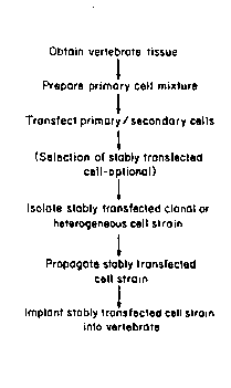
Qf Clonal or Heteroaenous Cell Strains
The method of the present invèntion is represented
schematically in Figure 4. As shown, vertebrate tissue is
first o~tained; this is carried out using known
procedures, such as punch biopsy or other surgical methods
of obtaining a tissue source of the primary cell type of
interest. For example, punch biopsy is used to obtain
skin as a source of fibroblasts or keratinocytes. A
mixture of primary cells is obtained from the tissue,
using known methods, such as enzymatic digestion or
explanting. If enzymatic digestion is used, enzymes such
as collagenase, ~yaluronidase, dispase, pronase, trypsin,
- 30 elastase and c~ymotrypsin can be used.
The resulting primary cell mixture can be transfected
directly or it can be cultured first, removed from t~e
culture plate and resuspended before transfection is
~ `
W093/09222 PCT/US92/096
2122991 30
carried out. Primary cells or secondary cells are
combined with exogenous D~A to be ~tably integrated into
their genomes And, optionally, DNA encoding a ~el~ctable
J~rker, and treated in order to accompli~h transfection.
The exogenous DNA and selectable marker-encoding DNA ~re
each on a ~eparate construct (e.g., pXGH5 and pcDNE0, see
Figures 1 and 2) or on a single construct (e.g., pXGH301
~nd pE3neoEP0, ~ee Figure 3 and Figure 6). An appropriate
quantity of DNA is used to ensure that at least one stably
transfected cell containing and appropriately expr~ssing
exogenous DNA is produced. In general, 0.1 to 500 ~g DNA
is used.
Using the present methods to introduce only a
electable marker qene, between 170 (1 in 588 starting
cells treated by electroporation, Example 6) and 2000 (1
in 49 starting cells treated by microinjection, Example 5)
stably transfected cells are generated per 100,000
starting cells. Using the present methods to introduce a
therapeutic gene as well as a selectable marker gene,
between 7 (1 in 14,705 starting cells treated by
electroporation, Example 6) and 950 (1 in 105 starting
cells treated by microinjection, Example 5) stably
transfected cells are generated per 100,000 starting
cells. Of these stable transfectants, from 43 to 90%
express the gene of therapeutic interest. Since only a
single appropriately expressing cell is required, it is
clearly possible to use substantially fewer starting
cells. Conversely, using transfection techniques which
are substantially less efficient than the present methods,
it would not be possible to obtain even a single such cell
unless large amount of the individual's tissue is used as
the source of starting cells.
In one embodiment of the present method of producing
transfected primary or secondary cells, transfection is
.
W093/09222 2 1 2 ~ ~ 9 1 PCT/US92/~627
effected by electroporation, as described in the Examples.
Electroporation i8 carried out at appropriate voltage and
cap~citAnce (and corresponding time constant) to result in
entry of the DNA construct(s) into the primary or
secondary cells. Electroporation can be carried out over
a wide range of voltages (e.g., 50 to 2000 volt~) and
corresponding capacitance. As described herein,
electropor~tion is very efficient if carried out at an
electroporation voltage in t~e range of 250-300 volts and
a capacitance of 960 ~Farads. Total DNA of approximately
O.1 to 500 ~g is generally used. As described in the
Ex~mples, total DNA of 60 ~g and voltage of 250-300 volts
. with capacitance of 960 ~Farads for a time constant 14-20
of msec. has been used and shown to be efficient.
In~another embodiment of the present method, primary
` or ~econdary cells are transfected using microin~ection.
;~ See, for ex~mple, Example 5. Alternatively, known metbods
sucb as calcium phosphate precipitation, modified calciu~
phosphate precipitation and polybrene precipitation,
liposome fusion and receptor-mediated gene delivery can be
used to transfect cells. A stably transfected cell is
isolated and cultured and subcultivated, under culturing
conditions and for sufficient time, to propagate the
stably transfected secondary cells and produce a clonal
cell strain of transfected secondary cells.
Alternatively, more than one transfected cell is cultured
and subcultured, resulting in production of a ~eterogenous
cell strain.
Transfected primary or secondary cells undergo a
sufficient number of doublings to produce either a clonal
cell strain or a heterogenous cell strain of sufficient
size to provide t~e therapeutic product (e.g~, EP0) to an
individual in effective amounts. In general, for example,
o 1 cm2 of skin is biopsied and assumed to contain loo,ooO
wo 93/0g222 PCr/US92/096
21229~1
-32-
cells; one cell is used to produce a clonal cell strain
and undergoes approximately 27 doublings to produce 100
million transfected secondary cells. If a heterogenous
cell strain is to be produced from an original transfected
population of approximately 100,000 cells, only 10
doublings are needed to produce 100 million transfected
cells.
The number of reguired cells in a transfected clonal
or heterogenous cell strain is variable and depends on a
variety of factors, whicb include but are not limited to,
the use of the transfected cells, the functional level of
the exogenous DNA in the transfected cells, the site of
i~plantation of the transfected cells (for ex~mple, the
number of cells that can be used is limited by the
anatomical site of implantation), and the age, surface
a~ea, and clinical condition of tbe patient. To put these
factors in perspective, to deliver therapeutic levels of
human qrowth hormone in an otherwise healthy 60 kg patient
with isolated growth hormone deficiency, approximately one
~; 2~ to five hundred million transfected fibroblasts would be
;~ necessary. This represents approximately the volume of
~ cells prosent on the very tip of the patient's thumb.
`~ ~pisomal Expression of Exoaenous DNA
DNA sequences that are present within the cell yet do
not integrate into the genome are referred to as episomes.
Recombinant episomes may be useful in at least three
settings: 1) if a given cèll type is incapable of stably
integrating the exogenous DNA; 2) if a given cell type is
adversely affected by the integration of DNA; and 3) if a
given cell type is capable of improved therapeutic
function with an episomal rather than integrated DNA.
Using the transfection and culturing approaches to
gene therapy described in the Examples, exogenous DNA in
` wo g3/09222 2 1 2 2 9 9 1 PCT/US92/09627
-33-
the form of episomes can be introduced into vertebrate
primary and secondary cells. Plasmid pXGH301 can be
converted into such an episome by the addition DNA
~guences for tbe Epstein-Barr virus origin o~ replication
and nuclear antigen [Yates, J.L. Nature ~19:780-78~3
(1985)~. Alternatively, vertebrate autonomously
replicating sequences can be introduced into the construct
(Weidle, U.H. Gene 1~(2):427-437 (1988). These and other
episomally derived sequences can also be included in DNA
~0 constructs without selectable-markers, such as pXGH5. The
epi~omal exogenous DNA is then introduced into primary or
secondary vertebrate cells as described in this
application (if a selective marker is included in the
episome a selective agent is used to treat the transfected
cells).
ImDlantation of Cional Cell Strains or Hetero~enous Cell
Strains of Transfected Secondary Cells
; ~ The tranæfected cells produced by the methods
described above and in the Examples that follow, are
introduced into an individual to whom the therapeutic
product is to be delivered, using known methods. The
clonal cell strain or heterogenous cell strain is then
introduced into an individual, using known methods, using
various routes of administration and at various sites
(e.g., renal subcapsular, subcutaneous, central nervous
system ~including intrathecal), intravascular,
intrahepatic, intrasplanchnic, intraperitoneal (including
intrao~ental), or intramuscular implantation). Once
implanted in the individual, the transfected cells produce
the therapeutic product encoded by the exogenous DNA or
are affected by the exogenous DNA itself. ~or example, an
- individual who has been diagnosed with Hemophilia B, a
bleeding disorder that is caused by a deficiency in Factor
W093/Os222 PCT/US92/~6
~ 12~991
-34-
IX, a protein normally found in the blood, is a candidate
for a gene therapy cure. The patient has a small skin
biopsy performed; this is a simple procedure which can be
performed on an out-patient basis. The piece of ~kin,
approximately the size of a matchhead, is taken, for
example, from under the arm and requires about one minute
to remove. The sample is processed, resulting in
isolation of the patient's cells (in this case,
fibroblasts) and genetically engineered to produce the
~i~sing Factor IX. ~ased on the age, weight, and clinical
condition of the patient, the required number of cells are
grown in large-scale culture. The entire process usually
. reguires 4-6 weeks and, at the end of that time, the
appropriate number of genetically-engineered cells are
introduced into the individual, once again as an
out-patient (e.g., by injecting them back under the
patient's skin). The patient is now capable of producing
his or her own ~actor IX and is no longer a hemophiliac.
; ~ A similar approach can be used to treat othe~
conditions or diseases. For example, short stature can be
treated by administering human growth hormone to an
individual by implanting primary or secondary cells which
express human growth hormone, or anemia can be treated by
implanting primary or secondary cells which express EP0.
2S In addition to the previous examples, transfected
cells, produced as described above, which contain
insulinotropin-encoding DNA are delivered into an
individual in whom insulin production, secretion, function
and/or sensitivity is compromised. They are introduced
into the individual by known methods and at various sites
o~ administration ~e.g., renal, subcapsular, subcutaneous,
central nervous system (including intrathecal), intra-
vascular, intrahepatic, intrasplanchnic, intraperitoneal
(including intraomental) or intramuscular implantation)~
` W O 93/09222 - 2 1 2 2 9 ~ 1 PC~r/US92/09627
-35-
Once implanted in the individual, the transfectedcells
produce insulinotropin encoded by the exogenous DNA. For
example, an individual in whom insulin production,
secretion or Eiensitivity i8 impaired can receive therapy
or preventive treatment through the implantation of
transfected cells expressing exogenous DNA encoding
insulinotropin produced as described herein. The cells to
be genetically engineered ~re obtained as described above,
processed in a similar manner to produce sufficient
numbers of cells, and introduced back into the individual.
As these examples suggest, the cells uæed will
generally be patient-specific genetically-engineered
cells. It is possible, however, to obtain cells from
another individual of the same species or from a different
species. Use of such cells might require administration
of an immunosuppressant, alteration of hOstocompatibility
antigens, or use of a barrier device to prevent rejection
of the implanted cells.
In one embodiment, a barrier device is used to
prevent rejection of implanted cells obtained from a
source other than the recipient ~e.g., from another human
or from a non-human mammal such as a cow, dog, pig, goat,
sheep or rodent). In this embodiment, transfected cells
of the present invention are placed within tbe barrier
device, which is made of a material (e.g., a membrane such
as Amicon XM-50) which permits the product encoded by the
exogenous DNA to pass into the recipient's circulation or
tissues but prevents contact between the cells and the
recipient's immune system and thus prevents an immune
resonse to (and possible rejection of) the cells by the
recipient. Alternatively, DNA encoding hGH, EPO or
insulinotropin can be introduced into an individual by
direct injection, such as into muscle or other appropriate
site. In this embodiment, the DNA construct includes
W093/09222 PCT/US92/096 -
~ !1 9 1
-36-
exogenous DNA encoding the therapeutic product (e.g., EP0,
insulinotropin) and sufficient regulatory sequences for
expression of the exogenous DNA in recipient cells. After
injection into the individual, the DNA construct is taken
up by some of the recipient cells. The DNA can be
injected alone or in a formulation which includes a
physiologially compatible carrier (e.g., a physiological
buffer) and, optionally, other components, such as agents
which allow more efficient entry of the DNA construct into
cells, stabilize the DNA or protect the DNA from
degradation.
For many diseases, this will be a one-time treatment
and, for others, multiple gene therapy treatments will be
required.
Uses of Transfected Primarv and Secondary Cells and Cell
Strains
Transfected primary or secondary cells or cell
strains as described herein have wide applicability ~s a
vehicle or delivery systen for therapeutic products, such
as enzymes, hormones, cytokines, antigens, antibodies,
clotting factors, anti-sense RNA, regulatory proteins,
transcription proteins, receptors, structural proteins,
ribozymes, novel (non-naturally occurring) proteins and
nucleic acid products, and engineered DNA. For example,
transfected primary or secondary cells can be used to
supply a therapeutic protein, including, but not limited
to, Factor VIII, Factor IX, erythropoietin, alpha-l
antitrypsin, calcitonin, glucocerebrosidase, growth
hormone, low density lipoprotein (LDL), apolipoprotein E,
receptor IL-2 receptor and its antagonists, insulin,
globin, immunoglobulins, catalytic antibodies, the
interleukins, insulin-like growth factors, superoxide
dismutase, immune responder modifiers, parathyroid hormone
2122991
W O 93/09222 PC~r/US92/09627
-37-
and interferon, nerve growth factors, tissue plasminogen
activators~ and colony stimulating factors.
Alternatively, transfected primary and secondary cells can
be used to immunize an individual (i.e., as a vaccine).
S The wide variety of uses of cell strains of the
present invention ran perhaps most conveniently be
summarized as shown below. The cell strains can be used
to deliver the following therapeutic products.
1. a secreted protein with predominantly systemic
effects;
2. a secreted protein with predominantly local effects;
3. a membrane protein imparting new or enhanced
cellular responsiveness;
4. membrane protein facilitating removal of a toxic
lS product;
5. a membrane protein marking or targeting a cell;
6. an intracellular protein;
7. an intracellular protein directly affecting gene
expression;
8. an intracellular protein with autolytic effects;
9. gene product-engineered DNA which binds to or
seguesters a regulatory protein;
10. a ribozyme; and
11. antisense-engineered RNA to inhibit gene expression.
The transfected primary or secondary cells of the
present invention can be used to administer therapeutic
proteins (e.g., hormones, enzymes, clotting factors) which
are presently administered intravenously, intra-muscularly
- or subcutaneously, which require patient cooperation and,
often, medical staff participation. When transfected
primary or secondary cells are used, there is no need for
extensive purification of the polypeptide before it is
W093/09222 PCT/US92~09
38-
administered to an individual, as is generally necessary
with an isolated polypeptide. In addition, transfected
primary or secondary cells o~ the present invention
produce the therapeutic product as it would normally be
produced.
An advantage to the use of transfected primary or
~econdary cells of the present invention is that by
controlling the number of cells introduced into an
individual, one can control the amount of the product
delivered to the body. In addition, in some cases, it is
possible to remove the transfected cells if there is no
longer a need for the product. A further advantage of
treatment by use of transfected primary or secondary cells
of the present invention is that production of the
therapeutic product can be regulated, such as through the
administration of zinc, steroids or an agent which affects
translation or transcription of a protein, product or
nucleic acid product or affects the stability of a nucleic
acid product.
Glucagon-like peptide 1 (GLP-l) and glucagon-like
peptide 1 derivatives (GLP-l derivatives) are additional
molecules that can be delivered therapeutically using the
in vivo protein production and delivery system described
in the present invention. GLP-l derivatives include
truncated derivatives GLP-1(7-37), GLP-1(7-36), GLP-1(7-
35) GLP-1(7-34) and other truncated carboxy-terminal
amidated derivatives and derivatives of GLP-l which have
amino acid substitutions, deletions, additions or other
alterations (e.g., addition of a non-amino acid component)
which result in biological activity or stability in the
- blood which is substantially the same as that of a
truncated GLP-1 derivative or enhanced bioloqical activity
or stability in the blood (greater than that of a
truncated GLP-l derivative). As used herein, the term
2122991
. W O 93/09222 P(~r/US92/09627
-39- -
GL*-l derivative includes al~ of the ~bove-described
molecules. The term GLP-l related peptide, as used
herein, includes GLP-l and GLP-l derivatives. GLP-1
derivatives, also known as insulinotropins or incretins,
~re normally secreted into the circulation by celli in the
gastrointestinal tract. In vivo studies have demonstrated
th~t these peptides function to stimulate insulin
secretion and inhibit glucagon secretion from the
endocrine pancreàs, as well as increase insulin
10 sensitivity in peripheral tissues lGoke, R. et ~ 1991)
Eur. J. Clin. Inv. ~:135-144; Gutniak, M. et ~ 1992)
New Enal. J. Med. 326:1316--13221. Patients with non-
insulin dependent diabetes mellitus (NIDDM) are often
treated with high levels of insulin to compensate for ;
their decreased insulin sensitivity. Thus, the
stimulation of insulin release and the increase in insulin
sensitivity by G~P-l derivatives would be beneficial for
NID~DM p~tients. Of particular importance is the fact that
` the insulinotropin-induced stimulation of insulin
~;20 secretion is strongly dependent on glucose levels,
suqgesting that tbese peptides act in response to
increases in blood glucose in vivo to potentiate insulin
release and, ultimately, lower blood glucose.
Re~lacement of a Requlatorv Seouence of a Gene bv
Hom~LoqQus ~ecombina~ion
As taught herein, gene targeting can be used to
replace a gene's existing regulatory region with a
regulatory sequence isolated from a different gene or a
novel regulatory sequence synthesized by genetic
~30 engineering methods. Such regulatory sequences may be
comprised of promoters, enhancers, Scaffold-attachment
regions, negative regulatory elements, transcriptional
initiation sites, regulatory protein binding sites or
W093lO9222 PCT/USg2/096
~ .t ~ 40-
combinations of said sequences. (Alternatively, sequences
which affect the structure or stability of the RNA or
protein produced may be replaced, removed, added, or
otherwi~e modified by targeting, including poly~denylation
signals, mRNA stability elements, splice cites, leader
sequences for enhancing or modifying transport or
secretion properties of the protein, or other sequences
which alter or improve the function or stability of
protein or RNA molecules).
Several embodiments are possible. First, the
targeting event may be a simple insertion of tbe
regul~tory sequence, placing the gene under the control of
; the new regulatory sequence (for example, inserting a new
' promoter or enhancer or both upstream of a gene). Second,
the targeting event may be a simple deletion of a
regulatory element, such as the deletion of a tissue-
specific negative regulatory element. Third, the
targeting event may replace an existing element; for
example, a tissue-specific enhancer can be replaced by an
enhancer that has broader or different cell-type
specificity than the naturally-occurring elements. In
this embodiment t~e naturally occurring sequences are
deleted and new sequences are added. In all cases, the
identification of the targeting event may be facilitated
by the use of one or more selectable marker genes that are
contiguous with the targeting DNA, allowing for the
selection of cells in which the exogenous DNA has
integrated into the host cell genome. The identification
of the targeting event may also be facilitated by the use
of one or more marker genes exhibiting the property of
negative selection, such that the negatively selectable
marker is linked to the exogenous DNA, but configured such
that the negatively selectable marker flanks the targeting
sequence, and such that a correct homologous recombination
:
.
2122991
W093/09222 PCT/US92/09627
-41-
event with sequences in the host cell genome does not
result in the stable integration of the negatively
selectable marker. Markers useful for this purpose
include the Herpes Simplex Virus thymidine kinase (TK)
gene or the bacterial xanthine-guanine phosphoribosyl-
transferase (gpt) gene.
The present invention will now be illustrated by the
following examples, which are not intended to be limiting
in any way.
EXAMPLES
~` , EXAMPLE 1. ISOLATION OF FIBROBLASTS -
a. Source of Fibroblasts
Hu~an fibroblasts can be obtained from a variety of
tissues, including biopsy specimens derived from liver,
kidney, lung and skin. The procedures presented here are
optimized for the isolation of skin fibroblasts, which are
readily obtained from individuals of any age with minimal
discomfort and risk (embryonic and fetal fibroblasts may
be isolated using this protocol as well). Minor modifica-
tions to the protocol can be made if the isolation offibroblasts from other tissues is desired.
Human skin is obtained following circumcision or
punch biopsy. The specimen consists of three major
components: the epidermal and dermal layers of the skin
itself, and a fascial layer that adheres to the dermal
layer. Fibroblasts can be isolated from either the dermal
or fascial layers.
b. Isolation of Human ~ascial ~ibroblasts
Approximately 3 cm2 tissue is placed into
approximately 10 ml of wash solution (Han~'s Balanced Salt
Solution containing 100 units/ml penicillin G, lO0 ~g/ml
W O 93/09222 PC~r/US92/096
~ ~ ~ 9 9 1 42-
streptomycin sulfate, and 0.5 ~g/ml Fungisone) andsubjected to gentle agitation for a total of three
10-minute washes at room temperature. The tissue is then
transferred to a 100 mm tissue culture dish containing
10 ml digestion solution (wash solution containing
0.1 units/ml collagenase A, 2.4 units/ml grade II
Dispase).
Vnder a dissecting microscope, the skin is adjusted
such that the epidermis i8 facing down. The fascial
tissue is separated from the dermal and epidermal ti~sue
by b}unt dissection. The fascial tissue is then cut into
small fragments ~less than 1 mm) and incubated on a
rotating platform for 30 min at 37C. The enzyme/cell
suspension is removed and saved, an additional 10 cc of
digestion solution is added to the remaining fragments of
tissue, and the tissue is reincubated for 30 min at 37C.
The enzyme/cell suspensions are pooled, passed through a
15-gauge needle several times, and passed through a
Cellector Sie~e (Sigma) fitted with a 150-mesh screen.
~; 20 The cell suspension is centrifuged at 1~00 rpm for 15 min
at room temperature. The supernatant is aspirated and the
disaggregated cells resuspended in 10 ml of nutrient
medium (see below)~ Fibroblast cultures are initiated on
tissue culture treated flasks (Corning) at a density of
approximately 40,000 cells/cm2.
c. Isolation of Human Dermal Fibroblasts
Fascia is removed from skin biopsy or circumcision
specimen as described above and the skin is cut into small
fra~ments less than 0.5 cm2. The tissue is incubated with
0.25% trypsin for 60 min at 37C ~alternatively, the
tissue can be incubated in trypsin for 18 hrs at 4C).
Vnder the dissecting microscope, the dermis and epidermis
` wo 93/og222 2 1 2 2 9 9 1 PCT/US92/09627
-43-
are separated. Dermal fibroblasts are then isolated as
described above for fascial fibroblasts.
d. Isolation of Rabbit Fibroblasts
The procedure is essentially as described above.
Skin should be removed from areas that have been ~haved
and washed with a germicidal solution surgically prepared
using ~ccepted procedures.
EXAMPLE 2. CULTURING OF FIBROBLASTS
~. Culturina of Human_Fibroblasts
When confluent, the primary culture is trypsinized
using standard methods and seeded at approximately 10,000
cells/cm2. The cells are cultured at 37C in humidified
air containing 5% C02. Human fibroblast nutrient medium
(containing DMEM, high glucose with sodium pyruvate,
10-15% calf serum, 20 mM HEPES, 20 mM L-glutamine, 50
units/ml penicillin G, snd 10 ~/ml streptomycin sulfate)
is changed twice weekly.
b. C~l~ g_Qf Rabbit Fibroblasts~
The cells are trypsinized and cultured ~s described
for human fibroblasts. Rabbit fibroblast nutrient medium
consists of a 1:1 solution of MCDB-110 tsigma) with 20%
calf serum and conditioned medium. Conditioned medium is
essentially human fibroblast nutrient medium (with 15%
calf serum) removed from ra~bit fibroblasts grown in mass
culture for 2-3 days.
EXAMPLE 3. CO~STRUCTION OF A PLASMID ~XGH3011 CONTAIN-
ING BOTH THE HUMU GROWTH HORMONE A~D NEOMYCI~
RESISTANCE GENES
pXGHi301 was constructed Dy a two-step procedure. The
SaII-ClaI fragment from pBR322 (positions 23-651 in
WOg3/09222 PCT/US92/096
44-
pBR322) was isolated and inserted into SaII-ClaI digested
pcD NEO, introducing a BamHI site upstream of the SV40
early promoter region of pcDNEO. This plasmid, pBNEO was
digested witb BamHI, and the 2.1 kb fragment containing
the neo gene under the control of the SV40 early promoter,
w~s isolated and inserted into BamHI digested pXGH5. A
plasmid with a single insertion of the 2.1 kb BamHI
fragment was iso~ated in which neo and hGH are transcribed
in the same direction relative to each other. Thi~ -
plasmid was designated pXGH301 ~Figure 3).
.
EXAMPLE 4. CONSTRUCTION OF A PLASMID (DXEPOl~ CONTAINING
THE_HUMAN ERYTHROPOIETIN GENE UNDER THE
CONTROL OF THE MOUSE METALLOTHIONEIN PROMOTER
The expression plasmid pXEPOl has the hEPO gene under
the transcriptional control of the mouse metallothionein
(mMT) promoter. pXEPOl is constructed as follows: Plasmid
pUCl9 ~ATCC ~37254) is digested with ~pnI and BamHI and
ligated to a 0.7 kb KpnI-BgIII fragment containing the
mouse metallothionein promoter [Hamer, D.H. and Walling,
20 M., J. NO1. A~D1~ Gen1:273-288 (1982). ~hiS fragment
can also be isolated by ~nown methods from mouse genomic
DNA using PCR primers designed from analysis of mMT
sequences available from Genbank; i.e. MVSMTI, MUSMTIP,
MUSMTIPRM]. The resulting clone is desiqnated pXQM2.
The hEPO gene was isolated by from a bacteriophage
lambda clone containing the entire hEPO gene. This
bacteriophage was isolated by screening a human Sau3A--
partial genomic DNA library (Stratagene) constructed in
the lambda vector LAMBDA DASH with 0.77 kb fragment of the
buman gene. This 0.77 kb fragment was amplified from
human genomic DNA using the primers shown below in the
polymerase chain reaction (PCR).
2122991
WO 93/Og222 Pcr/uss2/os627
HUMAN EPO PCR PRIMERS:
Oligo hEPO-l: 5'G m GCrCAGCTTGGTGCTTG ( æEQ ID NO 1)
(positions 2214-2234 in the Genbank HUMERPA sequence)
.
Oligo ~EPO-2: 5'TCAAGTTGGCCCTGTGACAT (SEQ ID NO 2)
S (positions 2986-2967 in the Genbank HUMERPA sequence)
The ~mplified fragment, encompassing exons 4 and 5 of
the human EPO gene, wa8 r~diolabelled and used to ~creen
the human genomic DNA library. Phage with a 5.4 kb
HindTII-8amHI fragment containing the entire human EPO
gene were assumed to contain the entire gene, based on
pùblished DNA sequence and restriction enzyme mapping data
Lin, F-K., et al., Proc. Natl. Acad. Sci. USA.
82:7580-7584 (1985)].
; A 4.8 kb BstEII-BamHI fragment (BstE~I site îs at
15~ pos~ition 580 in Genbank HUMERPA sequence; the BamHI site
4.8 kb 3' of this site, outside of tbe sequenced
règion) was isolated from the bacteriophage clone. The
purified fragment is made blunt-ended by treatment with
the Klenow fragment of E. ÇQli DNA polymerase and ligated
to HincII digested pXQM2, which cuts in the pUCl9-derived
polylinker ~djacent to the 3' side of the subcloned mMT
promoter. One orientation, in which the ablated BstEII
site is proximal to the mMT promoter, was identified by
restriction mapping and designated pXEPO1 (Figure 5).
EX~MPLE 5. STABLE TRANSFECTION OF PRIMARY HUMAN
FI8ROBLASTS BY MICROIN~ECTION
Direct injection of D~A into cell ~uclei is another
method for stably transfecting cells. ~ne ability of
primary and secondary human foreskin ~ oblasts to be
stably transfected by this method has not been previously
reported. The 8 ~b HindIII fragment from plasmid RV6.9h
............
W093/09222 PCT/US92/096
2 ~22~91 -46-
(Zheng, H. et al., Proc. Natl. Acad. Sci. USA 88:18 8067- -~
8071 (1991)) was purified by gel electrophoresis and
paQsage through an anion exchange column (QIAGEN Inc.).
DNA at (10 ~g/ml) was in~ected into primary or ~econdary
buman foreskin fibroblasts using 0.1 ~m outer diameter
- glass n~edles. 41 G418r clones were isolated ~fter
in~ection of 2,000 cells (1 in 49 starting cell~
hGH expressing clones were also generated by micro-
injection. Plasmid pXGH301 was linearized by ScaI
digestion (w~ich cuts once within t~e ampr gene in the
pUC12 backbone), purified by passage t~rough an anion
exchange column ~QIAGEN Inc.), and injected into ~econdary
um~n foreskin fibroblasts using o.i ~m outer diameter
glass needles. Several DNA concentrations were used,
ranging from 2.5-20 ~g pXGH301/ml. Twenty G418 resistant
clones were isoIated after microinjection into 2,100 cells
(1 in 105 starting cells). The fraction of G418 cells, is
approximately 1% of all cells treated. Nine of 10 clones
analyzed were expressing hGH, with averaqe hG~ expression
being 0.6 ~g/10 cells/24 hr for clones isolated in t~is
experiment, and 3 clones were expanded for studying
long-term expression of hGH. All 3 were expressing ~GH
stably, with hGH still being produced throug~ 33, 44, and
73 mpd for the 3 strains, respectively.
The methods described above may be used to obtain
stable transfection of primary human fibroblasts wit~
other DNA constructs, for example pE3neoEPO.
EXAMPLE 6. TRANSFECTION OF PRIMARY A~D SE~ONDARY
FIBROBLASTS WITH EXOGENOUS DNA AND A
SELECTABLE MARKER GENE BY ELECTROPORATION
Exponentially growing or early stationary phase
fibroblasts are trypsinized and rinsed from the plastic
surface wit~ nutrient medium. An aliquot of the cell
2122991
- W093/09222 PCT/US92/09627
-47-
suspension is removed for counting, and the remaining
cells are subjected to centrifugation. The supern~tant is
~spir~ted and the pellet is resuspended in 5 ml of
electroporation buffer (20 mM HEPES pH 7.3, 137 mM NaCl, s
S mM KCl, 0.7 mM Na2HP04, 6 mM dextrose). The cells are ;;
recentrifuged, the supernatant aspirated, and the cells
reQuspended in electroporation buffer containing 1 mg/ml
~cetylated bovine serum albumin. The final cell
~uspension cont~ins approximately 3 x 1o6 cells/ml.
Electroporation should be performed immediately following
resu~pen~ion.
Supercoiled plasmid DNA is added to ~ sterile cuvette
with a 0.4 cm electrode gap (Bio-Rad). The final DNA `~
concentr~tion is generally at least 120 ~g/ml. 0.5 ml of
the cell suspension (containing approximately 1.5 x 1o6
cells) is then ~dded to the cuvette, and the cell
suspension and DNA solutions are gently mixed. Electro-
~ poration is performed with a Gene-Pulser apparatus
-~ ~Bio-Rad). Capacitance and voltage are set at 960 ~F and
250-300 V, respectively. As voltage increases, cell
survival decreases, but the percentage of surviving cells
that stably incorporate the introduced DNA into their
genome increases dramatically. Given these parameters, a
pulse time of approximately 14-20 mSec should be observed.
Electroporated cells are maintained at room temper-
ature for approximately 5 min, and the contents of the
cuvette are then gently removed with a sterile transfer
pipette. The cells are added directly to 10 ml of
prewarmed nutrient media (as above with 15% calf serum) in
a 10 cm dish and incubated as described above. The
following day, the media is aspirated and replaced with 10
ml of fresh media and incubated for a further 16-24 hrs.
Subculture of cells to determine cloning efficiency and to
select for G418-resistant colonies is performed the
~ .
W093/09222 PCT/USg2/09~
~ 9 1 -48-
followinq day. Cells are trypsinized, counted and plated;
typically, fibroblasts are plated at 103 cells/10 cm dish
for the determination of cloning efficiency and at 1-2 x
104 cells/10 cm dish for G418 selection.
Human fibroblasts are selected for G418 resistance in
medium consisting of 300-400 ~g/ml G418 (Geneticin,
disulfate salt with a potency of approximately 50%; Gibco)
in fibroblasts nutrient media (with 15% calf serum).
Cloning efficiency is determined in the absence of G418.
The plated cells are incubated for 12-14 days, at which
time colonies are fixed with formalin, stained with
crystal violet and counted (for cloning efficiency plates)
or isolated usinq cloning cylinders (for G418 plates).
Electroporation and selection of rabbit fibroblasts is
performed essentially as described for human fibroblasts,
with the exception of the selection conditions used.
Rabbit fibroblasts are selected for G418 resistance in
medium containing 1 gm/ml G418.
Fibroblasts were isolated from freshly excised human
foreskins. Cultures were seeded at 50,000 cells/cm in
DMEM ~ 10% calf serum. W~en cultures became confluent
fibroblasts were harvested by trypsinization and trans-
fected by electroporation. Electroporation conditions
were evaluated by transfection with the plasmid pcDNE0. A
representative electroporation experiment using near
optimal conditions (60 ~g of plasmid pcDNE0 at an
electroporation voltage of 250 volts and a capacitance
- setting of 960 ~Farads) resulted in one G418 coloney per
588 treated cells (0.17% of all cells treated), or one
G418 colony per 71 clonable cells (1.4%).
When nine separate electroporation experiments at
near optimal conditions (60 ~g of plasmid pcDneo at an
electroporation voltage of 300 volts and a capacitance
setting of 960 ~arads) were performed, an average of one
' ~
W093/09222 2 1 2 2 9 9 1 PCT/US92/~6~
-49-
G418 colony per 1,899 treated cells (0.05%) was observed,
with a range of 1/882 to 1/7,500 treated cells. This
corresponds to an average of one G418 colony per 38
clonable cells (2.6~).
Low passage primary human fibroblasts were converted
to hGH expressing cells by co-transfection with plas~ids;
pcDNEO and pXGH5. Typically, 60 ~g of an egui~olar
~ixture of the two plasmids were transfected at ncar
optim~l conditions (electroporation voltage of 300 volts
and a capac~tance setting of 960 ~Farads). ~he results of
such an experiment resulted in one G418 colony per ~4,705
treated cells.
hGH expression data for these and other cells
isolated under identical transfection conditions are
summarized below. Ultimately, 98% of all G418r colonies
could be expanded to generate mass cultures.
Number of G418r Clones
Analyzed 154
Number of G418r/hGH
Expressing Clones 65
Average hGH Expression
Level 2.3 ~g hGH/106 Cells/24 hr
Maximum hGH Expression
Level 23.0 ~g hGH/106 Cells/24 hr
Stable transfectants also have been qenerated by
electroporation of primary or secondary human fibroblasts
with pXGH301, a DNA construct in which the neo ~nd hGH
genes are present on the same plasmid molecule (Example
3). For example, 1.5 x 10⁶ cells were electroporated wit~
60 ~g pXGH301 at 300 volts and 960 ~Farads. G418
resistant colonies were isolated from transfected
secondary fibroblasts at a frequency of 652 G418 resistant
.
'
W093/09222 PCT~US92/09
99 1 -50-
colonies per 1.5 x 10 treated cells (1 per 2299 treated
cells). Approximately 59% of these colonies express hGH.
EXAMPLE 7. ISOLATION OF TRANSFECTANTS IN THE ABSENCE OF
SELECTION
Stable transfection of primary fibroblasts with the
plasmid pXGH5 renders recipient fibroblasts capable of
secreting human growth hormone (hGH) into the surrounding
medium. Therefore, a radioimmunoassay for hGH (or for any
expressed protein) can be used as a simple and rapid
~0 screen for transfectants as an alternative to the use of
selective markers and selective agents. In addition, it
should be possible to determine the true frequency of
stable integration of exogenous DNA using a screening
method such as PCR which does not necessarily rely on gene
expression.
The results of experiments to be discussed below
demonstrated that it was indeed possible to isolate
transfectants stably expressing hGH without selection of a
cotransfected plasmid. These experiments have been
successful for primary human foreskin fibroblasts and
primary rabbit skin fibroblasts. The frequency of stably
expressing transfectants was approximately 1 in 10
colonies.
1. Human Foreskin Tissue
Approximately 2.0 x 10⁶ human cells that were freshly
dissociated from tissue were electroporated with 60 ~g of
pXGH5 at 300 volts, 960 ~Farads. The cells were plated
immediately in a 100 mm tissue culture dish containing
10 ml of prewarmed medium and incubated at 37C in a
humidified 5% C02 atmosphere. Two days following
transfection, 5 x 10³ cells were subcultured into a 24
well cloning plate (Bellco Glass Co.~. Each well of the
WO 93/09222 2 1 2 2 9 ~ 1 PCT/US92/09627
--51--
24 well plate contained 16 smaller wells (384 wells/
plate). Eight days later 10 of 24 large wells were
~creened for hGH expression via radioimmune assay. All
ten wells exhibited significant hGH expression. The media
was aspirated, replaced with fresh media and assayed for
hGH 24 hours later. Again all ten wells were expre~ing
~ignificant levels of hGH. Colony counts were perforoed
for each of the ten wells at this time. An average of
11.5 colonies per large well was observed. If one assumes
a minimum of one stable transfectant per well, a lower
li~it frequency of transfection of approximately 8-9% of
clonable cells can be calculated.
Individual colonies in each of the 16 small wells
' within one of these larger wells were trypsinized and
transferred to 16 wells of a 96 well plate. Three days
lster each of these wells was assayed for hGH expression.
-
one of the 16 was found to contain cells expressing hGH.
The cells in this well were expanded in culture. These
cells, HF26-19M, were producing 260 ng hGH/106 cells/24 hr
after 42 mpd in culture.
T~e above experiment was repeated using another
primary human foreskin fibroblast culture. From t~is
experiment a second clone expressing hGH was isolated.
After 24 mpd in culture these cells, H~24-GHl, were
producing 60 ng hGH/10 cellsl24 hr. hGH production
continued until the cells reached 45 mpd, at which point
the cells were frozen away. The lower limit transfection
freguency was similar to the first experiment (6-7% of
clonable cells).
2. Primary Rabbit Fibroblasts
Primary rabbit skin cells were transfected with
- pXGH5. The electroporation conditions were identical to
the human tissue electroporation described above. 1 x 10³
W093/D~222 ` PCT/USg2/09~
2122~
-52-
cells were subcultured into a 384 well plate. Seven d~ys
later, hGH assays were performed for 10 of the 24 larger
wells. Nine of the ten wells were expreæsin~ hGH. One of
the~e wells was chosen for colony isolation. This well
contained nine colonies dispersed among the 16 small
wells. Cells in each of the 16 small wells were
trypsinized and transferred to 16 wells of a 96 well
plate. Subsequent hGH assays showed that one of these 16
wells was expressing hGH. This clone was expanded in
culture. hGH production was at 58 ng/10 cells/24 hr after
35 mpd in culture. The estimated transfection frequency
for this experiment was 1 colony expressing hGH out of
nine total colonies (11%).
EXAMPLE 8. LONG TERM IN VITRO hGH PRODUCTION BY CELL
STRAINS DERIVED FROM TRANSFECTED PRIMARY HUMAN
SKIN FIBROB~A$TS
Fibroblasts were isolated ~rom freshly excised human
skin fibroblasts and cultured in DMEM + 10% calf serum.
Electroporation t250 volts, 960 ~Farads) with 60 ~g of an
equimolar mixture of pcDNEO and pXGH5 was performed and
treated cells were selected in G418-containing medium
(300 ~glml G418). Colonies were is;olated and expanded
usi~g standard methods, and the resulting primary cell
strains were subcultured repeatedly in order to monitor
the stability of hGH expression as a function of time in
culture. Data derived from two such strains, HF9~-11 and
HFg6-23, are shown in Figure 9. Cells were maintained in
a low level of G418 (75 ~g/ml G418) in DMEM + 10% calf
serum and subcultured at a seeding density of 10,000
cells/cm2. Culture medium was changed 24 hr prior to
harvesting the cells for passaging. At the time of
passage an aliguot of the culture medium was removed for
hGH assay and the cells were then harvested, counted, and
2122991
WOg3/09222 PCT/US92/09627
-53-
reseeded. hGH concentration in the medium was determined
using a commercially available immunoassay. hGH levels
(expressed as ~g hGH/106 cells/24 hr) were plotted for
various culture passage numbers from 9 to 28 passages.
S Results of these assays show that hGH expression remains
remarkably constant throughout this extended ~n vitro
culture interval (for example, data derived from two
different strains show that HF96-11 at passage 28 reached
69 mpd, while HF96-23 at passage 27 reached 76 mpd).
EXa~LPLE 9. LONG TERM IN VITRO hGH PRODUCTION BY CELL
STRAINS DERIVED FROM TRANSFECTED PRIMARY AND
SECONDARY RABBIT SKIN FIBROBLASTS
Fibroblasts were isolated from freshly excised rabbit
skin and cultured in DMEM ~ 10% calf serum. Electropora-
tion ~250 volts, 960 ~Farads) with 60 ~g of an equimolar~ixture of pcD NEO and pXGHS was performed and treated
cells were selected in G418-containing medium ~1 mg/ml
G418). Colonies were isolated using cloning cylinders and
expanded using standard methods, and the resulting prim~ry
cell strains were subcultured repeatedly in order to
monitor the stability of hGH expression as a function of
time in culture. Data derived from one such strain,
RF40/1-3 are shown in Figure 10. RF40/1-3 cells were
maintained in the absence of selection in rabbit
fibroblast nutrient medium and subcultured at a seeding
density of 10,000 cells/cm2. Culture medium was changed
24 hr prior to harvesting the cells for passaging. At the
time of passage an aliquot of the culture medium was
removed for hGH assay and the cells were then harvested,
counted, and reseeded. hGH concentration in the medium
was determined using a commercially available immunoassay.
tNichols Institute) hGH levels (expressed as ~g hGH/106
cells/24 hr) are plotted for culture passage numbers, 4,
W093/0s222 PCT/US92/09~
?~12~9~1
-54-
8, 10, 13, 16, 19, and 22, corresponding to culture mean
population doubling (mpd) levels of approximately 28
through 84. The results of these assays demonstrate that
hGH expression remains remarkably constant throughout this
S extended ~n vitro culture interval and virtually identical
to the levels observed at approximately 20 mpd in culture
(47 ~g hGH/106 cells/24 hr).
EXAMPLE 10. ~ONG-TERM EXPRESSION OF HUMAN GROWTH HORMONE
IN MICE
The nude mouse provides a valuable system to study
imp}ants of genetically engineered cells for their ability
to deliver therapeutically useful proteins to an animal's
' general circulation. The relative immune-incompetence of
these animals may allow certain primary and secondary
ra:bbit f~broblasts to survive n yivo for extended
periods.
~; For implantation of cells into the subrenal capsule,
~`; mice are given intraperitoneal injection of Avertin (a
solution of 2% w/v 2.2.2 tribromoethanol and 2% v/v
2-met~yl, 2-butanol.) at a dose of 0.017 ml/g body weight.
The kidney tgenerally the left kidney) is approached
througb an 8-10 mm incision made approximately 3 mm below
the rib cage. The skin, abdominal musculature, perito-
neum, and perirenal fascia are retracted to expose the
kidney. A small forcep is used to pull the kidney out of
the abdominal cavity. A 27-gauge hypodermic needle is
used to make a small opening in the renal capsule. Using
a 20-gauge I.V. catheter, cells to be implanted
(typically ~ million cells in a volume of 5-10 ~1) are
withdrawn into a 1 ml syringe and slowly ejected under the
renal capsule. Care is ta~en to ensure t~at t~e cells are
released distal to the opening in the renal capsule. The
incision is closed with one staple through the musculature
W O 93/09222 2 1 2 2 9 9 1 P(~r/US92/09627
-55-
and the skin. Blood is collected after placing the mouse
in a large beaker containing methoxyflurane until liqht
anest~esia is achieved. The tip of a Pasteur pipette i8 `~
~ placed between the eye and the periorbital space to
collect blood from the orbital sinus. Serum hGH levels
are determined using a commercially available kit
(Nichol's Institute).
In our initial experiment, a nude mouse wa~ implanted
with 5 million transfected rabbit fibroblast cells (strain
R~20-11). ~his mouse has displayed detectable hGH in its
~erum for one year. The time course of expression is
s~own below.
Montb 1 2 3 4 6 12
Serum hGH (ng/ml): 0.7 0.7 0.8 0.6 0.7 0.6
A larger experiment was then performed in which
animals were implanted with a rabbit fibroblast strain
(RF40/1-3) expressing 96 ~g/10 cells/24 hr. Results from
this experiment are shown in Figure 11. The b~rs
represent standard errors, and N indicates the number of
mice bled at each time point. These results demonstrate
`:
that average serum hGH levels remained relatively constant
for over 14 months, averaging from 1-3 ng/ml since
implantation. No side effects of any type arising from
the implanted cells have been observed over this time
period.
EXAMPLE 11. RECULTURING hGH PRODUCING CELLS ~Q~ S~BRE~AL
CAPSULE IMPLANTS
A stringent test of the viability of implanted cells
is their ability to grow and display their preimplantation
properties when removed from animals and recultured in
vitro. RF40/1-3 cells (3 x 10⁶ cells per mouse)
W093/09222 PCT/US92/Og~`
~2~9l
-56-
expressing 24.5 ~g hGH/106 cells/24 hr were implanted
under the subrenal capsule of nude mice. These cells had
undergone 49 mpd in culture prior to implantation. After
either 5 or 10 weeks ~n YiVo, the animals were sacrificed,
kidneys were harvested, and cells in the vicinity of the
implant (clearly visible on the surface of the kidney)
were microdissected out. The resulting tiæsue was
subjected to a scaled-down version of a standard enzymatic
cell dispersion protocol (Example 6), and the single cell
~u~pension was placed in G418-containing medium. Cell
culture data was obtained from 5 recultured implants from
mice having 5-week-old implants, and 4 recultured implants
from mice having 10-week-old implants. The recovered
cells were propagated for 19 mpd in culture and the hGH
levels were analyzed at various times after placing in
culture, ranging from about 10 to about 50 days in
culture. Results from these experiments are shown in
Figure 12. Each point in Figure 12 represents the average
(with standard deviation) of at least four separate
recovery experiments. In experiments involving both the
5-week-old implants and the 10-week-old implants, G418r
cells expressing high levels of hGH were recovered, with
hGH expression being relatively stable over the 19 in
vitro population doublings post-recovery.
EXAMPLE 12. EXPRESSION OF HUMAN GROWTH HORMONE AND
PRODUCTION OF HIGH TITER ANTI-hGH ANTI-SERA IN
RABBITS ~MPLANTED WITH TRANSFECTED AUTOLOGOUS
CELLS
An experiment that explores all of the technical and
logistical requirements for performing autologous gene
therapy was performed in rabbits. First, skin biopsies
were taken from 8 living rabbits. Skin fibroblasts were
isolated and placed in primary culture. After one passage
2122991
W093/09222 PCT/USg2/09627
-57-
vitro, cells were transfe~7ted with either pXGH301 or
cotransfected with pXGH5 an~ pcDNEo. G418 resistant
clones were isolated and analyzed for hGH expression.
Clones expressing greater than 10 ~g/106 cells/day were
S expanded into roller bottles. Finally, cells from each
clone were harvested from roller bottles and prepared for
either SRC implantation or IP injection into the same
rabbit used as the donor for the skin biopsy.
To isolate rabbit skin fibroblasts, the rabbit is
anesthetized with an intramuscular injection of Xetamine-
HCl (50 mq/kg body weight) and Xylazine-HCl (5 mg/kg body
; weight) until sedation i8 achieved. The animal is shaved
-~ in the area, for example, above the right pelvic joint and
prepped for surqery using accepted procedures. Two
arc-shaped incisions are made approximately 3 cm apart
which are joined at the dorsal and ventral areas to form -
an oval-shaped patch. A serrated forcep is used to remove
the epidermis and a small scissor is used to remove tbe
underlying fascia. The dermis and fascia are placed in
;- 20 culture medium and the incision is closed with a 3-0 nylon
suture. Rabbits are placed on a heating pad for recovery.
Fascial fibroblasts are cultured, transfected, and
selected as described in Example 1/ 2, and 4.
For implantation of cells into the renal capsule, the
rabbits are anesthetized as described above. The animal
is placed in a right side lateral recumbent position
exposing the area below the left rib cage. The animal is
shaved and prepped according to accepted procedures. The
left kidney is approached through a 6 cm dorso-ventral
incision approximately 2 cm below the twelfth rib. The
incision is carried through the abdominal musculature,
peritoneum, and peri-renal fat and fascia. A Balfour
- retractor is used to maintain the abdominal opening while
dissection of peri-renal fat and fascia is performed.
WO93/Og222 PCT/US92/09
2 12 2 9 9 1 -58- `~
Using an ll-gauge surgical blade, a 2-3 mm incision is
made at the surface of the renal capsule. Using a
20-gauge I.V. catheter, cells to be implanted (typically
100-200 million cells in a volume of 165-660 ~1) are drawn
into a 1 ml syringe ~nd slowly ejected under the renal
capsule. Care is taken to ensure that the cells are
released distal to the opening in the renal capsule. The
- peritoneal c~vity is closed by suturing the peritoneum and
abdominal muscle with 3-0 adsorbable chromic gut The skin
is closed with a 3-0 nylon suture and a sterile gauze
bandage and ~ntibiotic ointment (Bactrin) is applied to
the wound. Rabbits are placed on a heating pad for
recovery. Systemic antibiotics (Tribrissin) are
` ~ administered after skin biopsy and implantation. ~or
introducing cells by the intraperitoneal (IP) route, cells
; (7Q0-1100 million in a volume of 3.5-5.5 ml~ are injected
t~rough a 22-gauge needle. Blood is collected from the -
middle ~uricular artery in restrained animals using a
22-gauge needle.
The relevant information on hGH expression levels and
numbers of cells introduced into animals is listed in
~; Table 1. In all eight animals, serum hGH levels were
readily detectable. ~y day 14, no hGH could be detected
in any animal. The eventual disappearance of hGH in the
serum of these animals was expected, since human growth
hormone is known to be antigenic in rabbits. Serum
samples were therefore assayed for anti-hGH antibodies
(quantified by an anti-rabbit IgG ELISA). In each case,
the decrease in serum hGH levels coincides with a rise in
anti-hGH antibodies.
~ :
:
2122~91
W093/09222 PCT/US92/09627
-59-
TABLE 1
AUTOLOGOUS IMPLAN~ATION OF hGH EXPRESSING
FIBROBLASTSI NTO R~BBITS
Experiment/ # Cells ~mplant In Vitro
Rabbitr xl OE6) Site _ _ Exression
TKR5 120 SRC 14.0
TKR6 160 SRC 9.9
TKR10 120 SRC 8.0
TXR13 127 SRC 2.8
TKR9 900 IP 8.0
TKRll 1089 IP 0.7
TKR12 840 IP 3.6
TKR14 726 IP 6.7
Experiment/ SERUM hGH tng/ml)
Rabbit _ 1 dav 2 dav 4 dav 7 day 10 day 14 day
TKRS 1.9 0.8 0.3 0.9<0.1 <0.1
TKR6 7.4 3.1 0.7 0.60.2 <0.1
TKR10 4.7 1.4 0.3 <0.1<0.1 <0.1
TKR13 0.6 0.6 0.2 0.20.5 <0.1
TKR9 34.53.4 0.9 0.8<0.1 <0.1
TKRll 3.6 0.3 0.2 0.1<0.1 <0.1
TKR12 7.1 1.6 0.4 0.30.1 <0.1
TK~14 95.028.3 6.3 <0.1<0.1 <0.1
W093/09222 PCT/US92/09~ '
2122~1
-60-
These results indicate that there appears to be no
technical barrier to performing autologous gene therapy in
rabbits using transfected skin fibroblasts. bG~ delivery
by genetically engineered autologous cells was succe~sful
in all 8 experimental animals. As expected, serum hGH
levels decreased concomit~ntly with a rise in anti-hGH
antibodies. High titers tl:40,000) were not uncommon in
animals with IP or SRC implants, consistent with the
proposal that oontlnuous delivery of prot~ins by a single
~; ~ 10 implantation treatment can be an efficient method for
~~ vertebrate vaccination and the production of hi~h titer
anti~era against protein antiqens.
,
~AMPLE 13. IN VITRO hEPO PRODUCTION BY TRANSFECTED
SECONDARY HUMAN AND RABBIT SKIN FIBROBLASTS
l. ,Human^Skin Fibroblasts
; Fibroblasts were isolated from freshly excised human
skin fibroblasts and cultured in DMEM + 15~ calf serum.
Electroporation (250 volts, 960 ~Farads) with 60 ~g of an
equimolar mixture of pcDNEO and pXEPOl was performed on
passage 1 cells and treated cells were selected in G418-
containing medium (300 ~g/ml G418). Colonies were isolated
and expanded using standard methods. Data derived from an
analysis of fifty-six individual clones is shown in Table
2. Cells were maintained in G418 (300 ~g/ml G418) in DMEM
25 ~ 15% calf serum and subcultured at a seeding density of '
10,000 cells/cm2. Culture medium was changed 24 hr prior
to harvesting the cells for passaging. At the time of
passage, an aliquot of the culture medium was removed for
hEPO assay and the cells were then harvested, counted, and
reseeded. hEPO concentration in the medium was determined
using a commercially available ELISA (R & D Systems).
hEPO levels are expressed as mU/106 cells/24 hr., and
expression levels ranged from 69 to 55,591 mU/106 cells/24
2122391
WO93/Og222 PCT/US92/09627
hr. 19% of all G418-resistant colonies expressed
detectable levels of hEPO.
TABLE
h~_ ~p~SSIoN IN FIFTY-SIX I~D~PENDE~T SE~ONDARY
~pMAN FIRROBLAST CLONES ISOLATED BY CO-~RANSFECTIO~
WITH pcDNEO AND ~XEPO1
HEPO Expression Level
(m~/106cells/24 hr~ Number of Clones
<1,000 10
1,OOO-lO,O00 28
10,~00-50,000 17
~50,000
Clonally derived human fibroblasts isolated by
co-transfection with pcDneo and pXEPOl were analyzed for
the glycosylation state of secreted hEPO. Media was
collected from hEPO producing cells and immunoprecipitated
with a mouse monoclonal antibody tGenzyme Corporation)
specific for human erythropoietin. The immunoprecipitated
material was subject to electrophoresis on a 12.5% poly-
acrylamide gel and transferred to a PVDF membrane(~illipore). The membrane was probed with the same anti-
hEPO monoclonal antibody used for immunoprecipitation and
was subsequently treated with an HRP-conjugated sheep
anti-mouse IgG antisera (Cappel), followed by luminescent
detection ~ECL Western blotting detection kit; Amersham)
to visualize hE~O through the production of a fluorescent
product.
Western blot analysis of hEPO secreted by normal
~ human fibroblasts co-transfected with pEXPOl and pcDNEO
demonstrated that a molecule with a molecular mass of
W093/09222 PCT/US92/096
2~22~9.1 -62-
approximately 34 kd reacts with an antibody specific for
human erythropoietin. This is the expected size for
naturally occurring, fully glycosylated human
erythropoietin.
hEP0 produced by transfected buman fibroblast clones
~as further analyzed to deter~ine if the secreted material
had both N- and 0-linked glycosylation characteristic o~
n~tural human erythropoietin isolated from urine or
r combinant hEP0 produced by chinese hamster ovary cells.
Western blot analysis was performed on the supernatent
fro~ a clonal strain of nor~al human fibrobl~sts co-
transfected with pXEP01 and pcDNE0. Samples of the
supernatent were first treated with either endoglycosi-
dase-F ~New England Nuclear), neuraminidase Genzyme), or
with O-glycanase (Genzyme). Treatment with endoglycosi-
dase-F rësults in a shift in molecular weight from 34 kd
to approximately 27 kd. Treatment with neuraminidase
results in a barely detectable shift in band position,
while treatment with 0-glycanase further shifts the size
of the immunoreactive band down to approximately 18.5 kd.
These results are indistinguishable from those obtained
with natural human erythropoietin isolated from urine or
recombinant hEP0 produced by Chinese hamster ovary cells
(Browne, J.K. et al., Cold Spring ~arbor Symp. Quant.
Biol. 51:693-702 (1986)).
2. Rabbit Fibroblasts
Fibroblasts were isolated from freshly excised rabbit
skin and cultured in DMEM 10% calf serum. Electroporation
(250 volts, 960 ~Farads) with 60 ~g of an equimolar
mixture of pcDNE0 and pXEP01 was performed and treated
cells were selected in G418-containinq rabbit fibroblast
growth medium (1 mg/ml G418; Example 2). Colonies were
isolated and expanded`using standard methods, and t~e
2122~1
W O 93/09222 P~r/US92/09627
-63-
resulting secondary cell strains were analyzed for hEPO
expression. Data derived ~rom forty-nine independent
rabbit fibroblast clones is shown in T~ble 3. Expression
levels in these clones ranged from 43 to 2,900,000 mU/106
cells/24 hr., and 72% of all G418-resistant clones
expressed detectable levels of hEPO.
TABLE 3
hEPO EXPRESSION IN FORTY-NINE INDEPE~DENT SECONDARY
RABBIT FIBROBLAST CLONES ISOLA~EP BY
CO-TRANSFECTION WITH ~cDNEO AND ~EEPO
hEPO Expression Level
~mU/106 cellsl24 hr) Number of Clones
<1,000
1,000-10,000 3
10,000-50,000 7
50,Q00-500,000 19
~500,000 19
EXAMPLE ~4~ CONSTRUCTION OF A PLASMID CONTAINING BOTH THE
HUMAN EPO GENE AND THE NEOMYCIN RESISTANCE
2Q GENE
A 6.9 kb HindIII fragment extending from positions
11,960-18,869 in the HPRT sequence ~Genbank entry
HUMHPRTB; Edwards, A. et al., Genomics, 6:593-608 (1990)~
and includin~ exons 2 and 3 of the HPRT gene, is subcloned
into the HindIII site of pVC12. The resulting clone is
cleaved at the unique Xhol site in exon 3 of the HPRT gene
fragment and the 1.1 kb SaII-XhoI fragment containing the
neo gene from pMClNEO (Stratagene) is inser~ed, disrupting
- the coding sequence of exon 3. One orientation, with the
direction of neo transcription opposite that of HPRT
wo93iog222 PCT/US92/09~
2 1~299l -64-
transcription was chosen and designated pE3Neo. pE3neo
has a unique XhoI site at the junction of HPRT sequences
and the 5' side of tbe neo gene. pE3neo is cut with XhoI
and made blunt-ended by treatment with the Xlenow fragment
S of E. ÇQ~i DNA polymerase.
To insert the hEPO gene into the neo selection
plasmid pE3Neo, a 5.1 kb EcoRI-HindIII fragment was
isolated from plasmid pXEPOl (Example 3; Figure 5). The
EcoRI site is l~cated adjacent to the 5' side of the mMT
promoter, and the HindIII site is located 5.1 kb away, 3'
to the hEPO coding region. The purified Fragment is made
blunt-ended by treatment with Klenow fragment of E coli
DNA polymerase and ligated to the XhoI digested and
blunt-ended pE3neo fragment described above. After
transformation into E. coli, a plasmid with one copy of
the mMT-hEPO fragment inserted into pE3neo was identified
by restriction enzyme analysis in which the hEPO gene is
transcribed in the same orientation as the adjacent neo
gene. This plasmid was designated pE3neoEPO. In addition
to allowing direct selection of hEPO expressing G418r
clones, this fragment may also be used in gene targeting
to direct the integration of the hE~o gene to the human
HPRT locus.
EXAMPLE 15. ISOLATION OF HUMAN FIBROBLAST CLON~S
EXPRESSIIJG THE hEPO GEt~E AND A SELECTABI~E
M~RXER ( pE3 neoEPO
Fibroblasts were isolated from freshly excised human
skin fibroblasts and cultured in DMEM I 15% calf serum.
Electroporation (250 volts, 960 ~Farads) with 60 ~g of
30 supercoiled pE3neoEPO was performed on passage 1 cells and
treated cells were selected in G418-containing medium (300
~g/ml G418). Colonies were isolated and expanded using
standard methods. Data derived from an analysis of
2122391
W093/09222 PCTIUS9~/09627
-65-
twenty-six individual clones is shown in Table 4~ Cells
were maintained in G418 t300 ~g/ml G418) in DNEM ~ 15%
calf ~erum and subcultured at a seeding density of 10,000
cells/cm2. Culture medium was changed 24 hr prior to
harvesting the cells for passaging. At the time of
passage an aliquot of the culture medium was removed for
hEPO assay and the cells were then harvested, counted, and
reseeded. hEPO concentration in the medium was determined
using a commercially available ELISA ~R and D Systems).
hEPO levels are expressed as mU hEPO/l06 cells/24 hr, and
expression levels ranged from 240 to 961,620 mU/106
cells/24 hr. 89% of all G418-resistant clones expressed
detectable levels of hEPO.
TABLE 4
'hEPO EXPRESSION IN TWENTY-$IX INDEPENDENT
SECON~ARY HUMAN_FIBROBLAST CLONES ISQLATED
BY TRANSFECTION WITH pE3neo-EPO
hEPO Expression Level
(mUll06 cells~24 hr) Number of Clones
~0 <1,000 2
1,000-10,000 2
10,0~0-50,000 9
50~000-500,00~ 12
>500,0Q~ 1
hEPO expressing human fibroblast clones are also
isolated by electroporation with 60 ~g of HindIII diges~ed
pE3neoEPO. hEPO expressin~ rabbit fibroblast clones are
isolated using plasmid pE3neoEPO under identical
transfection conditions, with the exception that rabbit
fibroblast clones are selected in rabbit fibroblast growt~
medium (Example 2) containing l mg/ml G418
WO 93/09222 PCr/US92/09t
21229~1 -66-
XAMPLE 16. EXPP~ESSION OF BIOLOGICALLY ACTIVE HUM~N
ERYTHROPOIETIN IN ~ICE
The mouse provides a valuable system to study
implants of genetically engineered cells for their ability
to deliver therapeutically useful proteins to an animal's
general circulation. The relative immuneincompetence of
nude mice allow xenogeneic implants to retain biologic
function and may allow certain primary and secondary
rabbit fibroblasts to survive in vivo for extended
periods.
For implantation of cells into the subrenal capsule,
~ice are given intraperitoneal injection of Avertin at a
dose of 0.0175 ml/g body weight. The kidney (generally
`' the left kidney) is approached through an 8-10 D incision
~ade approximately 3 mm below the rib cage. The skin,
abdomina~l musculature, peritoneum, and peri-renal fascia
are retracted to expose the kidney. A small forcep is
used to pull the kidney out of the abdominal cavity. A
27-gauge hypodermic needle is used to make a small opening
in the renal capsule. Using a 20-gauge I.V. catheter,
cells to be implanted (typically 3 million cells in a
volume of 5-10 ~1) are withdrawn into a 1 ml syringe and
slowly ejected under the renal capsule. Care is taken to
ensure that the cells are released distal to the opening
in the renal capsule. The incision is closed with one
staple through the musculature and the skin. Blood is
collected after placing the mouse in a large beaker
containing methoxyflurane until light anesthesia is
achieved. The tip of a Pasteur pipette is placed between
the eye and the periorbital space to collect blood from
the orbital sinus. Serum hEPo levels are determined using
a commercially available kit tR and D Systems). An
aliquot of blood is also drawn into EDTA coated capillary
W O 93/09222 2 1 2 2 ~ 9 1 PC~r/US92/09627
-67-
tubes (Statspin, Norwood, MA) for determination of
hematocrit levels.
A clonal strain of rabbit skin fibroblasts was
isolated by the methods described in Example 13. One
clone, designated RF115-D4, was determined to be stably
transfected with the human EPO gene and expressed approx-
imately 786,000 mU hEPO/106 cells/24 hr. Three million
cells were i~planted into the subrenal capsule in each of
15 six nude mice. Approximately 400 ~l of blood was drawn
on days l and 7 after implantation and on every other day
t~ereafter until day 21. During t~is time an egual volume
of saline solution was injected after bleeding to prevent
hypotonic shock. Blood was drawn weekly thereafter until
day 63. An identical bleeding schedule was used on ten
mice that had no cells implanted. Figure 13a shows the
effect of these treatments on blood hematocrit (HCT), a
commonly used indicator of red blood cell number, was
measured in implanted and control animals. In control
animals, HCT drops dramatically by day 7, followed by a
return to approximately normal levels by day 15. In
contrast, animals receiving implants of the hEPO
expressing cells showed elevated HCT levels by day 7. HCT
remained elevated through day 63, reaching a peak of 64%,
or 1.4 times higher than the day 1 level of 45%, on day 35
after implantation. As shown in Figure 13b, immuno-
reactive hFPO was readily detectable in the blood of
implanted animals (the sensitivity of the hEPO ELISA has
been determined to be 2 mU/ml by the kit's manufacturer (R
and D Systems) and endogenous mouse EPO shows no cross-
reactivity with the antibodies used in the ELISA kit).hEPO levels in the implanted animals dropped gradually,
from 29 to 9 mU/ml, from days 7 to 63 postimpl~ntation
This Example clearly demonstrates that normal skin
fibroblasts that have been genetically engineered to
W093/09222 PCT/US92/Og~
2122991
-68-
express and secrete hEPO can: 1) survive in vivo to
deliver hEPO to an animals systemic circulation for up to
2 months, and 2) the hEPO produced is biologically
functional, serving to prevent the drop in hematocrit
observed in the frequently bled control an$mals, and
resulting in a net increase in HCT even when ~nimals were
cballenged with ~ bleeding schedule that produces an
~nemic response in control animals.
EXANPLE 17. EXPRESSION OF GLP-1(7-37) FROM SECONDARY HUMAN
SKIN FIBROBLAS~ STRAINS AFTER TRANSFECTION
WITH A GLP-1~7-37) EXPRESSION PLASMI~
, The portion of GLP-l from amino acid residues 7 to 37
tGLP-1(7-37); encoded by base pairs 7214 to 7306 in
Genbank~sequence HUMGLUCG2~ has been demonstrated to have
insulinotropin activity ~n vivo. Plasmid pXGLPl is
constructed such that the GLP-1(7-37) moiety is fused at
it- N-terminus to a 26-amino acid signal peptide derived
` from human growth hormone for efficient transport through
tne endoplasmic reticulum. The fusion protein is cleaved
immediately C-terminal to residue 26 prior to secretion,
such that the secreted product consists solely of residues
7-37 of GLP-l. Expression of the signal peptide:
GLP-1(1-37) fusion protein is controlled by the mouse
metallothionein promoter.
Plasmid pXGLPl is constructed as follows: Plasmid
PXGH5 ~Selden, R.F. et al., Mol. Cell. Biol. 6:3173-3179
(1986)~ is digested with SmaI and ligated to a double-
stranded oligonucleotide containing a BgIII site (BgIII
linkers; New England Biolabs). The ligated product is
digested with BgIII and EcoRI and the 0.32 kb fragment
corresponding to the 3'-untranslated region of the human
growth hormone gene is isolated (With a BgIII linXer
attached to the SmaI site lying at position 6698 in
: wo g3/09222 2 1 2 2 9 9 ~ PCT/US92/09627
-69-
Genbank entry HUMGHCSA). The hGH fragment can also be
isolated by known methods from human genomic DNA using PCR
primers designed to amplify the sequence between positions
6698 to 7321 in Genbank entry HUMGHCSA. A 1. 45 EcoRI-
BgIII fragment containinq the mouse metallothionein (mMT)promoter tHamer, D.H. and Walling, M., J. Mol. ADD1. Gen.,
~:273- 288 (1982)] is next isolated. The mouse
~etallothionein promoter ~ay be ~solated by known ~ethods
from mouse genomic DNA using PCR primers designed from
analysis of ~MT seguences available from Genbank (i.e.
Genbank entries NUSMTI, MUSMTIP, and NUSMTIPRM). Plasmid
pUCl9 (ATCC #37254) is di~ested with EcoRI and treated
with bacterial alkaline phosphatase. The treated plasmid
' is ligated with the hGH and mMT fragments described above.
T~e resulting plasmid has a single copy of each the mouse
~et~llothionein promoter and the 3'untranslated re~ion of
hGH joined at a BgIII site.
This plasmid, designated pXl is digested with BgIII
and the full-length linear product is purified by gel
electrophoresis. Oligonucleotides 11.1 and 11.2 are used
to ampli~y a DNA sequence encoding amino acids 7-37 of
GLP-l from ~uman genomic DNA by PCR. The amplified product
(104 bp) is purified and mixed with pXGH5 and oligonucleo-
tides 11.2, 11.3, 11.4, and 11.5 and subject to PCR.
Oligonucleotides 11.3 and 11.4 are complementary and
correspond to the desired junction between the hGH signal
peptide and GLP-1 amino acid residue 7. The 500 base pair
amplification product contains 5'-untranslated, exon 1,
intron 1, and part of exon 2 sequences from hGH
~nucleotides 5168 to 5562 in Genbank entry HUMGHCSA) 15
fused to a sequence encoding GLP-l residues 7-37 followed
by a stop codon. The fragment, by design, is flanked on
- both ends by BamHI sites.
W093/09222 PCT/US92/09
2 7~ 9 1 ~70-
The fragment is cleaved with BamHI and ligated to the
BgITI digest of pX1 described above. Ligation products
are analysed to identify those with one copy of the
hGH-6LP-1(7-37) fusion product inserted ~t the BgIII site
S separating the mMT promoter and the 3'-untranslated bGH
seguence in pXl, such that GLP-l residue 37 is distal to
the mMT promoter.
TABLE 5
OLIGONUCLEOTIDES FOR AMPLIFICATION OF
hGH-GLP-1(7-37~ FUSION GENE
11.1 5'CATGCTGAAG GGACC m AC CAGT (SEQ ID NO 3)
11.2 5'TTGGATCCTT ATCCTCGGCC TTTCACCAGC CA
(SEQ ID NO 4) BamHI
11.3 5'~GGCTTCAAGA GGGCAGTGCC CATGCTGAAG GGACCTTTAC CAGT
(SEQ ID NO 5)
11.4 5'ACTGGTAAAG GTCCCTTCAG CATGGGCACT GCCCTCTTGA AGCC
(SEQ ID NO 6)
11.5 5'AAGGATCCCA AGGCCCAACT CCCCGAAC (SEQ ID NO 7)
BamHI0 11.6 5'TTGGATCCTT A~CGGCC TTTCACCAGC CA (SEQ ID NO 8)
BamHI
Alterr.atively, the small sizes of the signal peptide
and GLP-l moieties needed allow complete fusion genes to
be prepared synthetically. DNA encoding the signal
peptides of the LDL receptor (amino acid residues 1-21),
preproglucagon (amino acid residues 1-20), or human growth
hormone (amino acid residues 1-26) may be synthesized by
known methods and ligated in vitro to similarly
synthesized D~A encoding amino acids 7-37 or 7-36 of GLP-l
~followed immediately by a stop codon). The sequences
necessary to design and synt~esize these molecules are
212~991
WOg3/09222 PCT/US92/~627
-71-
readily available in Genbank entries HUMLDLR01 (human LDL
receptor), HUMGLUCG2 (~uman GLP-l and glucagon sequences)~
and HUMGHCSA (human growtb hormone). The ligated product
- may be inserted into a suitable ma alian expression
vector for use in human fibroblasts. Plasmid pMSG
(Pharmacia LKB Biotechnology, Piscataway, NJ) is suitable
for thi~ purpose, having 5' and 3'untranslated ~equences,
a ~plice site, a polyA addition site, and an enhancer and
promoter for use in human skin fibroblasts. Alternatively,
the ligated product may be synthesized with an ~ppropriate
5~-untranslated sequence and inserted into plasmid pXl
described above.
A ~econd insulinotropic GLP-l derivative, GLP-1(7-
36), can be expressed by substituting oligonucleotide 11.6
for oligonucleotide 11.2 described above. All subsequent
cloning operations described above for construction of
pXGLPl are followed, such that the final product is
lacking the C-terminal glycine residue characteristic of -
GLP-1(7-37). Alternatively, this terminal glycine residue
may be removed n vivo by t~e activity of a peptidyl-
glycine alpha-amidating enzyme to produce the insulino-
tropin G W -1(7-36) amide.
Plasmid pXGLPl is co-transfected into primary human
skin fibroblasts with plasmid pcDNE0 exactly as described
for pXEPOl and pcDNEO in Example 13. Clones are selected
in G418 containing medium, transferred to 96-well plates,
and assayed for GLP-lt7-37) activity or immunoreactivity `~
in cell supernatants. GLP-1(7-37) activity is determined
by incubation of cell supernatants with rat insulinoma
RINmSF cells and measuring the ability of the supernatants
to induce insulin secretion from these cells using a
commercially available insulin radioimmuno-assay tCoat-a-
Count Insulin, DPC, Los Angeles, CA). GLP-1(7-37) antigen
is determined using a commercially available antisera
W093/09222 PCT/US92/09
2122~ 72-
against GLP-l (Peninsula Laboratories, Belmont, CA).
GLP-1(7-37) positive clones are expanded for implantation
into nude mice as described in Example 16 and blood
samples are taken to monitor serum human GLP-1(7-37)
levels.
In vivo ~ctivity is monitored in fasting animals by
determininq the insulinogenic index after intraperitoneal
injection of glucose (1 mg glucose per gram of body
weight). Typic~lly, implanted and non-implanted groups of
32 mice are fasted overnight, and 28 are injected with
~lucose. After in~ection, the 28 mice are arbitr~rily
assigned to seven ~roups of four, and blood sampling ~for
serum glucose ~nd insulin) is performed on each group at ~-
5, 10, 20, 30, 45, 60, or 90 minutes post-injection, with
t~e non-glucose injected group serving as a fasting
control.- Increases in the postinjection insulinogenic
index ~the ration of insulin to glucose in the blood) in
animals receiving GLP-1(7-37) expressing cells over
non-implanted animals provides in vivo support for the
insulinotropic activity of the expressed peptide.
~ EXAMPLE 18. PRODUCTION OF TRANSFECTED CELL STRAINS 8Y GENE
:
TARGETING
Gene targeting occurs when transfecting DNA either
integrates into or partially replaces chromosomal DNA
sequences through a homologous recombinant event. While
such events may occur in the course of any given transfec-
tion experiment, they are usually masked by a vast excess
of events in which plasmid DNA integrates by non-
homologous, or illegitimate, recombination.
2122991
W093/09222 PCT/US92/09627
~-73-
a. GENERATION OF A CONSTRUCT USEFUL FOR SELECTION 0
G~E TARGETI~G EVENTSI N HUMAN CELLS
One approach to selecting the targeted events is by
genetic ~election for the loss of a gene function due to
the integration of transfecting DNA. The human HPRT locus
encodes the enzyme hypoxanthine-phosphoribosyl
transferase. hprt cells can be selected for by growth in
~edium containing the nucleoside analog 6-thioguanine
(6-TG): cells with the wild-type (HPRT+) allele are killed
by 6-TG, while cells with mutant (hprt~) alleles can
survive. Cells hasboring targeted events which di~rupt
HPRT gene function are therefore selectable in 6-TG
medium.
To construct a plasmid for targeting to the HPRT
locus, the 6.9 kb HindIII fragment extending from
positions 11,960-18,869 in the HPRT sequence (Genebank
name ~UMHPRTB; Edwards, A. et al., Genomics 6:593-608
;(1990)) and including exons 2 and 3 of the HPRT gene, is
, ~
~ subcloned into the HindIII site of pUC12. The resulting
~, ~
2Q cIone is cleaved at the unique XhoI site in exon 3 of the
HPRT gene fragment and tt.e 1.1 kb SalI-XhoI fragment
containing the neo gene from pMClNeo (Stratagene) is
inserted, disrupting the coding sequence of exon 3. One
orientation, with the direction of neo transcription
opposite that of HPRT transcription was chosen and
designated pE3Neo. The replacement of the normal HPRT
exon 3 with the neo-disrupted version will result in an
~prt~, 6-TG resistant phenotype. Such cells will also be
G418 resistant.
WO 93/09222 PCI/US92/09f
--74--
21 2~91 GENE TARGETING IN ~N ESTABLISHED HUMAN FIBROBLAS~
CELL LINE
As a demonstration of targeting in immortalized cell
lines, and to establish that pE3Ne~ functions properly in
gene targeting, the human fibrosarcoma cell line HT1080
(ATCC CCL 121) was transfected with pE3Neo by
electroporation.
HT1080 cells were maintained in HAT (hypoxanthine/
aminopterin/xanthine) supplemented DMEM with 15% calf
serum (Hyclone) prior to electroporation. Two days before
electroporation, the cells are switched to the same medium
without aminopterin. Exponentially growing cells were
trypsinized and diluted in DMEM/15% calf serum,
centrifuged, and resuspended in PBS (phosphate buffered
saline) at a final cell volume of 13.3 million cells per
ml. pE3~eo is digested with HindII~, separating the 8 kb
HPRT-neo fragment from the pUC12 backbone, purified by
phenol extraction and ethanol precipitation, and
resuspended at a concentration of 600 ~g/ml. 50 ~1 (30 ~g)
was added to the electroporation cuvette (0.4 cm electrode
gap; Bio-Rad Laboratories), along with 750 ~1 of the cell
suspension (10 million cells). Electroporation was at 450
volts, 250 ~Farads (Bio-Rad Gene Pulser; Bio-Rad
Laboratories). The contents of the cuvette were
immediately added to DMEM with 15% calf serum to yield a
cell suspension of 1 million cells per 25 ml media. 2S ml
of the treated cell suspension was plated onto lS0 mm
diameter tissue culture dishes and incubated at 37C, 5%
C02. 24 hrs later, a G418 solution was added directly to
the plates to yield a final concentration of 800 ~g/ml
G418. Five day5 later the media was replaced with
DMEM/15% calf serum/800 ~g/ml G418. Nine days after
electroporation, the media was replaced with DMEM/15% calf
serum/800 ~g/ml G418 and 10 ~M 6-thioguanine. Colonies
W093/09222 2 1 2 2 9 ~ 1 PCT/US92/09627
-75-
resistant to G418 and 6-TG were picked using cloning
cylinders 14-16 days after the dual selection was
initiated.
The results of five representative targeting
experiments in HT1080 cells are shown in Table 6.
Number of Number of G418r
Transfection Treated Cells 6-TGr Clones
.. . . _
1 1 x 10⁷ 32
2 1 x 10~ 28
3 1 x ~0~ 24
4 1 x 10~ 32
1 x 10~ ~6
For transfection 5, control plates designed to
determine the overall yield of G418r colonies indicated
that 33,700 G418r colonies could be generated from the
initial 1 x 10~ treated cells. Thus, the ratio of
targeted to non-targeted events is 66/33,700, or 1 to 510.
In the five experiments combined, targeted events arise at
a frequency of 3.6 x 10⁶, or 0.00036% of treated cells.
Restriction enzyme and Southern hybridization
experiments using probes derived from the neo and HPRT
genes localized the neo gene to the HPRT locus at the
predicted site within HPRT exon 3.
c. GENE TARGETING IN PRIMARY AND SECONDARY HUM~N SKIN
FIBROBLASTS
pE3Neo is digested with HindIII, separating the 8 kb
HPRT-neo fragment from the pUC12 backbone, and purified by
W093/09222 PCT/US92/09
2 1 2 ~ ~ 9 1 -76-
phenol extraction and ethanol precipitation. DNA was
resuspended at 2 mg/ml. Three million secondary human
foreskin fibroblasts cells in a volume of 0.5 ml were
electroporated at 250 volts and 960 ~Farads, with 100 ~g
of HindIII pE3Neo (50 ~1). Three separate transfections
were performed, for a total of 9 million treated cells.
Cells are processed and selected for G418 resistance as
described in Example 6, except that 500,000 cells per
150 mm culture dish are plated for G418 selection. After
10 days under selection, the culture medium is replaced
with human fibroblast nutrient medium containing 400 ~g/ml
G418 and 10 ~M 6-TG. Selection with the two drug
combination is continued for 10 additional days. Plates
are scanned microscopically to localize human fibroblast
colonies~ resistant to both drugs. The fraction of G418r
t-TGr colonies is 4 per 9 million treated cells. These
colonies constitute 0.0001% (or 1 in a million) of all
cells capable of forming colonies. Control plates
designed to determine the overall yield of G418r colonies
indicated ~hat 2,850 G418r colonies could be g,enerated
from the initial 9 x 10⁶ treated cells~ Thus, the ratio
of targeted to non-targeted events is 4/2,850, or 1 to
712. Restriction enzyme and Southern hybridization
experiments using probes derived from the neo and HPRT
genes were used to localize the neo gene to the HPRT locus
at the predicted site within HPRT exon 3 and demonstrate
that targetinq had occurred in these four clonal cell
strains. Colonies resistant to both drugs have also been
isolated by transfecting primary cells (1/3.0 x 107).
The results of several pE3Neo targeting experiments
are summarized in Table 7. HindIII digested pE3Neo was
either transfected directly or treated with ex~nuclease
III to generate 5' single-stranded overhangs prior to
transfection (see Example 18c). DNA preparations with
2122~91
. W093/09222 PCT/US92/Og627
.
-77-
single-stranded regions ranging from 175 _o 930 base pairs
in length were tested. Using pE3neo digested with HindIII
alone, 1/799 G418-resistant colonies were identified by
restriction enzyme ~nd Southern hybridization ~nalysis ~s
having a targeted insertion of the neo gene at the HPRT
locus (a total of 24 targeted clones were isolated).
Tarqeting was maximally ~.timu~ated (approximately 10-fold
~timulation) when overhangs of 175 bp were used, with 1/80
G418r colonies displaying restriction fragments that ~re
diagnostic for targeting ~t HPRT (a total of 9 targeted
clones were isolated). Thus, using the conditions and .~.
recombinant DNA constructs described here, targeting is
readily observed in normal human fibroblasts and the
overall targeting frequency (the number of targeted clones
divided by the total number of clones stably transfected
to G418-resistance) can be stimulated by transfection with
targeting constructs containing single-stranded
overhanging tails, by the method as described in Example
18e.
TABLE 7
TARGETING TO THE HPRT LOCUS IN HUMAN FIBROBLASTS
pE3neo Number of Number Tar~eted Total Number of
Treatment Ex~eriments Per G418r Colony Targeted Clone
HindIII digest 6 1/799 24
175 bp overhang 1 1/80 9
350 bp overhang 3 1/117 20
930 bp overhang 1 1/144
W093/09222 PCT/US92/09
-78-
Z 1 2 ~ 9 ~ ENERATION OF A CONSTRUCT ~OR TARGETED IN~ERTION QF
GENE OF THE~APEUTIC INTEREST INTP THE HUMAN GENOME
AND ITS USE IN GENE TARGETING
A variant of pE3Neo, in which a gene of therapeutic
S interest is inserted within the HPRT coding region,
adjacent to or near the neo gene, can be used to target a
gene of therapeutic interest to a specific position in a
recipient primary or secondary cell genome. Such a
variant of pE3Neo can be constructed for targeting the hGH
gene to the HPRT locus.
pXGH5 is digested with ~coRI and the 4.1 kb fragment
containing the hGH gene and linked mouse metallothionein
(mMT) promoter is isolated. The EcoRI overhangs are
filled in with the Klenow fragment from E. coli DNA
polymerase. Separately, pE3Neo is digested with XhoI,
which cu~s at the junction of the neo fragment and HPRT
exon 3 (the 3' junction of the insertion into exon 3).
The XhoI overhanging ends of the linearized plasmid are
filled in with the Klenow fragment from ~. coli DNA
polymerase, and the resulting fragment is ligated to the
4.1 kb blunt-ended hGH-mMT fragment. Bacterial colonies
derived from the ligation mixture are screened by
restriction enzyme analysis for a single copy insertion of
the hGH-mNT fragment and one orientation, the hGH gene
transcribed in the same direction as the neo gene, is
chosen and designated pE3Neo/hGH. pE3Neo/hGH is digested
with HindIII, releasing the 12.1 kb fragment containing
HPRT~ neo and mMT-hGH sequences. Digested DNA is treated
and transfected into primary or secondary human
fibroblasts as described in Example 18c. G418r TGr
colonies are selected and analyzed for targeted insertion
of tbe mMT-hGH and neo sequences into the HPRT gene as
described in Example 18c. Individual colonies are assayed
2122'~1
w o 93/09222 P(~r/us92/09627
-79-
for hGH expression using a commercially available
immunoassay (Nichols Institute).
Second~ry human fibroblasts were transfected with
pE3Neo/hGH and thioguanine-resistant colonies were
analyzed for stable hGH expreæsion and by restriction
enzyme and Southern hybridization analysis. Of thirteen
TGr colonies analyzed, eight colonies were identified wit~
~n insertion of the hGH gene into t~e endogenous HPRT
locus. All eight strains stably expressed significant
quantities of hGH, with an average expression level of
22.7 ~g/106 cellsl24 hours.
e. MODIFICATION OF DNA TERMINI TO ENHANCE TARGET~NG
Several lines of evidence suggest that 3'-overhanging
ends are involved in certain homologous recombination
pat~ways of E. ÇQli, bacteriophage, S. cerevisiae and
Xenopus laevis. In Xenopus laevis oocytes, molecules with
3'-overhanging ends of several hundred base pairs in
length underwent recombination with similarly treated
mo,ecules much more rapidly after microinjection than
molecules with very short overhangs ~4 bp) generated by
restriction enzyme digestion. In yeast, the generation of
3'-overhanging ends several hundred base pairs in length
appears to be a rate limiting step in meiotic recombina-
tion. No evidence for an involvement of 3'-overhanging
ends in recombination in human cells has been reported,
and in no case have modified DNA substrates of any sort
been shown to promote targeting (one form of homologous
recombination) in any species. In human cells, the effect
of 3'-overhanging ends on targeting is untested. The
experiment described in the following example suggests
that 5'-overhanging ends are most effective fo- targeting
in primary and secondary human fibroblasts.
W0~3/09222 PCT/US92/09;
2122~ 80-
There bave been no reports on the enhancement of
targeting by modifying the ends of the transfecting DNA
molecules. This example serves to illustrate tbat
modification of the ends of linear DNA molecules, by
conversion of the molecules' termini from a double-
stranded form to a single-stranded form, can stimulate
t~rgeting into the genome of primary and secondary human
fibroblasts.
1100 ~g of plasmid pE3Neo (Example 18a) is digested
with HindIII. This DNA can be used directly after phenol
extraction and ethanol precipitation, or the ~ kb HindIII
fragment containing only HPRT and the neo gene can be
s~parated away from the pUC12 vector seguences by gel
electrophoresis. ExoIII digestion of tbe HindIII digested
DNA results in extensive exonucleolytic digestion at each
end, initiating at each free 3' end, and leaving 5'-
overhanging ends. ~he extent of exonucleolytic action
and, hence, the length of the resulting 5'-overbangings,
can be controlled by varying the time of ExoIII digestion.
ExoIII diqestion of 100 ~g of HindIII digested pE3Neo is
carried out according to the supplier's recommended
conditions, for times of 30 sec, 1 min, 1.5 min, 2 min,
2.5 min, 3 min, 3.5 min, 4 min, 4.5 min, and 5 min. To
monitor the extent of digestion an aliquot from each time
point, containing 1 ~g of ExoIII treated DNA, is treated
with mung bean nuclease (Promega), under conditions
recommended by the supplier, and the samples fractionated
by gel electrophoresis. The difference in size between
non-treated, HindlII digested pE3Neo and the same
molecules treated with ExoIII and mung bean nuclease is
measured. This size difference divided by two gives the
average length of the 5'-overhang at each end of the
molecule. Using the time pcints described above and
21229~1
W093/09222 PCT/US92/09627
-81-
digestion at 30O, the 5'-overhangs produced should range
from 100 to 1,000 bases.
60 ~g of ExoIII treated DNA (total HindIII digest of
pE3NEO) from each time point is purified and
electroporated into primary or secondary human fibroblasts
under the conditions described in Example 18c. The degree
to which targeting is enhanced by each ExoIII treated
preparation is quantified by counting the number of G418r
6-TGr colonies and comparing these numbers to targeting
with HindIII digested pE3Neo that was not treated with
ExoIII.
The effect of 3'-overhanging ends can also be
quantified usinq an analogous system. In this case
HindIII digested pE3Neo is treated with bacteriophase T7
gene 6 exonuclease (United States Biochemicals) for
varying time intervals under the supplier's recommended
conditions. Determination of the extent of digestion
(average length of 3'-overhang produced per end) and
electroporation conditions are as described for ExoIII
treated DNA. The degree to which targeting is enhanced by
each T7 gene 6 exonuclease treated preparation is
quantified by counting the number of G418r 6-TGr colonies
and comparing these numbers to targeting with HindIII
digested pE3~EO that was not treated with T7 gene 6
exonuclease.
Other methods for generating 5' and 3' overhanging
ends are possible, for example, denaturation and annealing
of two linear molecules that partially overlap with each
other will generate a mixture of molecules, each molecule
having 3'-overhangs at both ends or 5'-overhangs at both
ends, as well as reannealed fragments indistinguishable
from the starting linear molecules. The lenath of the
overhangs is determined by the length of D~A that is not
in common between the two DNA fragments.
W093/09222 PCT/US9~/09~
2~22991 -82-
f. CONSTRUCTION.OF TARGETING PLASMIDS FOR PLACING THE
HUMAN ERYTHROPOIETIN GENE UNDE~ THE CONIE~OL OF TH~
MOUSE METALLOTHIONEIN PRO~OTER IN PRIMARY AND
SECONDARy HUMAN FIBROBLASTS
The following serves to illustrate one embodiment of
the present invention, in which the normal positive and
negative regulatory seguences upstream of the human
erythropoietin (EPO) gene are altered to allow expression
of human erythropoietin in primary or secondary human
fibroblast strains, which do not express EPO in
significant quantities as obtained.
A region lying exclusively upstream of the human EPO
coding region can be amplified by PCR. Three sets of
primers useful for this purpose were designed after
analysi~ of the published human EPO ~enbank designation
HUMERPA; Lin, F-K., et al., Proc. Natl. Acad. Sci.. USA
82:7580-7584 tl985)]. These primer pairs can amplify
fragments of 609, 603, or 590 bp.
- 2~22~91
W093~0922~ PCT/US92/09627
-83-
TABLE 8
HUMERPA
Primer Coordinate Sequence Fragment Size
~1 2 1 20 5' AGCTTCTGGGCTTCCAGAC
(SEQ ID N0 9)
R2 610 ~ 595 5' GGGGTCCCTCAGCGAC 609 bp
(SEQ ID N0 10)
F2 8 ~ 24 5' TGGGCTTCCAGACCCAG
(SEQ ID N0 11)
R2 610 - 595 5' ~GGGTCCCTCAGCGAC 603 bp
F3 ^21 ~ 40 5' CCAGCTACTTTGCGGAACTC
(SEQ ID N0 12)
R2 610 - 595 5' GGGGTCCCTCAGCGAC 590 bp
The three fragments overlap substantially and are
interchangeable for the present purposes. The 609 bp
fragment, extending from -623 to -14 relative to the
translation start site (HUMERPA nucleotide positions 2 to
610), is ligated at both ends with ClaI linkers. The
resulting ClaI-linked fragment is digested with ClaI and
inserted into the ClaI site of pBluescriptIISK/+
(Stratagene), with the orientation such that HUMERPA
nucleotide position 610 is adjacent to the SalI site in
the plasmid polylinker~. This plasmid, p5'EP0, can be
cleaved, separately, at the unique ~spI or SfiI sites in
the EP0 upstream fragment (HUMERPA nucleotide positions
150 and 405, respectively) and ligated to the mouse
metallotheionein promoter. Typically, the 1.8 kb
W093/09222 PCT/US92/096
2 t~29 91 -84-
EcoRI-BglII from the mMT-I gene ~containing no mMT coding
seguences; Hamer, D.H. and Walling M., J. Mol. Ap~l. Gen.
1:273 288 (1982); this fragment can also be isolated by
known methods from mouse genomic DNA using PCR primers
designed from analysis of mMT sequences available from
Genbank; i.e., MUSMTI, MUSMTIP, MUSMTIPRM] is made blunt-
ended by known methods and ligated with SfiI digested
(also made blunt-ended) or FspI digested pS'EP0. The
orientations of resulting clones are analyzed and those in
w~ich the former mMT BglII site is proximal to the SalI
~ite in the plasmid polylinker are used for targeting
prim~ry and secondary human fibroblasts. This orientation
directs mMT transcription towards HUMERPA nucleotide
position 610 in the final construct. The resulting
plasmids are designated p5'EP0-mMTF and p5'EP0-mMTS for
the ~MT insertions in the FspI and SfiI sites,
respectively.
Additional upstream sequences are useful in cases
where it is desirable to modify, delete and/or replace
negative regulatory elementc or enhancers that lie
upstream of the initial target sequence. In the case of
EP0, a negative regulatory element that inhibits EP0
expression in extrahepatic and extrarenal tissues
~Semenza, G.L. et al., Mol. Cell. 8iol. 10:930-938 (1990)]
can be deleted. A series of deletions within the 6 kb
fragment are prepared. The deleted regions may be
replaced with an enhancer with broad host-cell activity
[e.g. an enhancer from the Cytomegalovirus (CMV)~.
The orientation of the 609 bp 5'EP0 fragment in the
pBluescriptIISK/+ vector was chosen since the HUMERPA
sequences are preceded on their S' end by a BamHI tdistal)
and HindIII site (proximal). Thus, a 6 kb BamHI-HindIII
fragment normally lying upstream of the 609 bp fragment
[Semenza, G. L. et al., Mol. Cell~ Biol. 10:930-938
2122~91
WO93/Os222 PCT/US92/09627
-85-
(1990)~ can be isolated from genomic DNA by known methods.
For example, a bacteriop~age, cosmid, or yeast artificial
chromosome library could be screened with the 609 bp PCR
~mplified fragment as a probe. The desired clone will
S have a 6 kb BamHI-HindIII fragment and its identity can be
confirmed by comparing its restriction map from a
restriction map around the human EP0 gene determined by
known methods. Alternatively, constructing a restriction
map of the buman genome upstream of the EP0 gene using the
609 bp fragment as a probe can identify enzymes which
gener~te a fragment originating between HUMERPA
coordinates 2 and 609 and extending past the upstream
BamHI site; this fragment can be isolated by gel
electrophoresis from the appropriate digest of human
genomic DNA and ligated into a bacterial or yeast cloning
vector. The correct clone will hybridize to the 609 bp
5'EP0 probe and contain a 6 kb BamHI-HindIII fragment.
The isolated 6 kb fragment is inserted in the proper
orientation into pS'EP0, pS'EP0-mMTF, or p5'EP0-mMTS (such
that the HindIII site is ad;acent to HUNERPA nucleotide
position 2). Additional upstream sequences can be
isolated by known methods, using chromosome walking
techniques or by isolation of yeast artificial chromosomes
hybridizing to the 609 bp 5'EP0 probe.
The cloning strategies described above allow
sequences upstream of EP0 to be modified ~n vi~ro for
subsequent targeted transfection of primary and secondary
human fibroblasts. The stratagies describe simple
insertions of the mMT promoter, as well as deletion of the
negative regulatory region, and deletion of the negative
regulatory region and replacement wit~ an enhancer wit~
broad host-cell activity.
WOg3/092~2 PCT/US92/096
2 12299 1 -86-
g. TARGETING TO SEOUENCES FLANKING THE HUMAN EPO GENE
AND ISOLATION OF TARGETED PRIMARY. SECONDARY AND
IMMORTALI&ED~H~MAN FIBROBLASTS BY SCREENING
For targeting, tbe plasmids are cut with restriction
enzymes that free the insert away from the plasmid
backbone. In the case of p5~EPO-mMTS, HindIII and SaII
digestion rele~ses a targeting fragment of 2.4 kb,
co~prised of the 1.8 kb mMT promoter flanked on the 5' and
~` 3' sides by 405 bp and 204 base pairs, respectively, of
DNA for targeting this construct to the regulatory region
of the EPO gene. This DNA or the 2.4 kb targeting
fragment alone is purified by phenol extraction and
. ethanol precipitation and transfected into primary or
secondary ~uman fibroblasts under the conditions described
in Example 18c. Transfected cells are plated onto 150 mm
dishes in human ~ibroblast nutrient medium. 48 hours
late~r the cells are plated into 24 well dishes at a
density of 10,000 cells/cm2 tapproximately 20,000 cells
per well; if targeting occurs at a rate of 1 event per 106
clonable cells (Example 18c, then about 50 wells would
need to be assayed to isolate a single expressing colony~.
Cells in which the transfecting DNA has targeted to the
~omologous region upstream of EPO will express FpO under
the control of the mMT promoter. After 10 days, whole
2s well supernatants are assayed for EPO expression using a
commercially available immunoassay kit (Amgen). Clones
from wells displaying EPO synthesis are isolated using
known methods, typically by assaying fractions of the
heterogenous populations of cells separated into
individual wells or plates, assaying fractions of these
positive wells, and repeating as needed, ultimately
isolating the targeted colony by screening 96-well
miGrotiter plates seeded at one cell per well. DNA from
entire plate lysates can also be analyzed by PCR for
2122991
W093/09222 PCT/US92/09627
-87-
~mplification of a fragment using a mMT specific primer in
conjunction with a primer lying upstream of HUMERPA
- nucleotide position 1. This primer pair should amplify a
~NA fr~gment of a size precisely predicted based on the
DNA sequence. Positive plates are trypsinized and
replated at successively lower dilutions, and the DNA
preparation ~nd PCR steps repeated as needed to isolate
t~rgeted cells.
The targeting schemes herein described may also be
used to activate hGH expression in immortalized human
cells (for example, HT1080 fibroblasts, HeLa, MCF-7 breast
cancer cells, K-562 leukemia cells, KB carcinoma cells or
2780AD ovarian carcinoma cells) for the purposes of
producing hGH for conventional pharmacologic delivery.
h. TARGETING TO SEOUENCES FLANKING THE HUMAN EPO GENE
AND ISOLATION OF TARGETED PRIMARY. SECONDARY AND
IMMORTALIZED HUNAN FIBROBLASTS ~Y A PoSITIVE OR A
COMBINED POSITIVE/NEGATIVE SELECTION SYSTEM
The strategy for constructing p5'EPO-mMTF,
pS'EPO-mMTS, and deri~atives of such with the additional
upstream 6 kb BamHI-HindIII fragment can be followed with
the additional step of inserting the neo gene adjacent to
the mMT promoter. In addition, a negative selection
marker, for example, gpt ~from pNSG (Pharmacia) or another
suitable source~, can be inserted adjacent to the HUMERPA
sequences in the pBluescriptIISK/+ polylinker. In the
former case, G418r colonies are isolated and screened by
PCR ampli~ication or restriction enzyme and Southern
hybridization analysis o DNA prepared from pools of
colonies to identify targeted colonies. In the latter
case, G418r colonies are placed in medium containing
6-thioxanthine to select against the integration of the
gpt gene ~Besnard, C. et al., Mol. Cell~ Biol. 7:4139-4141
W093/09222 PCT/US92/09~
2122~91 -88-
(1987)~. In addition, the HSV-TK gene can be placed on
the opposite side of the insert as gpt, allowing selection
for neo and against both gpt and TK by grawing cells in
human fibroblast nutrient medium containing 400 ~g/ml
G418, 100 ~M 6-thioxanthine, and 25 ~g/ml gancyclovir.
The double negative selection should provide a nearly
absolute ~election for true targeted events and Southern
blot ~nalysis provides an ultimate confirmation.
The targeting schemes herein described may also be
used to activate bEPO expression in immortalized human
cell~ (for ex~mple,. HT1080 fibroblasts, HeLa, MCF-7 breast
cancer cells, K-562 leukemia cells, KB carcinoma cells or
2780AD ovarian carcinoma cells) for the purposes of
producing hEPO for conventional pharmacologic delivery.
i; CONSTRUCTION OF TARGETING PLASMIDS FOR PLACING THE
~ y~N GROWTH HORMONE GENE UNDER THE CONTROL QF_~HE
- MOUSE METALLOTHIONEIN PROMOTER IN PRIMARY HUMAN
FIBROBLASTS
The following example serves to illustrate one
embodiment of the present invention, in which the normal
regulatory sequences upstream of the human growth hormone
gene are altered to allow expression of human growth
hormone in primary or secondary human fibroblast strains.
Targeting molecules similar to those described in
Example ~8f for targeting to the EPO gene regulatory
region are generated using cloned DNA fragments deriv~d
from the 5' end of the human growth hormone N qene. An
approximately 1.8 kb fragment spanning HUNGHCSA (Genbank
Entry) nucleotide positions 3787-5432 (the positions of
two EcoNI sites which generate a convenient sized fragment
for cloning or for dia~nostic digestion of su~clones
involving this fragment) is amplified by PCR primers
designed by analysis of the HUMGHCSA sequence in this
212299l
W O 93/09222 P ~ /US92/09627
-89-
region. This region extends from the middle of hGH gene N
intron 1 to an upstream position approximately 1~4 kb 5'
to the translational start ~ite. pUC12 is digested with
EcoRI ~nd BamHI, treated with Klenow to generate blunt
ends, and recircularized under dilute conditions,
resulting in plasmids which have lost the EcoRI and BamHI
sites. This plasmid is designated pUC12XEB. HindIII
linkers are ligated onto the amplified hGH fragment and
the resulting fragment is digested with HindIII and
ligated to HindIII digested pUC12XEB. The resulting
plasmid, pVC12XEB-5'hGH, ~s digested with EcoRI and BamHI,
to remove a 0.5 kb fragment lying immediately upstream of
the hGH transcriptional initiation site. The digested DNA
is ligated to the 1.8 kb EcoRI-BglII from the mMT-I gene
tcontaining no mMT coding sequences; Hamer, D.H. and
Walling, M., J. Mol. Appl. Gen. 1:273-288 (1982); the
fragment can also be isolated by known methods ~rom mouse
genomic DNA using PCR primers designed from analysis of -
mMT sequences available from Genbank; i.e., MUSMTI,
MUSMTIP, MUSNTIPRM~. This plasmid p5'hGH-mMT has the ~MT
promoter flanked on both sides by upstream hGH sequences.
The cloning strategies described above allow
sequences upstream of hGH to be modified n vitro for
subsequent targeted transfection of primary and secondary
human fibroblasts. The strategy described a simple
insertion of the mMT promoter. Other strategies can be
envisioned, for example, in which an enhancer with ~road
host-cell specificity is inserted upstream of the inserted
mMT sequence.
W O 93/09222 PC~r/US92/096
2 1~`~5~ -90-
j. TARGETING TO SEOUENCES FLANKING THE ~UMAN hGH GENE
AND ISOLATION OF ~ARGETED PRIMARY,_SECONDARY AND
IMMORTALIZED HUMAN_FIBROBLASTS BY SCREENING
For targeting, the plasmids are cut with restriction
enzymes that free the insert away from the plasmid
backbone. In the case of p5'hGH-mMT, HindIII digestion
releases a targeting fragment of 2.9 kb, comprised of the
1.8 kb mMT promoter flanked on the 5' end 3' sides by DNA
for targeting this construct to the regulatory region of
the hGH gene. This DNA or the 2.9 kb targeting frag~ent
alone is purified by phenol extraction and ethanol
precipitation and transfected into primary or secondary
human fibroblasts under the conditions described in
Example 6. Transfected cells are plated onto 150 mm
dishes in human fibroblast nutrient medium. 48 hours
later the cells are plated into 24 well dishes at a
density of lQ,OOO cells/cm2 ~approximately 20,000 cells
per well; if targeting occurs at a rate of 1 event per 106
clonable cells (Example 18c), then about SO wells would
need to be assayed to isolate a single ~xpressing colony].
Cells in which the transfecting DNA has targeted to the
homologous region upstream of hGH will express hGH under
the control of the mMT promoter. After 10 days, whole
well supernatants are assayed for hGH expression using a
commercially available immunoassay kit (Nichols). Clones
from wells displaying hGH synthesis are isolated using
known methods, typically by assaying fractions of the
heterogenous populations of cells separated into
individual wells or plates, assaying fractions of these
positive wells, and repeating as needed, ultimately
isolated the targeted colony by screening 96-well
microtiter plates seeded at one cell per well. DNA from
entire plate lysates can also be analyzed by PCR for
amplification of a fragment using a mMT specific primer in
W093/09222 2 1 2 2-9 9 1 PCT/US92/09627
--91--
conjun¢tion with a primer lying downstream of HUMGHCSA
nucleotide position 5,432. This primer pair should
amplify a DNA fragment of a size precisely predicted based
on the DNA sequence. Positive plates are trypsinized and
replated at successively lower dilutions, a~d the DNA
preparation and PCR steps repeated as needed to isolate
targeted cells.
The targeting schemes herein described may al~o be
used to activate hHGH expression in immortalized human
cells tfor example, HT1080 fibroblasts, HeLa, MCF-7 breast
cancer cells, K-562 leukemia cells, XB carc~noma cells or
2780AD ovarian carcinoma cells) for the purposes of
producing hGH for conventional pharmacologic delivery.
k. TAR~ETI~G TO SEOUENCES FLANKING THE HUMAN hGH GENE
AND ISOLATION OF TARGETED PRIMARY. SECONDARY AND
IMMORTALIZED HUMAN ~IBROBLAS~S BY A POSITIVE OR A
COMBINEP_POSITIVE/NEGATIVE SELECTION SYSTEM
The strategy for constructing p5'hGH-mMT can be
followed with the additional step of inserting the neo
gene adjacent to the mMT promoter. In addition, a
negative selection marker, for example, gpt [from pMSG
~Pharmacia) or another suitable source], can be inserted
adjacent to the HUMGHCSA sequences in the pUC12 poly-
linker. In the former case, G418r colonies are isolated
and screened by PCR amplification or restriction enzyme
and Southern hybridization analysis of DNA prep~red from
pools of colonies to identify targeted colonies. In the
latter case, ~418r colonies are placed in medium
containing thioxanthine to select against the integration
of the gpt gene (Besnard, C. et al., Mol. Cell. Biol. 7:
4139-4141 (1987)~. In addition, the HSV-TK gene can be
placed on the opposite side of the insert as gpt, allowing
selection for neo and against both gpt and TK by growing
W093/09222 PCT/US92/096
2 ~ 2 5 ~ 9 ~ -92-
c ls in human fibroblast nutrient medium containing
400 ~g/ml G418, 100 ~M 6-thioxanthine, and 25 ~g/ml
gancyclovir. The double negative selection should provide
a nearly absolute selection for true targeted events.
Southern hybridization analysis is confirmatory.
The targeting schemes herein described may also be
used to activate hGH expression in immortalized human
cells (for example, HT1080 fibroblasts, HeLa, MCF-7 breast
cancer cells, K-5~2 leukemia cells, KB carcinoma cells or
2780AD ovarian carcinoma cells) for the purposes of
producing hGH for conventional pharmacologic delivery.
EXANPLE_19. CONSTRUCTION OF TARGETING PLASMIDS WHICH
RESULT IN CHIMERIC TRANSCRIPTION UNITS_IN
~ WHICH HUMAN GROWTH HORMONE AND ERYTHROPOIETIN
$EOUENCES ARE FUSED
The following serves to illustrate two further
embodiments of the present invention, in which the normal
regulatory sequences upstream ~f the human EPO gene are
altered to allow expression of hEPO in primary or
secondary fibroblast strains w~ich do not express EPO in
detectable quantities in their untransfected state as
obtained. In these embodiments, the products of the
targeting events are chimeric transcription units in which
the first exon of the human growth hormone gene is
positioned upstream of EPO exons 2-5. The product of
transcription, splicing and translation is a protein in
which amino acids 1-4 of the hEPO signal peptide are
replaced with amino acid residues 1-3 of hGH. The two
embodiments differ with respect to both the relative
positions of the foreign regulatory sequences that are
inserted and the specific pattern of splicing that needs
to occur to produce the final, processed transcript.
2122g91
W093/09222 PCT/US92/09627
-93-
Plasmid pXEPo-10 is designed to replace exon 1 of
hEP0 with exon 1 of hGH by gene targeting to the
- endogenous hEP0 gene on human chromosome 7. Plasmid
pXEPO-10 is constructed as follows. First, the
intermediate plasmid pT163 is constructed by inserting the
6 kb HindIII-BamHI fragment (see Example 18f) lying
upstream of the hEP0 coding region into HindIII-BamHI
digested pBluescriptII SX+ (Stratagene, LaJolla, CA). The
product of this ligation is digested with XhoI and HindIII
and ligated to the 1~1 kb HindlII-XhoI fragment from
pMClneoPolyA [Tbomas, K. R. and Capecchi, M. R. Cell ~1:
S03-512 (1987) available from Strategene, LaJolla, CA] to
create pT163. Oligonucleotides 13.1 - 13.4 are utilized
in polymerase chain reactions to generate a fusion
fragmen~ in which the mouse metallothionein 1 (mMT-I)
pro~oter - hGH exon 1 sequences are additionally fused to
hEP0 intron 1 seguences. First, oligonucleotides 13.1 and
13.2 are used to amplify the approximately 0.73 kb mNT-I
promoter - hGH exon 1 fragment from pXGH5 (Figure 1).
Next, oligonucleotides 13.3 and 13.4 are used to amplify
the approximately 0.57 kb fragment comprised predominantly
of hEP0 intron 1 from human genom:ic DNA. Finally, the two
amplified fragments are mixed and further amplified with
oligonucleotides 13.1 and 13.4 to generate the final
fusion fragment (fusion fragment 3) flanked by a SalI site
at the 5' side of the mMT-I moiety and an XhoI site at the
3' side of the hEP0 intron 1 sequence. Fusion fragment 3
is digested with XhoI and SalI and ligated to XhoI
digested pT163. The ligation mixture is transformed into
E. coli and a clone containing a single insert of fusion
fragment 3 in which the XhoI site is regenerated at the 3'
side of hEP0 intron 1 sequences is identified and
designated pXEP0-10.
W093/09222 PCT/US92/09(
2122~91
13.1 5' AAAAGTCGAC GGTACCTTGG TTTTTAAAAC CAGCCTGGAG
SalI kpnI
(SEQ ID NO 13)
13.2 5' CCTAGCGGCA ATGGCTACA~ GTGAGTACTC GCGGGCTGGG CG
(SEQ ID NO 14~
13.3 5' CGCCCAGCCC GCGAGTACTC AC~TGTAGCC ATTGCCGCTA GG
(SEQ ID N0 15)
13.4 5' ~TTTCTCGAG CTAGAACAGA TAGCCAGGCT GAGAG
XhoI
tSEQ ID NO 16)
The non-boldface region of oligo 13.1 is
identical to the mMT-I promoter, wit~ the
natural KpnI site as its 5' boundary. The
boldface type denotes a SalI site tail to
convert the 5' boundary to a SalI site. The
boldface region of oligos 13.2 and 13.3 denote
hGH sequences, while the non-boldface regions
are intron 1 sequences from the hEPO gene. The
non-boldface region of oligo 13.4 is identica~
to last 25 bases of hEPO intron 1. The boldface
region includes an XhoI site tail to convert the
3' boundary of the amplified fragment to an XhoI
site.
Plasmid pXEPO-11 is designed to place, by gene
targeting, the mMT-I promoter and exon 1 of ~GH upstream
of the hEP0 structural ~ene and promoter region at the
endogenous hEP0 locus on human chromosome 7. Plasmid
pXEPO-ll is constructed as follows. Oligonucleotides 13.1
and 13.5 - 13.7 are utilized in polymerase chain reactions
to generate a fusion fragment in which the mouse
metallot~ionein I (mMT-I) promoter - hGH exon ' sequences
are additionally fused to hEP0 sequences from -1 to -630
2122991
W093/09222 PCT/US92/~K27
-95-
relative to the hEPo coding region. First, oligonucleo-
tides 13.1 and 13.5 are used to amplify the approximately
0.73 kb mMT-I promoter - hGH exon 1 fragment from pXGH5
~Figure 1). Next, oligonucleotides 13.6 and 13.7 are used
to amplify, from human genomic DNA, the approximately 0.62
kb fragment comprised predominantly of hEP0 ~equences from
-1 to -620 relative to the hEP0 coding region. Both
oligos 13.5 and 13.6 contain a 10 bp linker sequence
located at the hGH intron 1 - hEP0 promoter region, which
corresponds to the natural hEP0 intron 1 splice donor
site. Finally, the two amplified fragments are mixed ~nd
further amplified with oligonucleotides 13.1 ~nd 13.7 to
, -generate the final fusion fr~gment (fusion fsagment 6)
flanked by a SalI site ~t the 5' side of the mMT-I moiety
and an X~oI site ~t the 3' side of the hEP0 promoter
region. Fu~ion fragment 6 is digested with XhoI and SalI
nd ligated to XhoI digested pT163. The ligation mixture
,
is transformed into E. coli and a clone containing a
single insert of fusion fr~gment 6 in which the XhoI site
is regenerated at the 3' side of ~EP0 promoter seguences
is identified and designated pXEP0-11.
13.5 5' CCTAGCGGCA ATGGCTACAG GTGAGTACTC AAGCTTCTGG
~indIII
GCTTCCAGAC CCAG (SEQ ID NO 17)
13. 6 5' CTGGGTCTGG AAGCCCAGAA GCTTGAG~AC TCACCTGTAG
HindIII
CCATTGCCGC TAGG (SEQ ID NO 18)
13.7 5' TTTTCTCGAG CTCCGCGCCT GGCCGGGGTC CCTC
X~oI
30(SEQ ID NO ~9)
::~
W093/09222 PCT/US92/09~
2 i 22931
-96-
The boldface regions of oligos 13.5 and 13.6
denote hGH sequences. The italicized regions
correspond to the first 10 base pairs of hEP0
intron 1. The remainder of the oligos
correspond to hEP0 sequences from -620 to -597
relative to the hEP0 coding region. The non-
boldface region of oligo 13.7 is identical to
bases -1 to -24 relative to the hEP0 coding
region. The boldface region includes an XhoI
site tail to convert the 3' boundary of the
amplified fragment to an XhoI site.
Plasmid pXEP0-10 can be used for gene targeting by
digestion with BamHI and XhoI to release the 7.3 kb
fragment containing the mMT-I/hGH fusion flanked on both
sides by hEP0 sequences. This fragment (targeting
fragment 1) contains no hEP0 coding sequences, having only
sequences lying between -620 and approximately -6620
upstream of the hEPO coding region and hEP0 intron 1
sequences to direct targeting to the human EP0 locus.
Targeting fragment 1 is transfected into primary or
secondary human skin fibroblasts using conditions similar
to those described in Example 18c. G418-resistant
colonies are picked into individual wells of 96-well
plates and screened for EP0 expression by an ELISA assay
(R&D Systems, Minneapolis MN). Cells in which the
transfecting DNA integrates randomly into the human genome
cannot produce EP0. ~ells in which the transfecting DNA
has undergone homologous recombination with the endogenous
hEP0 intron 1 and hEP0 upstream sequences contain a
chimeric gene in which the mMT-I promoter and non-
transcribed sequences and the hGH 5' untranslated
sequences and hGH exon 1 replace the normal hEP0 promoter
and hEP0 exon 1 (see Figure 7). Non-hEP0 sequences in
~ 2122991
W093/09222 PCT/US~2/Og627
-97-
targeting fragment 1 are joined to hEPO sequences down-
stream of hEPO intron 1. The replacement of the normal
~EPO regulatory region with the mMT-I promoter will
~ctivate the EPO gene in fibroblasts, which do not
normally express EPO. The replacement of hEPO exon 1 with
hGH exon 1 results in a protein in which the first 4 ~mino
acids of the ~EPO signal peptide are replaced with ~mino
acids 1-3 of hGH, creating a functional, chimeric signal
peptide which is removed by post-translation processing
from the mature protein and is secreted from the
expressing cells.
Plasmid pXEPO-ll can be used for gene targeting by
digestion with BamHI and XhoI to release the 7.4 kb
fragment containing the mMT-I/hGH fusion flanked on both
sides by hEPO sequences. This fragment (targeting
fragment 2) contains no hEPO coding sequences, having only
sequences lying between -1 and approximately -6620
upstream of the hEPO coding region to direct targeting to
the human EPO locus. ~argeting fragment 2 is transfected
into primary or secondary human skin fibroblasts using
conditions similar to those described in Example 18g.
G418-resistant colonies are picked into individual wells
of 96-well plates and screened for EPO expression by an
ELISA assay (R&D Systems, Minneapolis, MN). Cells in
which the transfecting DNA integrates randomly into the
human genome cannot produce EP0. Cells in which the
transfecting DNA has undergone homologous recombination
with the endogenous hEPO promoter and upstream sequences
contain a chimeric gene in which the mMT-I promoter and
non-transcribed sequences, hGH 5' untranslated sequences
and hGh exon 1, and a 10 base pair linker comprised of the
first 10 bases of hEPO intron 1 are inserted at the
HindIII site lying at position -620 relative to the hEPO
coding region (see Eigure 8). The localization of the
W093/09222 PCT/US92/09~
2 1 2 ~ 98-
mMT-I promoter upstream of the normally silent hEP0
promoter will direct the synthesis, in primary or
secondary skin fibroblasts, of a message reading (5' to
3') non-translated metallothionein and hGH sequences, hGH
exon 1, 10 bases of DNA identical to the first 10 base
pairs of hEP0 intron 1, and the normal hEP0 promoter and
hEP0 exon 1 (-620 to +13 relative to the EP0 coding
sequence). The 10 base pair linker sequence from hEP0
intron 1 acts as a splice donor site to fuse hGH exon 1 to
the next downstream splice acceptor site, th~t lying
immediately upstream of hEP0 exon 2. Processing of the
resulting transcript will therefore splice out the hEP0
promoter, exon 1, and intron 1 sequences. The replacement
of hEP0 exon 1 with hGH exon 1 results in a protein in
which the first 4 amino acids of the hEPO signal peptide
are replaced with amino acids 1-3 of hGH, creating a
functional, chimeric signal peptide which is removed by
post-translation processing from the mature protein and is
secreted from the expressing cells.
A series of constructs related to pXEP0-10 and pXEP0-
11 may be constructed, using known methods. In these
constructs, the relative positions of the mMT-I promoter
and hGH sequences, as well as the position at which the
mMT-I~hGH sequences are inserted into hEP0 upstream
sequences, are varied to create alternatiYe chimeric
transcription units that facilitate gene targeting, result
in more efficient expression of the fusion transcripts, or
have other desirable properties. Such constructs will
give similar results, such that an hGH-hEP0 fusion gene is
placed undèr the control of an exogenous promoter by gene
targeting to the normal hEP0 locus. For example, the 6 kb
HindIII-BamHI fragment upstream of the hEP0 gene (See
Example 18f) has numerous restriction enzyme recognition
sequences that can be utilized as sites for insertion of
W093/09222 2 1 2 2 9 ~ 1 PCT/US92/09627
_~g_
the neo gene and the mMT-I promoter/hGH ~usion fragment.
One such site, a BglII site lying approximately 1.3 kb
upstream of the HindIII site, is unique in this region and
can be used for insertion of one or more selectable
markers and a regulatory region derived from another gene
that will serve to activate EPO expression in primary,
secondary, or immortalized ~uman cells.
First, the intermediate plasmid pT164 is ~onstructed
by inserting the 6 kb HindIII-BamHI fragment (Ex~mple 18f)
lying upstream of the hEPO coding region into HindIII-
BamHI digested pBluescriptII SK+ (Stratagene, LaJolla,
CA). Plasmid pMClneoPolyA [Thomas, K.R. and Capecchi,
M.R. Cell ~1:503-512 (1987); available from Stratagene,
LaJolla, CA~ is digested with BamHI and XhoI, made blunt-
ended by treatment with the Klenow fragment of E . coli DNApolymerase, and the resulting 1.1 kb fragment is purified.
pT164 is digested with BglII and made blunt-ended by
tr~atment with the Klenow fragment of E. coli DNA
polymerase. The two preceding blunt-ended fràgments are
ligated together and transformed into competent E. coli.
Clones with a single insert of the 1.1 kb neo frag~ent are
isolated and analyzed by restriction enzyme analysis to
identify those in which the BglII site recreated by t~e
fusion of the blunt XhoI and BglII sites is localized
1.3 kb away from the unique HindIII site present in
plasmid pTI64. The resulting plasmid, pT165, can now be
cleaved at the unique BglII site flanking the 5' side of
the neo transcription unit.
Oligonucleotides 13.8 and 13.9 are utilized in
polymerase chain reactions to generate a fragment in whic~
the mouse metallothionein I (mMT-I) promoter - hGH exon 1
sequences are additionally fused to a 10 base pair
fragment comprising a splice donor site. The splice donor
site chosen corresponds to the natural hEPO intron 1
WO 93/Og222 PCI'/IIS92/09~
21229~1
--100--
splice donor site, although a larger number of splice
donor sites or consensus splice donor sites may be used.
The oligonucleotides (13.8 and ~3.9) are used to ~mplify
the approximately 0.73 kb mMT-I promoter - hGH exon 1
S fr~gment from pXGH5 (Figure 1). The amplified fragment
(fF~gment 7) is digested with BglII and ligated to BglII
digested pT165. The lig~tion mixture is transformed into
E. coli ~nd ~ clone, containing a single insert of
fr~goent 7 in which the KpnI site in the mMT-I promoter i~
~d~acent to the 5' end of the neo gene and the ~MT-I
promoter is oriented such that transcription is directed
tow~rds the unique HindIII site, is identified and
designated pXEPO-12.
13 . 8 5 ' A~ 9a~ ÇÇ~ÇS~GG TTTTTAAAAC CAGCCTGGAG
BglII KpnI
(SEQ ID NO 20)
The non-boldface region of oligo 13 . 8 is
identical to the mNT-I promoter, with the
natural KpnI site as its 5' boundary. The
boldface type denotes a BglII site tail to
convert the 5' boundary to a BglII site.
13.9 5' TTTTAGATCT GA~ACTCAC CTGTAGCCAT TGCCGCTAGG
BglII
~SEQ ID NO 21)
The boldface region of oligos 13.9 denote hGH
sequences. The italicized region corresponds to
the first 10 base pairs of hEPO intron 1. The
underlined BglII site is added for plasmid
construction purposes.
~ W093/09222 2 1 2 2 ~ 9 ~ PCT/US92/09627
--101--
Plasmid pXEPO-12 can be used for gene targeting by
digestion with BamHI and HindIII to release the 7.9 kb
fragment containing ~he neo gene and the mMT-I/hGH fusion
flanked on both sided by hEPO sequences. This fragment
(targeting fragment 3) contains no hEPO coding sequences,
~aving only sequences lying between approximately -620 and
approximately -6620 upstream of the hEPO coding region to
direct targetin~ upstream of the human EPO locus.
T~rgeting fragment 3 is transfected into primary,
secondary, or immortalized human skin fibroblaæts using
conditions similar to those described in Examples 18b and
18c. G418-resistant colonies are picked into individual
wells of 96-well plates and screened for EPO expression by
an ELISA assay (R&D Systems, Minneapolis MN). Cells in
which t~e transfecting DNA integrates randomly into the
human genome cannot produce EP0. Cells in whicb the
transfecting DNA has undergone homologous recombination
with the endogenous hEPO promoter and upstream sequences
contain a chimeric gene in which the mMT-I promoter and
non-tr~nscribed sequences, hGH 5' untranslated sequences,
and hGH exon 1, and a 10 base pair linker comprised of the
first 10 bases of hEP0 intron 1 are inserted at the BglII
site lying at position approximately -1920 relative to the
hEPO coding reqion. The localization of the mMT-I
promoter upstream of the normally silent hEPO promoter
will direct the synthesis, in primary, secondary, or
immortalized human fibroblasts (or other human cells), of
a message reading: (5' to 3') nontranslated
metallothionein and hGH sequences, hGH exon 1, 10 bases of
DNA identical to the irst 10 base pairs of hEPO intron 1,
and hEPO upstream region and hEP0 exon 1 (from
approximately -1920 to ~13 relative to the EP0 coding
sequence). The 10 base pair linker sequence from hEPO
intron 1 acts as a splice donor site to fuse h~H exon 1 to
W093/09222 PCT/US92/096
2~2~991 -102-
a downstream splice acceptor site, that lying immediately
upstream of hEPO exon 2. Processing of the resulting
tran~cript will therefore splice out the hEPO upstream
seguences, promoter region, exon 1, and intron 1
sequences. When using pXEPO-10, -11 and -12, post-
transcriptional processing of the message may be improved
by using ~n vitro mutagenesis to eliminate splice acceptor
sites lying in hEPO upstream sequences between the mMT-I
promoter and hEPO exon l, which reduce level of productive
~plicing event~ needed create the desired message. The
replacement of hEPO exon l with hGH exon l results in a
protein in which the first 4 amino acids of the hEPO
signal peptide are replaced with amino acids 1-3 of hGH,
creating a functional, chimeric signal peptide which is
removed by post-translation processing from the mature
protein and is secreted from the expressing cells.
EXAMPLE 20. STABLE TRANSFECTION OF NORMAL HUMAN MAMMARY
EPITHELIAL CELLS WITH PLASMIDS CONTAINING THE
hGH AND/OR neo GENES
Human mammary epithelial cells ~HMEC) are obtained by
digestion of ductal fragments dissociated from breast
tissue. Breast tissue is trimmed of fat and minced into 2
- 4 mm3 pieces. Minced tissue is washed with Hanks Basic
Salt Solution (HBSS) and then placed in enzyme solution in
trypsinizing flasks (Bellco, Vineland, NJ) at
approximately 50 ml enzyme solution per 10 g tissue.
Enzyme solution consists of Collagenase A (Boehringer
Mannheim, Indianapolis, IN) at l mg/ml and hyaluronidase
(Sigma, St. Louis, MO) at 100 units/ml dissolved in
Mammary Epithelial Basal Nedia (MEBM, Clonetics, San
Diego, CA) supplemented with 5~ calf serum, ~% penicillin-
streptomycin (Gibco, Grand Island, NY), and lO ~g/ml
bovine insulin (Sigma). Digestion is carried out at 37C
2122991
W093/09222 PCT/US92/0962
-103-
and 5~ C02 on an orbital shaker (180 rpm) overnight.
Digested tissue is further dissociated by repeated passage
through a wide bore pipette. Ductal fragmen~s are washed
in 3 changes of HBSS (10 - 40 ml) by spinning at 1000 rpm
for 10 minutes in 15 or 50 ml conical centrifuge tubes.
Tbe pellet is either resuspended in freezing media (MEBM
20% calf serum ~ 10~ DMS0) and frozen under liquid
nitrogen or further digested to obtain single cells. To
obtain ~ingle cells, trypsin ~0.05% trypsin-EDTA, Gibco)
is added to t~e ductal pellet (10 ml trypsin per 0.5 ml
pellet) and incubated on an orbital shaker at 37C for 30
minutes (200 rpm). Trypsinization is stopped with the
addition of 2 ml calf serum and cells are spun down at
1200 rpm for 10 minutes. The cell pellet is resuspended
in growth media, counted and seeded into uncoated culture
vessels at 10,000 cells/cm2. HMEC are routinely cultured
in serum-free growth media, NEGM, composed of MEBM
supplemented with bovine insulin ~10 ~glml, Sigma),
epidermal growth factor (10 ng/ml, Collaborative Research,
::
Waltham, MA), bovine transferrin (5 ~g/ml, Sigma),
hydrocortisone ~0.14 ~M, Sigma), and bovine pituitary
extract (3S ~g/ml, Sigma).
Subconfluent HMEC at early passage are trypsinized,
resu~pended in HBSS ~ 15% calf serum, and counted. Cells
are then pelleted, resuspended in 10 ml of electroporation
buffer (EPB) and pelleted once again. Cells are
resuspended at 3 x 10⁶ cells/ml EPB + bovine serum albumin
(BSA, Sigma). Supercoiled plasmid DNA is added to a
sterile cuvette with a 0.4 cm electrode gap (Bio Rad,
Melville, NY). 0.5 ml of the cell suspension is added to
the cuvette and mixed with t~e DNA using a sterile
transfer pipette (Costar, Cambridge, MA). For
electroporation, a Gene Pulser apparatus (Bio Rad,
~ *elville, NY) is set at a capacitance of 960 ~F and 150 -
::
W093/09222 PCT/USg2/Og;
2 1229 9 1 -104-
400 V. Following electroporation, cells are removed with
a transfer pipette and added to 15 ml polypropylene
conic~l centrifuge tubes containing MEGM. The cell
~uspension is added to t~ssue culture dishes at 1. 5 X 10⁶
S cells per 100 mm dish for celection. Cells are re-fed
~fter 48 hours with MEGM containing 50 ~g/ml G418
(Geneticin, Gibco). Selection dishes are incubated for 14
- 28 days until distinct colonies of 2 mm2 or larger are
~ observed. During the ~election period cultures are re-fed
;~ 10 twice weekly with MEGM I 50 ~g/ml G418 after one week
without refeeding. Colonies are isolated with cloning
-~; cylinders and transferred into 96 or 24 well plates.
HMEC were transfected with pXGH301 (120 ~g), pcDNEO
(5 - 60 ~g), or cotransfected with pXGH5 and pcDNEO (100
; 15 ~g each) at voltages ranging from 150 - 400 V. Aver~ge
hGH expression for 23 hGH expressing clones was 2.2 ~g
hGH/106 cells/24 hours. Table 9 summarizes the number of
population doublinqs achieved for a sample of transfected
and non-transfected clones. As the data indicate, stably
transfected human mammary epithelial cell clones can be
propagated for more than 20 population doub~ings and may
be used further as cellular implants for in v vo protein
delivery ~nd gene therapy.
TABLE 9
POPU~ATION DOUBLING CAPACITY OF HUMAN
MAMMARY EPITHELIAL CELL CLONES
Condition Number of clones Achievina:
10 - 20 mpd > 20 mpd
Non-transfected 12 10
30 pcDNEO 2 13
pXGH301 1 5
pcDNEO ~ pXGH5 12 15
' ^ W093~09222 2 1 2 2 9 9 1 PCT/US92/09627
-105-
EXAMPLE 21. T~RGETING AND ACTIVATION OF ~HE H~AN EPO
LOCUS IN AN IM~ORTALIZED YUMAN FIBROBLAST LINE
The targeting construct pXEPO-13 was made to test the
hypothesis that the endogenous hEPO gene could be
activated in a human fibroblast cell. First, plasmid
pT22.1 was constructed, containing 63 bp of genomic hEPO
~eguence upstream of the first codon of the hEPO gene
fused to the mouse metallothionein-l promoter (mMT-I).
Oligonucleotides 22.1 to 22.4 were used in PCR to fuse
mMT-I and hEPO sequences. The properties of tbese primers
are as follows: 22.1 is a 21 base oligonucleotide
homologous to a segment of the mMT-I promoter beginning 28
bp upstream of the mMT-I KpnI site; 22.2 and 22.3 are 58
nucleotide complementary primers which define the fusion
of hEPO and mMT-I sequences such that the fusion contains
~8 bp of hEPO sequence beginning 35 bases upstream of the
first codon of the hEPO gene, and mMT-I sequences
beginning at base 29 of oligonucleotide 22.2, comprising
the natural BglII site of mMT-I and extending 30 bases
into mMT-I sequence; 22.4 is 21 nucleotides in length and
is homologous to hEPO sequence beginning 725 bp downstream
of the first codon of the hEPO gene. These primers were
used to amplify a 1.4 kb DNA fragment comprising a fusion
of mMT-I and hEPO sequences as described above. The
resulting fragment was digested with KpnI (the PCR
fragmPnt contained two XpnI sites: a single natural KpnI
site in the mMT-I promoter region and a single natural
KpnI site in the hEPO sequence), and purified. The
plasmid pXEPOl (Figure 5) was also digested with KpnI,
releasing a 1.4 kb fragment and a 6.4 kb fragment. The
6.4 kb fragment was purified and ligated to the 1.4 kb
KpnI PCR fusion fragment. The resulting construct was
called pT22.1. A second intermediate, pT22.2, was
constructed by ligating the approximately 6 kb HindIII-
WO g3/Og222 PCr/USg2/Og6,
,29~-
-106-
BamHI fragment lying upstream of the hEPO structural gene
(see Example 18f) to BamHI and HindIII digested pBSIISK~
(Stratagene, LaJolla, CA). A third intermediate, pT22.3,
was constructed by first excising a 1.1 kb XhoI/BamHI
S fragment from pNCINEOpolyA ~Stratagene,, LaJolla, CA)
containing the neomycin phosphotransferase gene. The
fragment was then made blunt-ended with the Klenow
fragment of DNA polymerase I (New England Biolabs). This
fragment was then ligated to the HincII site of pBSIISX~
(si~ilarly made blunt with DNA polymerase I) to produce
pT22.3. A fourth intermediate, pT22.4, was made by
purifying a 1.1 kb XhoI/HindIII fragment comprising the
neo gene from pT22.3 and ligating this fraqment to XhoI
and HindIII digested pT22.2. pT22.4 thus contains the neo
gene adjacent to the HindIII side of the BamHI-HindIII
upstream hEPO fragment. Finally, pXEPO-13 was gererated
~y first excising a 2.8 kb EcoRI/AccI fragment from
pT22.1. The EcoRI site of this fragment defines the 5'
oundary of the mMT-I promoter, while the AccI site of
this fragment lies within hEPO exon 5. Thus, the
AccI/EcoRI fragment contains a nearly compléte hEPO
expression unit, missing only a part of exon 5 and the
natural polyadenylation site. This 2.8 kb EcoRI/AccI
fragment was purified, made blunt-ended by treatment with
the Klenow fragment of DNA polymerase I, and ligated to
XhoI digested, blunt-ended, pT22.4.
HT1080 cells were transfected with PvuI-BamHI
digested pXEPO-13. pXEPO-13 digested in this way
generates three fragments; a 1 kb vector fragment
including a portion of the amp gene, a 1.7 kb fragment of
remaining vector sequences and an 8.9 kb fragment
containing hEPO, neo and mMT-I sequences. The 8.9 kb
BamHI/PvuI fragment contained the following sequences in
order from the BamHI site: 6.0 kb of upstream hEPO genomic
` 2l~299l
W O 93/09222 P(~r/US92/09627
-107-
sequence, the 1.1 kb neC> transcription unit, the 0 .7 kb
mMT-I promoter and the 2.8 kb hEPO coding sequence
truncated within exon 5. 45~g of pEXPO-13 digested in
this way was used in an electroporation of 12 million
cells (electroporation conditions were described in
Example 18b). This electroporation was repeated a total
of eight times, resulting in electroporation of a total of
96 million cells. Cells were mixed with media to provide
a cell dansity of 1 million cells per ml and 1 ml aliquots
were dispensed into a total of 96, 150mm tissue culture
plates (Falcon) each containing a minimum of 35 ml of
DMEM/15% calf serum. The following day, the media was
aspirated and replaced with fresh medium containing 0.8
mg/ml G418 (Gibco). After 10 days of incubation, the
I5 media of each plate was sampled for hEPO by ELISA analysis
(R & D Systems). Six of the 96 plates contained at least
10 mU/ml hEPO. One of these plates, num~er 18, was
selected for purification of hEPO expressing colonies.
each of the 96, 150 mm plates contained approximately 600
G418 resistant colonies (an estimated total~of 57,600 G418
resistànt colonies on all 96 plates). The approximately
600 colonies on plate number 18 were trypsinized and
replated at 50 cells/ml into 364 well plates (Sterilin).
After one week of incubation, single colonies were visible
at approximately 10 colonies per large well of the 364
well plates (Sterilin). After one week of incubation,
single colonies were visible at approximately 10 colonies
per large well of the 364 well plate (these plates are
comprised of 16 small wells within each of the 24 large
wells). Each well was screened for hEPO expression at
this time. Two of the large wells contained media with at
least 20 mU/ml hEPO. Well number A2 was found to contain
15 colonies distributed among the 16 small wells. The
contents of each of these small wells were trypsinized and
WOg3/09222 PCT/US92/~
~122g91
-108-
transferred to 16 individual wells of a 96 well plate.
following 7 days of incubation the media from each of
these wells was sampled for hEPO ELISA analysis. Only a
single well, well number 10, contained hEPO. This cell
strain was designated HT165-18A2-10 and was expanded in
culture for quantitative hEPO analysis, RNA isolation and
DNA i~olation. Quantitative measurement of hEPO
production resulted in a value of 2,500 milliunits/million
cell~/24 hours.
A 0.2 kb DNA probe extending from the AccI site in
hEPO exon S to the BglII site in the 3' untranslated
r~gion was used to probe genomic DNA from HT165-18A2-10
cells. The targeting construct, pXEPO-~3, truncated at
the AccI site in exon 5 does not contain these AccI/BglII
~equences and, therefore, is diagnostic for targeting at
t~e hEPO locus. Only cell strains that have recombined in
a homologous manner with natural hEPO sequences would
produce an hEPO mRNA ~ontainingosequence homologous to the
AccI/BglII sequences. HT16518A2-10 was found to express
an mRNA of the predicted size hybridizing with the 32-P
beled AccI/BglII hEPO probe on Northern b~ots.
Restriction enzyme and Southern blot analysis confirmed
that the neo gene and mMT-I promoter were targeted to on~
of the two hEPO alleles in HT165-18A2-10 cells.
These results demonstrate that homologous
recombination may be used to target a regulatory region to
a gene that is normally silent in human fibroblasts,
resulting in the functional activation of that gene.
22.1 5' CACCTAAAAT GATCTCTCTG G (SEQ ID NO 22)
22.2 5' CGCGCCGGGT GACCACACCG GGGGCCCTAG ATCT5GTGAA
GCTGGAGCTA CGGAGTAA (SEQ ID NO 23)
212~91
~ W O 93/09222 ' ` ` ~ PC~r/US92/09627
--lOg--
2~.3 5' TTACTCCGTA GCTCCAGCTT CACCAGATCT AGGGCCCCCG
GTGTGGTCAC CCGGCGCG (SEQ ID NO 24)
22.4 5' GTCTCACCGT GATATTCTCG G (SEQ ID NO 25)
E~uivalents
Those skilled in the art will recognize, or be able
to ~scertain using not more than routine experimentation,
many eguivalents to the specific embodiments of the
invention described herein. Such equivalents are intended
to be encompassed by the following claims.