Note: Descriptions are shown in the official language in which they were submitted.
3 ~
TEMPORARY INFLA~a~LE INTRAYA~CUh~ PROT~SIS
Technical Field
This invention is in the general field of
surgical instruments and relates speci~ically to an
inflatable prosthetic device suitablQ for use in
cardiovascular or endo~ascular procedures,such a~
angioplasty, to restore normal blood flow through the
vessel. The devic~ i8 generally a helically wound
balloon having a false lumen through its center which
is inflatable through it~ integral inflation lumen.
~3ackg~un~ of th~-2~yen~iQn
Blood vessels within the vasculature of the
human body may becomG disea~ed duQ to plaque deposit~
within the endothelial lining of the vesssl wall.
Progression o~ the diseasQ causQs narrowing of the
vessel segment thus limiting th~ blood Ylow acros~ th~
sQgment. Ultimately, blood coagulates in the diseased
segment causing cellular necrosi~ in the supply region
of the artery, often with fatal re3ults,
A common method of treatment used in
restoring normal blood flow through a disQased segment
of a blood vessel is balloon angioplasty. A3 is
described in U.S. Patent No. 4,909,252, th~ therapy
involves the U8Q of a balloon catheter, that i8, a
balloon fastened around the exterior o~ a hollow
catheter tube. The balloon $8 placed acro~ the
diseased vessel seg~ent and i~ inflated with
æufficient pressure to cause the deposit to compress
against the ve~sel wall.
r~
-~ 3 ~ i3
-2-
The above method of intervention, using
conventional balloon catheters, causes a completa
interruption of blood flow of the vassel while the
balloon is pressurized. This blood flow interruption
limits the inflation duration of the balloon. Keeping
the balloon inflated for an extended period cause~ a
risk of damage to the region nourished by the vessel -
- a region likely already weakened by insufÆicient
blood supply. Ideally, the situation trzlls for a
device that will compress the depo~it again3t tha
artery wall for an extended period while maintaining a
sufficient flow of blood through th~ ~egment. To
offset the limitation of the current conventional
devicea, a repeated cycl~ of balloon positioning,
balloon inflation, balloon deflation, balloon
withdrawal from the diYeased segment, and fluoroscopic
determination of lumen enlargement of the dilated site
i9 performed until sufficient blood flow is restored.
This method is tediou3, time con uming, cumbersom~ to
the phy~ician and inconvenient to the patient.
As an alternative to a conventional balloon
catheter, U.S. Patent No. 4 t 909 ~ 252 de~cribe~ a
perrusion balloon catheter that allows passage of
blood even when the balloon i8 fully inflatt2d. The
cylindrical balloon is substanti~lly donut shaped in
cross-sQction and permit3 the flow o~ blood through
the blood vessel.
Another type of balloon catheter is
described in U.S. Patent No. 4,762,130. That catheter
has a corXscrew-like balloon that can be inflated,
reportedly without possible perforation or abrasion of ~ -~
the vessel wall.
A similar devic~ i~ shown in W0 92/18195, to
Shturman et al. The device shown ther~ involves a
thin walled, helically coiled balloon suitable for use
as an angioplasty catheter. The successive turn~ of
the coil are ~oined by an adhesive, by longitudinal
straps, or ~y wire.
. ~.
". ,., , . .~. . , ,. ; ,.......... ; ., : . .. ~ ,: . ; . : , ~: :
--3--
The current invention involves the use of an
inflatable intravascular prosthesi~ to restore normal
blood flow to a diseased vessel. Unlike the balloon
catheters described above, the proRthesis is a
helically wound polymeric tube, the coil turns
preferably secured using longitudinal or helical
strips of polymer. The prosthesi~ i~ adapted to
remain in place inside the blood vessel until nor~al
blood ~low resumes and may then bQ removed. Mor~
perDanent use~ for intravascular stents are known in
the art. U.s. Patent No. 4,820,298 describe~ the use
of an internal vascular prosthe~is for clotting and
ingrowth o~ tissue to seal of~ an aneurysm. The
prosthesis i3 a single helix of thin-walled,
elliptical tubing that assumes a natural spiral
configuration when releasad into the ves~el. No
mention i~ made o~ a temporary prosthe~is ~or use in
opening a vessel and supporting the ves el wall during
angiopla~ty procedures. Such is also true ~or
copending U.S. patent application serial no.
07/011,480 ~iled 26 January 1993, dir~cted to an
endovascular in~latable stent for use in the treatment
o~ aneurysms, diseased blood vessels and other bodily
lumen.
In addition to plaque depo~it~ in the blood
ves~el, restriction of normal blood ~low through a
segment of the vasculature can be due to other causes.
Narrowing o~ the artery lumen can be caused by
vascular ~pasms. Spa~m ha~ been well documentsd ~or
example, on the segment o~ an artery proximal in which
an aneurysm has been clipped. For this type of
clinical case, prolonged dilation of the narrowed
segmen~ is highly desirabl~. A t~porary prosthe~
that will support the arterial wall and allow
sufficient blood flow is a preferred mode of therapy.
The temporary pro thesis o~ the pre3ent invention can
be kept in place for an extended period o~ tim~ and be
removed onc~ the spasm has sub~id~d.
--4--
SummarY o~ the Invention
The prei~ent inven~ion is a catheter assembly
for use in conjunction with an angioplasty procedur~
or interventional therapy for vascular spasm. The
S assembly- comprises an elongate polymeric catheter
body having a proximal end and a distal end and a
central lumen passing from ~h~ pro~imal end to the
distal end. The catheter body further comprises an
inflation lumen that is generally coaxial and colinear
with the central lumen, the inflation lumen pa~3ing
from the proximal end and co~municating with an
inflatable tip. The inflation lumen is located
proximally to the catheter bcdy distal end and has a
proximal and a distal end. The distal end of' the
inflatable tip is a helically wound tubing section
that is generally colinear with tho inflation lumen of
the catheter body. The inflatable tip proximal end is
adapted for fluid communication with the inflation
lumen and the inflatabl~ tip di~tal end ii~ iealad.
The inflatable tip may bo inflated and deflatsd.
In a second variation, the invention i8 an
inf'latable vascular pro~thQsi~. The pro~the~
comprises an elongate tube having a proximal and a
distal end, with a lumen ext~nding f'rom the proximal
end to the di~tal end and closinq at that distal end.
The distal portion of the tube is wound into a helix
to form a sQcondary lumen within the helix having a
def'lated outside diameter and a larger inf'latad
outside diameter.
Brief Descrition of the Drawinas
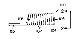
Figure 1 is an enlarged ~idevi~w o~ a
variation of the inventiv~ prosthesi~.
Figure 2 i9 an enlarged end view of' the
prosthesis ~hown in Figure l.
Figure 3-6 are enlargad sid~view~ of
variations of' the inventive pro~thesi~.
- - -5-
Figures 7 and 8 are enlarged sideview of
variations of the inventive ca~het~r assembly.
S
~etailed Description o~-3~L~ry~ Qn
The invention de~ice i8 an inf latable
prosthetic device suitable for temporary placement in
any body lumen, but primarily in a vascular lumen. It
is mado of two critical portions--an inflatabla tip
having a lumen along it~ axis and an inflation lu~en
or shaft. Th~ inflatablQ tip i~ al80 deflatable and
i8 made of a simple, hnlical windinq o~ poly~ric
tubing which is h~ld in th~ helical configuration by
tie strip~ which adhere to the tubing and "ti~" it
into the desired inflated shap~. Tha inflation lumen
or shaft carrie~ and re~ovea in~lation fluid from thQ
helical inflatzble tip.
Figure 1 showR an enlarged side view o~ on~
variation o~ the prosthetic devico (100). In thi8
variation th~ polymeric coils ~102) are tightly wound,
that is, the pitch Or the coil i~ the same a~ the
d~ameter of th~ polymeri¢ tubing m~king up th~ coil.
The out~ide diameter o~ the coil may be a~ much as
fivs times the ~ize Or the diam~ter Or thQ polymeric
material or may be fold~d or compresssd such that tha
out~ide diameter Or the coil i~ no gr~ater than about
twice the outer diameter o~ the ~nflation lumen or
shaft.
In th~ variation shown in Figur~ 1, thore
are no ~ie strips. The coil shape is maintainQd by
causing the coil turn~ to adher~ to each othQr. Thi~
adherence may be accompli hed by choosing a polymer ~-
which after winding Or the coil, may be haat treated
to allow adjacent coils to stick to each other. The
distal end of the polymeric tube making up the
inflatabla column is closed ~104) by crimping the
tubing and/or plugging it with a thermopla~tic f$11er
,
,
2~315
-6-
or the like. A radiopaque marker (106) may be placed
at the distal end and one (108) at the proximal end o~
the column. These markers are suitable for proper
placement of the prosthesis prior to in~lation and may
be any suitable radiopaque material, preferably metal.
Materials ~uch as the platinum series of metals
(platinum, palladium, etc.) and gold, silver, and
tantalum may be used as the~e markers. Certain
stainless steel are also suitable ~or use as markers.
Alternatively, the polymer used in the cath~ter body
may be radio-opaque or made so by addition o~ a ~iller
such as barium sulfate, bismuth trioxide, bismuth
carbonate, tungsten, tantalum, or th~ lika. ~-
The inflation lumen or sha~t (110) in thi
15 variation is common to the polymeric tubinq making up ~-
the coils (102).
Figure 2 is an enlarged end view of th2
prosthesis found in Figure l. The various coils
(102), distal end plug (104), and distal radiopaqua
20 marker (106) may be seen. ~-
Figure 3 shows a variation o~ th~ invention
device similar in design to that ~hown in Figures 1
and 2 but using a sizing strlp (302). Sizing strips
may be placed outside the helical column (304) as is
shown in Figure 3 and may be placed inside th~ lumen
o~ ths column. They may be used in con~unction with
or in~tead of th~ adhe~ive method~ di3cuss~d in
relation to Figure 4. ;~
The polymeric tubing making up the helical
tip o~ each of tha variation~ discussed abov~ and
below desirably i~ made o~ material which i8
relatively inelastic. That is to ~ay, the coil
inflates to a specific size when filled with an
in~lation ~luid, but do~s not in~late ~urthar or
"balloon". Elastomeric pro~these~ that ~urt~er
inflate become somewhat in~lexible and do not follow
vascular curves very readily.
.'
7-
Preferred polymers for the tubing include
polyolefins such as high and low density polyethylene,
polypropylene, and polybutylene a3 well as mixture
and interpolymers of these materials. Other suitable
polymers include polyacrylonitrile, polyurethanes,
polyethylene terephthalate and polybutylene
terephthalate, and "TEFLON" (tetrafluoroethylene).
These materials are substantially bioinert or, at
least, biocompatible. An especially suitable tubing
is made of polyethylene having a melting point of
about 350F. Such polyolefins may be ~hardened
through the use of irradiation if so desired.
The sizing strips may be of the same
polymeric materials or others of appropriate physical
characteristics. Especially suitable are the
polyolefins listed above. The sizing strips may be
glued to the helical tubing or made to adhere to the ~-
tubing in some other fashion. For instance, if a
tubing have a high melting temperature is used for the
helical column, sizing strips of a lower melting
temperature may be used. Placement of the sizing
strips against the tubing in a configuration such as
shown herein, and heat treated at a temperature
between the heat indice o~ the two polymers~ The
sizing strip~ will deform and adhere to the tubing
holding it in place.
Figure 4 shows another variation in which ;~
the sizing strips (402) are placed helically about the
column. As with the variations discussed above, the
strips may be placed either (or both) inside and
outside the column. It is desirable that the helix
formed by the sizing strips be of an opposite
"handedness" to that of the tubing helix. In this
way, the sizing strips maintain the shape o~ the
column upon inflation. If the sizing strips are wound
in the same direction, the coil will show a modest
tendency to unwind upon in~lation. As will be
.
~.
3 ~ 5
~; -8-
discussed below, in certain circumstance~, a diamQter
increase may be desirable.
Figure 5 showR a variation in which the
sizing strips (502) cross in a helical fashion. Thi~
variation usually ha~ the l~ast axial flexibility of
all of those discussed abova.
As may be apparent from th~ above discussion
of the structures of the variation~, each of the
variations will hava more or le88 compliance or
flexibility depending, in ma~or part, on the number
and placement of the sizing strips. For instanc~, the
Figure 1 and 2 variation has moderate flexibility wh~n
the coils adhere to each other. The Figure 3
variation, using only an inside or outside sizing
strip, will have significant compliance with th8
interiors o~ variou~ body lumens. That i~ to say that ~-
the Figure 3 variation could be adopted to ~orm a "C~
shape prosthesi~ when place in a region whera, rOr
instance, an artery make~ a larg~ curve. The ~-~
prosthesis region on th~ outRide Or the coil, as
placed, will have gaps betw~en the coil windings. The
compliance ~or the remainder o~ th~ variations should
be apparent.
The coil column need not be tightly wound,
as has been ~hown in tha ~igure~ discussed ~bove.
Depending in large measur~ upon the u8e to which the
prothe~is is placed, the coil column ne~d not be
liquid tight. Obviously, i~ the column coil is wound
with a pitch greater than the di3metox o~ the
tub$ng--see, rOr instance, Figure 6--there will bo
gaps between the adjacent coils. When treating
vascular spasms, a long prothQsi4 applylng a gentle
pressure over a significant distance o~ th~ vessel
lumen may be desirable.
Figure 6 shows a sido Yiew o~ a variation
(602) o~ the i~vention similar in most respects to the
tightly wound version (304) shown in Figure 3. The
coils (604) are, howevQr, not tightly wound. Tho
3 1 ~
~:`
.. :. g
distal end of the column is plugged (606). A distal
radiopaque marker (608) and a proximal radiopaque
marker (610) are also present. A sizing strip (612)
is shown a-~ placed on the outsidQ of tha column.
As is evident from the above discussion,
Figure~ 1-6 show the inventive prosthesis in their
inflated state. The device is inserted into and
removed ~rom the vasculature $n it3 uninflated state,
as indicated in Figure~ 7 and 8.
The other variation~ discussed abova, e.g.,
sizing strip~ placed inside the column lu~en, multiple
sizing strips, crossed sizing strips, etc., are all
applicable to the open wind version discussed here.
The inventive device may be used as a
portion of a single lumen or a double lumen catheter
assembly. For a single lumen catheter assembly, the
proximal end of the inflation lumen or sha~t ~e.g.,
110 in Figure 7) i~ fitted with a conventional ~itting
(701) to allow attachment to an inflation device, such
as a syrings, preferably having a pre~sure indicator.
The inventive device may be delivered within a guidlng
catheter (702) that coaxially surrounds tho lnvention
device, the helically wound coil (703) being in it~
relaxed, folded, or crushQd con~iguration. The
catheter assembly is maneuvered ~nto position in the
body lu~en prior to inflation. A guidewirQ may b~
inserted through the guiding catheter (702) and the
~alse lumen (704) o~ the helical coil to aid in
guiding the catheter assembly to thQ target ~ite. The
proximal end fitting (701) i8 then attached to the end
of the in~lation lumen (110). The prosthe~ then
in~lated such that the walls o~ the targeted ve~sal
are supported yet blood i8 allowed to ~low through the
lumen ~704) of the distal helically wound portion
(703). When the procedurc is completed, the device
may be deflated by applying vacuum to tha proximal end
~itting (710) and the device removed.
~ 1 ~ b ~
.~
1 0 -
Similarly, the invention device may be used
in a double lumen catheter assembly. In thi~
instance, the coil column (110) would include an
attachment (801~ adapted to attach the coil column
~110~ to the distal end of th~ catheter (802) in such
a way that the lumen of the coil column (110) is
generally colinear with the inner catheter luman -~-
(803). In this configuration, the outsidQ diameter of
the coil i~ the 8ize oP th~ outside diamater o~ the
cath~ter, or greater, usually about 20% greater.
A guidewire (804) may bs inserted through
the catheter outer lumen (805) and th~ Salse lumen of
the helically wound coil (704). Th~ guidewire (804)
is then used to maneuvsr the collapsed catheter
assembly to the desired target sita. The guidewire
(804) i~ then removed and an in~lation device attached
to the proximal end fitting (701). The prosthesis i8
inrlated such that the walls of the targeted vesYel
are supported yet blood is allowed to flow through the
lumen ~704) of the distal helically wound portion
(703). When the procedure is completed, the device
may be d~lated by withdrawing the included liquid
~rom the proximal end ~itting (701). The entire
device may then be removed.
Th~ embodiment~ fihown and described abovs
are only sxemplary. Various modlrication~ can be made
in the con3truction, material, and arrangement and
still be within the scope Or th~ invention round below
in the claims.
,
. ..
.
~`
