Note: Descriptions are shown in the official language in which they were submitted.
~ ~'~93/12723 ~ 2 ~ 4 4 2 PCT/US92/11311
-- 1 -- . . .
~ TR~O~R~C~S ~I~TER
!~
;, R~UND 0~ ln~ INVYNTION
The pre~ent invention relates to thrombosis filters.
To be more specific, it is about le~ dble thrombo~is filters
which can be securely fixed at a selected location in the
'! vascular sy~tem for trapping blood clot~, and removed when
desired such as when no longer reguired.
In recent year~, pl-l m~n~ry embolism has become an
increasingly cnmmo~ medical emergency. p11lm~n~ry embolisms
may be caused by venous thrombosis that in turn may be caused
i by blood flow retention, venou~ intimal damage or coagulation
abnormalities. Emergency or prophylactic treatments for these
~~ conditions include anticoagulant therapy, thrombolytic
; 15 therapy, thrombectomy and inferior vena cava blocking
'!' procedures~
;- ; Among these therapeutic options, when an inferior
; vena cava blocking procedure i8 #elected, one option is to
perform a laparotomy under general ane the~ia during which the
; ~ 20 inferior vena ca~a i5 Iigated, sutured and shortened, or
clipped. Another optio~ i~ to intravenously in3ert a
thrombosis filter into the inferior ~ena cava under local
~ir ~ ane~thesia. A laparotomy however, requires general anesthesia
~ and is susceptible to th~o,~o~is formation due to the
,~ 25 di~continuation of anticoagulant herapy prior to surgery.
For these reasons; percutaneou filter insertion i9 now more
~' widely employed~ ince it requires o~ly local anesthesia.
; Blood~ i8 ~returned to the heart from ~he lower part
of the~hl~m~n body thr~ough~the inferior ~e~a ~ava and from the
'~ 30 upper part of the body through the superior ~ena cava. The
neck uetn~ known as the jugular vein, leads to the superior
~ena ca~a which is a vein that enters the upper part of the
heart. A percutaneously inserted catheter that extends
through the jugular vein and the superior vena cava and then
into the heart can be manipulated to exit the heart through
, ~7~ .
the mouth of the inferior vena ca~a into the heart. The
, inferior ~ena cava, a ~ein that enters the lower part of the
r~':
.
:r.
~ . . I
WO93/1272~ PCT/US92/11311~
2 1 2 ~ 2 -
heart, can also be accessed through the patients femoral ~ein.
Thus, the inferior vena cava can be reached through two
endovenous routes, both of which are available percutaneously.
A currently u3ed percutaneous filter i8 shown in
S Figure 9 The filter has anchors, 43, at the end of 8iX
struts, 81, with a head or central hub or fixation part, 5.
One example of how this filter is used is to lead the filter
within a capsule with the head of the filter introduced first
if using the tran~femoral uein approach or with the tail of
it introduced first if using the transjugular ~ein approach.
This type of filter po~sesses hooks at the end of the struts
that impinge ~he inner wall of the blood ves~el in order to
ensure its firm fixati~on at the time of insertion. It is
released from the capsule and ex~ within the inferior vena
ca~a where it function as thrombosis filter.
Filters of the type shown in Figure 9 perform the
~ desired filtering function, requiring only local anesthesia
,~ for insertion. Thi~ ha ~contxibuted to the high usage of the
filter. A filter~of the type shown in Figure 9 i9 disclosed
in U.S. Patent No. 4,817,600. Approximately two to three
weeks after insertion of this type of filter, neointima grows
around the filter and its anchors to the inner wall of the
blood ~essel, which becomes an obstacle to the ~ v~l of the
filter~after its~function has~been fuIfilled. Furtkermore,
25 this filter w s~not~.designed or intended to be l~o~e~. Since
the need for a thrombo~is~filter, in many patients, i8
; t ~ orary,~it may be~desirable to l.~.~ve the filter when it is
no longer nee~A.~ In~the prior art devices, such removal can
be prefor~e~ surgi~cally~hich expo9e8~ the patient to the usual
risk and trauma~a~sociated~with surgery. Th~s there i~ a need
~ for a thrombosis fi}ter that can be removed without surge~.
-~ ; The subject invention fulfill~ the need of a medical
ilter which can be non-surgically removed even after
neointima has developed. As a result of this invention, a
,
~; 35~ medical filter has been developed whi~h includes insertion and
Le~..O~dl accessories that enable the goal to be achieved. The
filter~of this in~ention includes a filtering portion that is
,",,,, , ~ r ~ S
", ~V~) 93/12723 PCr/US92/11311
- 3
p~rm~hle to the blood flow but which will filter out emboli
and thrombus and will also hold the filter firmly in a
selected location and permit remo~al through an endovenous
route. The filtering and holding device of this medical~ S filter includes two filte~ units that face each other.and do
not have hooks that impinge the inner wall of the blood
vessel, in the same manner as the prior art filters, in order
to anchor the de~ice in the blood vessel.
The objective of this invention, therefore, i8 to
provide thrombosis filters with a favorable filtering function
which can be securely anchored at a desired location and be
!~. removed through an endovenous route even after the growth of
: neointima.
'':
S~MMARY OF T~E INVENTION
The above-mentioned objective is achieved by a
thrombosis filter which is characterized by the following
str:cture. The thrombosis filter of this invention includes a
first and a second filtering and holding unit each unit
!.' ~
~; 20 including a coupl~ing merh~n;~m in the form of a hook. Each
unit has a plurality of resilient struts which radiate from a
,,,~. ~,
, central hub and lies on the surface of a cone. The units are
~.
interconnected by a core~shaft which consist~ of a compre~sion
spring and a pa~r of core~wires. The term cG~ ession ~pring
25~ m~An~a~spring that~ will~:draw~the filtering and holding units
toward;each other~to~thereby m;ni~7e the overall longit
length of the filter. ~Inside the core shaft, there are core
wires that are anchored in the units and have free ends which
extend from their ~n~hored ends toward the other unit. The
30 8trut8 are resil~ient~and their~free ends bear a~-~nQt the
inner wall of the ~ein. Howe~er, the free ends do not include
~c~oring hooks, of the type disclosed in the prior art, that
impinge the wall of the vein as does the above discussed prior
art filter. The Anchoring of the filter, of this invention,
35 i8 accompl; ~h~ by orientating the two filter units in
opposite directions. The force from the compre~sion springs
pulls the hubs from the two units toward each other, which
, ~
.,~.,
. ~
212~4~2
~ WO93/12723 PCT/US92/11311~
.j ,
~5 tends to open the struts of both unit~ thus forcing their free
ends into the wall of the vein and holding the filter in its
!' selected location. More specifically, the core shaft of ~he
upper unit (as seen in Figure l) consists of multiple struts
which spread radially in an e~p~n~ive direction while the core
shaft of the lower unit consists of multiple strut which
spread radially. The struts from the first and 3econd units
cross each other between their hubs and free endsl which when
consideri~g the conical surfaces that the ~truts lie upon,
~'~, lO defines a V-~haped circular filtering grove. This V-shaped
i, i
circular filtering groove is perm~hle to the flow of blood
and performs an impro~ed filtering function.
Although materials of the present thrombosis filters
are not particularly restrictedj it is desirable to use a
flexible material that will return to it9 original shape after
~,~ being deformed such as identity elastic alloy wire, high
elastic alIoy wire ~uch as stairless steel, tungsten,
' platinum, piano wire, shape memory alloy wire, super elastic
,~ metal wire and chromium alloy. It is ~mportant that the~~ 20 coupling me~h~n;~m~ in the form of hooks and the cord be
con~tructed of material such a tung~ten, platinum or gold that
can be seen on a fluoroscope, to aid in the process of
securing the coupling merh~n;~m~.
Multipl4 number of strut~ or thrombosis filtering
wires u~ed for;the present invention are divided into two
., ~
units and are placed~and ~nrh~red around their corre~po~i n~
centra1 hub at regular intervals to the extent po sible. The
struts of each unlt lie on the surface of a cone with an
~ ~ anchoring part at the apex. The anchoring part iR connected
;~' 30 to and a part o~ the central hu~.
Inside the co,--~ es~ion pring, which con~titutes the
core shaft, a core wire e~tends from each of the filter unitQ
and function to reinforce the compression spring.
; ~ The angle betwee~ the radially spread struts and the
~ 35 core ~haft can be optional, although it is preferable to
~ .
~ maintain this angle within the range of 70 to 80 degrees.
,,j, .
'I
:,
,.
~' .
~1 ? 6 ~
~93/12723 PCT/US92/11311
. ~ .
The thickness of the wire constituting a strut is
preferably 0.05 to 0.2 mm in diameter.
The ideal outside diameter of the compression spring
is O.5 to 2 mm in diameter, but there is no particular
restriction to it. This diameter can of course be determ;ned
by the particular u~e int~n~PA for the filter.
As for the size of the thrombosis filters, there i8
i no particular restriction. The size can be changed at one's
discretion depending on the site of its application.
~0 The present invention enables thrombosis filtering
through multiple struts which ~pread r~ 1y. As a result
the filtering and holding device is flexible in its radial
" ~im~n~ion and can be compressed or collapsed radially for; insertion~ travel and removal. Since the struts do not have
hooks for impinging the inner wall of the blood veq~el, even
~$' when the strut~ are covered by neointima, they can be easily
,~;! removed by pulling them towards the head or central hub. The
compression spring, which serves a~ the core shaft, pro~ides a
core shaft that is flexible and facilitates maneuvering the
filter units into the recovery tubes for removal. The
compres io~ spring allows relative movement of the units along
the filter's longit~l~;n~l~axi6.
The core wires~within the compression spring
function to maintain the core shaft along a central axis
" : ~ :
~25 exten~n~ between~the~two filter units as well as to achieve a
; desired flexibiIity~of the ~o~,y~e~sion ~pring.
The co~upl~ing merh~n;Qm~ in the form of a hook
element provided~at~the~bead of each filter unit, functions to
connect ~he unit to: merhAn;~m that can be m~n;pulated through
~ 30 ~ an endo~enous route at~the time of ~.ov~l.
'~ As for the structure of the filter u~its facing each
other, thi~ arrangement serves to stabilize a~d anchor the
filter inside the~blood ves~el and to prevent unfa~orable
h~nnm~nQn ~uch ag the displacement of the filter inside the
ves el~.
. ~
:'.1
~,lf. ~
.~ .,
.~, . .
: 1!,
. ~ .
! '
WO 93/12723 2 1 2 l~ ~ 4 ~ 6 - . PCI'/US92/11311,~
,3 BRI13F DESCRIPTION OF ll~K DRAWINGS
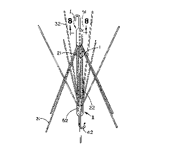
Figure 1 ic a perspective view ~howing the
thrombosis filter of thi~ invention.
Figure 2 is a perspective view showing a strut of
the thrombosis filter of this invention.
Figure 3 is an top view of filter unit I of the
~;, thrombosi~ fiiter of this in~ention.
Figure 4 i a cross-sectional view illustrating a
step in the method for removing the thrombosi~ filter of this
invention from the patient.
Figure S is a cross-sectional view illustrating a
step in the method for ~e.-G~ing the thrombosis filter of thi~
inventio~ from the patient.
Figure 6 is a cros~-sectional view illustrating a
: 15 8tep in the method for removing the thrombosis filter of this
,~ invention from the patient.
- ~ Figure 7 is~a croRs-~ectional view illustrating a
step in the ~ethod~for~removing the thrombo~i~ filter of this
invention from the patient.
Pi~ re 8 iB a ~cross-section view of the head portion
of the thrombosis filter of this i~vention taken along line 8-
8 of Figure 1. ~
Figure 9 is a~perspective view of a conventional
pri~r art thrombosis filter.
TT.Rn n~SCDTPTION OF T~B ~X~K~ EM~ODrMENT
The following~is a discussion of how the pre ent
in~ention can~be applied~to~a particular embo~im~nt . Thi~,
however,~ does not~mean~that the application of the present
invention is limited only~to the following u~e.
Figure 1 i a perspective view, of the filtering a~d
holding de~ice of the preferred~embo~m~nt of the present
invention. The filter is fabricated mainly from stainless
steel wires. me wire i8 0.05 to 0.2 mm in diameter. m e
filter con~ist~ of~twin units, I and II, that are ~paced from
~; each other along the filtering and holding devi~e's
: :
,
~ . '
! ~
~6~2
''~93/12723 PCT/US92/11311
- 7 -
longit~ n~l axis and which face each other and are connected
'fi by the compre~sion ~pring l.
The filter unit I includes the following structure.
~, The upper end of core wire 21, as ~iewed in Figure l, extend~
downwardly from hook 41. Strut~ 31 also exte~d downwardly
~ from hook 41 and spread r~ y in an eYp~n~ive direction,
j~ centering around the core wire 21. The upper end~ of struts ,~
31 are fixed in the central hub or fixation part 51.
Filter unit II has the following ~tructure. As
~iewed in Figure l, the core wire 22 and ~truts 32 extends
upwardly from hook 4~. Struts 32 spread radially at an angle
to the filtering and holding device's ~ongit~in~l axi~ in an
~xr~n~ive direction, centering around ~he core-wire 22. The
~, s~ruts 32 are anchored at, their lower end in central hub or
'~' 15 fixation part 52.
The free end~ of the struts for both units I and II
~ are thus ~pring biased outwardly and can be forced inwardly to
-~ decrease the diameter of the filtering and holding de~ice when
' ~ it is located in the insertion and remo~al tubes.
,: 20 As 8hall be discu ed in greater detail, a welding
or binder material i~ normally used for brazing the ends of
the hooks 41 and 42 the core wires 21 and 22 the struts 31 and
32 and the ends of spring 1 to the central hub or fixation
;,; parts 51 and 52.
;j ~ 25 The filter units I and II are spaced from each other
; along the filtering and holding de~ice's longit~1~;n~l axis
such that ~truts 3l and 32 cro~ed each other at abou~ their
mld poi~ts. ~oo~ 41 and 42 are constructed of tungsten or
stainles~ ~t~el wires that have been bent into the shape ~hown
~ 30 in Figure l.
7 Fir~t ends of core wires 21 and 22 are connected to
~ the central hub or fixation parts 51 and 52 along with the
., .
straight endR of ho~k~ 41 and 42. The core wire~ 21 and 22
extend through the lumen formed by compre~sion spring 1 and
function as a core shaft that enhances the flexibility of
,' compression spring l. The other ends of core ~ires 21 and 22
j~ are free.
;.
. ~ .
WO g3~12723 ~ 1 2 6 ~1~ 4 2 PCT/US92/1131~
, -- 8
" , .
," Figure 2 i~ a diagonal view of a strut 31 that
~,~. includes a head 33. Strut 32 is identical ~o strut 33 and
~",, thus is not illustrated in an isolated ~iew. The heads 33 of
,' struts 31 and 32 become a part of the core shaf~ by being
4~ 5 anchored in central hub or fixation parts 51 and 52
,/~ Figure 3 is a top view of filter unit I. ,As can be
,.
,~, seen in this view, struts 31 emana~e from the central core
," shaft and spread outwardly from thi~ center in e~ual amounts
into the surrounding area. Although 8iX struts 31 in each
filter unit have been found to be optimum, this numbex can be
varied within the range of three to ten.
~,,', Figure 8 i~cIudes two enlarged cross-sectional views
~' ? taken through the central hub or fixation part 51. A tube 53
~' is used to form the central hub or fixation part 51 of the
v 15 thrombosis filter. The straight end of hook 41, first end of
core wire 21 and the heads 33 of strut~ 31 are all inserted
into the tube 53. The ends o~ spring 1 could also be inserted
~ into the tube 53 and bonded to the tube 53 or ~he end3 could
i'" ~ be fixed to the tube by welding or adhe~i~e. A bonA~n~
~' 20 material~54 i8 then introduced, while, in a pliable stage, into
i~ tube 53 such that the ho~ng material 54 fill~ all the voids
between the end of the hook 41, core wire 21 and head~ 33
~q~ within tube 53. Whe~ the pliable material 54 harden~ the ends
of the hook 41,~core wire 21 and the heads 33 are all fixed in
the central hub or ~ixation part 51. The tube 53 could remain
as a perm~ent part of central hub or fixation part 51 or it
'~- could be ~ ov~d.
It should be noted that the compres~ion spring l,
;Y hooks 41 and 42~! core wire~ 21 and 22 and the ~trut~ 31 and 32
can all be fabricated from the 8~me material or each of these
' parts could be fabricated from different material that will
,'~ provide the desired characteri~tic~ for the particular part.
' The following is an explanation of how this
embo~im~nt of the ~hrombosis filter is u~ed. The thrombosis
~,~ 3S filter is ~m~n~ioned Buch that it can be stored in the distal
"~
': end of a thin tube (2 to 3 mm in diameter). This in~ertion
,~ tube is percutaneous1y inserted into the patient and follows
~ .
.
P~ .
...
, . ..
,.... ,; .
1, .. ,~ .
, ~.
2~';6~4 ~
~ " ~93/12723 PCT/US92/11311
. !,~ . _ . .
!' an endo~enous route into the patient's inferior vena cava.
This procedure is performed under local anesthe~ia. When the
~ distal end of this tube reaches the target site, the filtering
-j and holding device stored inside the tube i9 caused to exit
the di tal end of the tube where it become implanted in the
patient's inferior vena ca~a. A pu~her rod i8 extended
through the insertion tube and iB maneuverable from the
proximate end of the insertion tube for expelling the
filtering and holding device out the distal end of the
insertion tube. It should be noted that the insertion
procedure can be monitored on a fluoroscope.
The filtering and holding device after being
relea~ed from the tube into the patient's inferior vena cava,
i~ through the above descri~ed procedure, is in the form as shown
in figure l~ Within ~everal week after the filtering and
holding de~ice has been implanted in the inferior vena cava,
? the struts 31 and 32, which are in con~act wi~h the inferior
vena cava become covered by neointima.
When it becomes necessary or desirable to remove the
filtering and holding device, the following procedure is
followed. The term "medical filter" as usPd to describe and
claim this ~mho~m~nt includes in addition to the filtering
and holding device the above mentioned insertion tube and the
"., ~
recovery hardware necessa ~ to remo~e the filter. A recovery
'~ 25 device is provided that can be inserted percutaneously and
- threaded through the endovenous route to the filtering and
holding device. The recovery de~ice includes a first tube 64,
~i a second tube or sheath 61, a third tube 63 a~d a recovery
' tube or sheath 62. ~
Referring to figu~e 4, a doubled o~er cord 71
in~erted through the proY;mA1 end of the first tube 64 such
'~ that the cord ~.-E ye8 at the distal e~d of th~ t~be 64 and
~'' functions as a cot1rl;ng me~h~n;~m in the form of a loop. The
tube 64 then is pa~sed through the second tube or sheath 61
which has been percutaneously in~erted into the patient ~ia
~' the femoral vein such that its distal e~d is in the inferior
vena cava of the patient adjacent the filteri~g and holding
.
,
... .
, ~.
.. .. .. , . . . , , ,, .,, , .. ,, ~ .. . .. .. ... .... .
W093/12723 2 1 2 6 4 4 ~: PCT/VS92/1131~
~" - 1 0
.. device. The tube 64 i~ advanced, through tube or sheath 61,
to the filtering and holding de~ice. The tube 6~, that has
.. the loop formed in cord 71 emexging from its distal end, i8
~ manipulated such that the coupling me~h~n~sm in the form of a
:~ 5 loop is grasp by the complementary coupling mech~n; ~m in the
form of a hook 4l.
In ~he ~ame m~nner~ a doubled over cord 72 i8 passed
'~' through third tube 63 such that a ~oop that functions as a
coup~ing mechanism emerges from the distal end of the tube.
Third tube 63 i8 threaded through a recovery tube or sheath 62
.: which was percutaneously inserted through the right internal
jugular ~ein such that its distal end i9 in the patients
,~ inferior ~ena cava. The third tube 63 i~ manipulated such
that the coupling mechanism in the form of a loop is gra~ped
Al~' 15 by the complementary coupling mech~n;~m in the form of a hook
42. After cord 64 is connected to hook 41 and cord 72 is
connected to hook 42 the cords 64 and 72 are simultaneously
pulled in opposite directions, causing units I and II to mo~e
away from each other. me compression spring 1 eyr~n~ to
: 20 permit this relati~e movement of units I and II. A tension is
~ ~ ~ m~;nt~; nefl on the cords 64 and 72 to insure that the
connection~etween the cords 64 and 72 and the hook~ 4l and 42
maint~;ne~
~- : Cords 64 and 72 are made of or include radiopaque
material ~uch as stainless steel:so the at the coupling of the
loops with hook 41 and 42 can be monitored of a scope.
The relati~e:movement of units I and II has resulted
in a correspon~n~mo~ ~t of the-3truts 31 and 32 relatiYe
to the ~e~el wall~and::the filters have been freed from the
~; 30 neointima.
The next Qtep in the process of remo~ing the
: filtering and holding de~ice from the patient i8 illustrated
in Figure 5. The cord 71 i~ pulled in it~ proY~m~1 direction
: to thus pull the filter unit l into tuhe or sheath 61 for
temporary storage. Then tube 64 i~ adv~nc~ further through
~;0; : tube or sheath 61 to the point where its distal end is
~ i! .
,i, ~
~"
~ ~93/12723 ~ 12 ~ ~ 4 2 ' 'f ' PCT/US92/11311
adjacent the ends of struts 32 that are ~ecured in the
fixation member 51 of unit II.
~i As can be best seen in Figure 6, the next ~tep in
~ the removal procedure is to advance tube or sheath 62, in the
,~ 5 direction toward its~distal end, ~uch that unit II and the
distal end of tube or sheath 61 in which i~ stored unit I, are
- received within tube or sheath 62.
The entire flltering and holding device i8 now
stored in recovery tube or sheath 62. By pulling one strand
of cord 71, from~its~prox;m~l end, cord 71 i8 relea~ed from
hook 41 and can be~removed from the patient. The second tube
5~ or sheath 61 can then be~removed from the patient, and as
hown in Figure 7, and the entire filtering and holding device
~;~ is now stored in recovery tube or sheath 62.
The final step in the proce~ for removing the
filtering and holding device from its resting place in the
patient's inferior vena~cava is to pull out the recovery tube
..
or sheath 62. ~
When the~filters u~ed for the pre~ent invention are
employed in vessel~, ln;order to prevent the adhesion of
' thrombosis it is~preferable to coat the filters with
antithrombotic~agents ~(~such~as heparin, urok;n~e and
antithrombotic mater~ including h~d ~y methacrylate-styrene
co~olym~er).
~ ~ ~As a~result~of;~the thrombosis filters units I and II
~ h~ng~connected by~a~compression spring with the struts 31 and
!J''~ ' 32 ~YtenAi~n~ in~opposite directions,~ fixation of the filtering
and~h~ de~ice~at~a~selected~;location i8 accompli~he~ upon
release of the~filter from the tube. In addition, the
arrangemen~ of the~struts 31 and 32 exte~ng in opposite
directio~s and crossing provides a very effective thrombus
; filter.~Moreover~ the~ehrombus filter of this invention can
be removed when the patient no longer ha~ a need for it.
,, , ~
: ..... . . ..
.. . . ... .
C
:; :
'