Note: Descriptions are shown in the official language in which they were submitted.
E526
2127879 42/35
-
-- 1 --
DESCRIPTION
METHOD AND APPARATUS FOR OPERATION ON EYE CORNEA
TECHNICAL FIELD
This invention relates to a method of con-
ducting an operation on the cornea of an eye for cor-
recting an abnormal curve of the cornea of the eye,
such as myopia, hyperopia and astigmatism, or for
treating opacity of the cornea of the eye, and also
relates to an apparatus used in this method.
BACKGROUND ART
For correcting an abnormal curve of the
cornea of an eye, such as myopia, hyperopia and astig-
matism, or for treating opacity of the cornea of the
eye, there have heretofore been conducted surgical
operations in which the cornea of the eye is rubbed by
a spatula-like scalpel, or is ground by a rotating
file. However, in the case of using such a spatula-
like scalpel or a file, there have been encountered ~- -
drawbacks that it has been difficult to accurately form
the optical axis of the eye since the eyeball moves -~
during the operation, that the operation on the cornea
20 could not be performed smoothly and neatly, so that -~
satisfactory treatment effects could not be expected,
and that much time has been required for the curing -~
after the operation.
As a surgical operation on the cornea of an
212787~
eye replacing this, there is already known a method of
conducting an operation on the cornea of an eye by the
use of a laser knife, for example, from U. S. Patent
No. 4,718,418 and U. S. Patent No. 4,994,058. However,
even with this operation method using a laser, there
have been pointed out problems that adverse effects due
to photochemical heat such, for example, as an attack
on the tissue of the cornea, are intense, so that after
the operation, the cornea is liable to undergo a tissue
destruction, a burn, alternation, distortion, opaci~y
and so on, and that much time is required for the
curing after the operation.
To solve these problems, an operation on the
cornea has now been conducted using an ultraviolet
laser, particularly an excimer laser, which is said to
less suffer from the effects of photochemical heat
among lasers; however, even with the excimer laser, the
effects due to a photochemical thermal reaction can not
be entirely avoided.
When ablating the tissue of the cornea with
an excimer laser, the molecules of the cornea are cut,
and this is called ablation. When the excimer laser is
applied to the cornea, many free radicals are produced
with the ablation, and also it is thought that the
following phenomenon occurs. The moisture in the
corneal stroma is dissipated as steam upon laser beam
radiation, and also is heated to a temperature of
200~300 C to be formed into bubbles in the cornea, and
:, ,~ . I . . ' . ' ' '
.: ~ .- . " . ;
,, .. ~ ~ :
~" ' ' , ~ , ` ':
2127~7~
-- 3 --
moves actively to destroy the arrangement of collagen
in the corneal stroma. Further, the excimer laser
beam, when impinging on the collagen, cuts the bond of
the molecules thereof to produce a plume (mushroom-
shaped cloud) to produce a thermal imbalance conditionlocally, thereby generating a high temperature of
1000C. This high temperature diffuses to the neigh-
borhood to impart a thermal equilibrium condition to
the cornea in the vicinity of the laser beam-irradiated
portion, thereby causing a temperature rise of about
15C as a whole to impart a thermal trouble to the
corneal stroma, which creates the cause of the opacity.
Furthermore, when the excimer laser beam is applied to
the cornea, an impact sound is produced, and a high
15 pressure impulsively develops. As described above, -
even if the excimer laser is used, the temperature of
the cornea rises, and the arrangement of the collagen
of the cornea is destroyed, and stresses due to the
pressure increase are applied to the cornea, and par-
ticularly opacity develops immediately beneath the
surface layer of the cornea. Thus, these side effects
can not be avoided. Also, a collagen-like material is
secreted from the endothelial cells to Descemet's mem-
brane.
~5 It is an object of this invention to provide
a method of conducting an operation on the cornea of an
eye by the use of an ultraviolet laser, in which the
side effects caused by the photochemical thermal reac-
- ,,
: , .. ' . , : .... . , - ~
' '' ~ ,'' ':' , .',' ''"' . ., ~
2127~79
-- 4 --
tion can be suppressed as much as possible.
Another object of the invention is to provide
an apparatus which can be effectively used for the
above operation.
DISCLOSURE OF THE INVENTION
A method of conducting an operation on the
cornea of an eye according to the present invention is
characterized in that it comprises the steps of cooling
an operation-applying portion of the eye cornea to
suppress a photochemical thermal effect and also to
lower the activity of cells of the cornea; sprinkling
or spraying a liguid medicine, having such effects as
cornea remedy, cure promotion and resolution, onto the
cornea at predetermined times before, during and after
the operation; separating the epithelium of the cornea;
and removing the excess liquid medicine on the cornea,
and applying an ultraviolet laser beam to the opera-
tion-applying portion.
Apparatus for conducting an operation on the
cornea of an eye according to the present invention is
characterized in that it comprises an ultraviolet laser
beam source; control means for controlling an ultravio-
let laser beam emitted from the laser beam source; an
optical system for guiding the laser beam to an opera-
tion-applying portion of the cornea; means for cooling
that portion of the cornea to undergo the operation;
means for sprinkling or spraying a liquid medicine onto
the cornea; and means for removing the liquid medicine
2~278~9
-- 5 --
supplied onto the cornea cooler of the cornea.
Although the present invention can be exten-
sively applied to operations on the cornea of an eye
employing an ultraviolet laser, the most preferred
ultraviolet laser is an excimer laser.
In the present invention, before, during and
after the operation, the operation-applying portion of
the cornea of the eye is cooled so as to form 2 cooling
barrier, thereby alleviating side effects due to heat
produced by excimer laser radiation, and also suppress-
ing the activity of the organism and cells of the
cornea, thus suppressing an excessive reaction of the
organism against excimer laser radiation. Also, the
liquid medicine for remedy, protection, cure promotion
and resolution purposes is suitably sprinkled or
sprayed onto the operation-applying portion of the eye
cornea, thereby protecting the eye cornea, accelerating
the remedy, and preventing side effects such as
opacity. The excess liquid medicine sprinkled on the
cornea must be removed when the laser radiation is
applied, and the irradiation of the laser beam is
effected in an ON-OFF manner to produce pulses, and
therefore the control is effected in such a manner that
the sprinkling of the liquid medicine as well as the
blowing-off of the liquid medicine by gas is carried
out during a short time period (corresponding to the
pulse interval) when the laser radiation is OFF.
Instead of merely sprinkling the liquid medicine on the
? ~
2~ 27~79
cornea, it can be sprayed in an atomized condition, in
which case the effect is also achieved.
It is proper that the medicine for protecting
the cornea and for alleviating the side effects con-
tain, as ingredients, salts such as NaCl, CaCl2, KCland MgCl2, buffer salts such as NaH2PO4 and NaHCO3, and
an energy source such as glucose and glutatione, and
that the medicine be in the form of a solution having
an osmotic pressure of 305~310 mOsm and PH 7.2~7.6.
According to results of animal tests, good
results can be obtained when the cornea cooling temper-
ature is 0~10C. In some cases, the operation may be
conducted in a frozen condition of the cornea cooled to
a temperature of not more than 0C. In order to accu-
rately control the cooling temperature to a desired
value, it is necessary that a sensor for detecting the
temperature of the cooling means should be provided,
and that a temperature control means responsive to a
signal from this sensor for controlling the temperature
of the cooling means to a predetermined temperature
should be provided. This temperature control can be
effected quite simply and accurately by using Peltier
elements as the cooling means.
BRIEF DESCRIPTION OF THE DRAWINGS
Fig. 1 is a cross-sectional view of one
embodiment of a cornea cooling and liquid medicine
supply apparatus used for an operation on the cornea of
an eye by an ultraviolet laser;
.. ~ . . ' ~ -:
~ 27879
Fig. 2 iS a cross-sectional view of an em-
bodiment obtained by modifying part of the apparatus of
Fig. 1;
Fig. 3 is a front cross-sectional view of one
embodiment of a corneal epithelium separating device;
Fig. 4a and Fig. 4b are a front cross-sec-
tional view and a bottom view of another embodiment of
a corneal epithelium separating device, respectively;
Fig. 5 is a block diagram showing a control
system for the apparatus for conducting a corneal
operation according to the present invention;
Fig. 6 is an explanatory diagram showing the
timings of the operations used in the operation accord-
ing to the present invention;
Fig. 7 is a perspective view showing the
cross-section of a half of an embodiment of an appara-
tus for effecting the cooling of the cornea and the
spraying of a liquid medicine during the operation
where the cornea is deeply cut;
Fig. 8 is a perspective view of an embodiment
of an apparatus used for a slight-degree eye cornea
operation;
Fig. 9 is a perspective view showing the
cross-section of a half of an embodiment of an appara-
tus used for a medium- and high-degree eye cornea
operation, showing an internal structure thereof;
Fig. 10 is a cross-sectional view of an
embodiment of an apparatus for effecting the cooling of
2~2787~
-- 8 --
the cornea before and after an eye cornea operation, in
which a cooling medium is used as cooling means;
Fig. 11 is a cross-sectional view of an
embodiment of an apparatus for effecting the cooling of
the cornea before and after an eye cornea operation, in
which a Peltier element is used as cooling means;
Fig. 12 is a cross-sectional view of another
embodiment of an apparatus for effecting the cooling of
the cornea before and ater an eye cornea operation, in
which a Peltier element is used as cooling means;
Fig. 13 is a cross-sectional view of an
embodiment of an apparatus for effecting the cooling of
the cornea and the sprinkling of a liquid medicine
before and after an eye cornea operation;
Fig. 14 is a cross-sectional view of an
embodiment of an apparatus for effecting the cooling of
the cornea during an eye cornea operation;
Fig. 15 is a cross-sectional view of another
embodiment of an apparatus for effecting the cooling of
29 the cornea during an eye cornea operation;
Fig. 16a and Fig. 16b are a plan view and a
cross-sectional view, respectively, of a further em-
bodiment of an apparatus for effecting the cooling of
the cornea during an eye cornea operat~on in which a
Peltier element is used as cooling means; and
Fig. 17 is a schematic view of an embodiment
in which a peripheral portion of the cornea is cooled
by a cooling fluid during an eye cornea operation.
212787~
.
g
BEST MODE FOR CARRYING OUT THE INVENTION
An eye cornea operation according to the
present invention is initiated by starting the instil-
lation of a liquid medicine on an eye and an internal
administration of a medicine one week before the opera-
tion. This liquid medicine has the effect of alleviat-
ing troubles due to a photochemical thermal reaction
involved in the operation using a laser, and more
specifically has such effects as the safety of the
cornea treatment, a promoted remedy after the operation
and resolution, and it has been confirmed through
animal tests that the liquid medicine is extremely
effective when it contains as ingredients oxygltatione
+ salts + buffer salts + glucose + glutatione
(tradename: BSS PLUS), Rinderon (steroid), tarivid
(antibiotic agent) and Sikon + Toki (burn curing medi-
cine). By repeatedly instilling this liquid medicine
on the eye before the operation, the concentration of
the liquid medicine in the cornea is increased. It has
been found that to internally administer an antiphlo-
gistic, such as tatione and steroid or indacine, simul-
taneously with the instillation further enhances the
effect.
Before starting the operation, the cooling of
the cornea, as well as the sprinkling or spraying of a
liquid medicine onto the cornea, is effected. Although
the side effects are reduced only with the cooling, it
has been confirmed through animal tests that the use of
2~ 27879
, ~
-- 10 --
both the cooling and the liquid medicine further reduce
the side effects. The liquid medicine is the same as
that used for instillation. Sikon and Toki in this
liquid medicine are herb medicines, and the Sikon
serves to cure a burn, an inflammation and a tumefac-
tion and also to lower a local temperature, while the
toki serves to promote the effect of the Sikon.
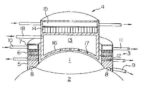
Fig. 1 shows one embodiment of an apparatus
for effecting the cooling of the cornea as well as the
sprinkling of the liquid medicine on the cornea. In
Fig. 1~ 1 denotes the cornea of an eye, and 2 denotes
the sclera thereof. The cornea-cooling and liquid
medicine-sprinkling apparatus comprises an outer tube 3
and an inner tube 4, and the outer tube 3 comprises
three-stage blocks 5, 6 and 7 integrally connected
together, and is adapted to be placed on the sclera 2.
A lower surface of the lower-stage block 5 is formed
into a curved surface conforming in configuration to
the surface of the sclera 2, and a downwardly-open
annular suction chamber 8 is formed in this lower
surface. The suction chamber 8 is connected to a
vacuum source via a tube 9 so as to create a negative
pressure therein, thereby holding the outer tube 3 on
the sclera 2 by suction. During the operation, the
eyeball is thus fixed in a stationary condition, and
the optical axis of the eyeball is accurately posi-
tioned. `
Peltier elements are incorporated in the
.
. ~
: ,, : : ~ ~ : -
,
2~27~7~
11
intermediate-stage block 6, and is arranged in such a
manner that their heat-absorbing side is directed
toward the lower-stage block 5 while their heat-gener-
ating side is directed toward the upper-stage block 7.
The upper-stage block 7 serves as a heatsink, and a
cooling medium, flowed thereinto from a tube 10, cools
the heat-generating side of the Peltier element in the
block 6, and flows out into a tube 11. Thus, the
temperature of the heat-absorbing side of the Peltier
element is further lowered. The cooling temperature by
the Peltier element can be controlled by electric
current flowing through the Peltier element, and there-
fore there is provided an advantage that the cooling
temperature can be easily controlled. Low-temperature
gas such as cooled carbon dioxide gas, liquid nitrogen,
liquid helium, Freon gas and the air, or a cooled
liquid such for example as any arbitrary cooling liquid
such as city water can be used as the cooling medium
flowing through the heatsink 7.
The lower-stage block 5 is preferably made of
metal such as platinum, gold, silver and stainless
steel, but other material than metal such as ceramics
can be used in so far as it has a high thermal conduc-
tivity. The lower-stage block 5 is cooled by the
Peltier element in the intermediate-stage block 6, and
cools the sclera 2 and the outer peripheral portion of
the cornea 1, so that the peripheral portion of the
cornea to undergo the operation is cooled from the
: . ,,
,~ .
.-.;....... .
,'
~ ~ : ., ,
2127879
- 12 -
periphery. Suction holes 12 are formed in the lower-
stage block 5 for removing the liquid medicine, sprin-
kled on the cornea 1, before laser radiation as de-
scribed later.
In the sequence from the bottom to the top, a
liquid medicine reservoir portion 13, a Peltier element
portion 14, and a heatsink 15 for cooling the Peltier
element portion 14 are provided at the inner tube 4. A
bottom wall 16 of the liquid medicine reservoir portion
13 is formed into a curved surface conforming in curva-
ture to the surface of the cornea 1, and has a number
of liquid medicine discharging holes 17. The liquid
medicine reservoir portion 13 has a surrounding wall
made of a thermally-conductive material similar to that
of the lower-stage block 5 of the inner tube 3, and is
cooled by the Peltier element 14 above it, so that the
liquid medicine therein is cooled, and also the bottom
wall 16 is cooled, thereby cooling the cornea 1 dis-
posed in contact with the bottom wall 15. 18 denotes a
tube for supplying the liquid medicine to the liquid
medicine reservoir portion 13. Incidentally, it is
desirable that a cooling device utilizing Peltier
elements be provided midway on the tube 18 so as to
beforehand cool the liquid medicine to be supplied to
the liquid medicine reservoir portion 13.
When the above apparatus is used for provid-
ing a pretreatment for the operation, the outer tube 3
is always kept fixed on the sclera 2 while the inner
~ .
- ~
.
~, . .. . .. .
. . . - - . ~
2127~7~
- 13 -
tube 4 is moved upward and downward between a position
where the inner tube 4 is in contact with the cornea 1
and a position where the inner tube is upwardly apart
slightly from the cornea 1, thereby effecting the
cooling of the cornea 1 and the supply of the liquid
medicine onto the cornea 1 alternately. After this
treatment, the liquid medicine on the cornea 1 is
finally removed by suction through the suction holes 12
formed in the outer tube 3.
Fig~ 2 shows a modification of the apparatus
of Fig. 1 which differs from the embodiment of Fig. 1
only in that a tube 18' for supplying the liquid medi-
cine to the liquid medicine reservoir portion 13 passes
through a central portion of the inner tube 4, and
extends upwardly. The inner tube 4 can be withdrawn
from the outer tube 3 as in the embodiment of Fig. 1.
The cooling of the cornea and the sprinkling
of the li~uid medicine on the cornea are carried out as
described above before the operation, and thereafter
the epithelium of the cornea is separated. The follow-
ing devices can be used for separating the epithelium.
Fig. 3 shows one example of separation device in which
an annular blade 20, projecting from a lower surface of
a disk 19 at a peripheral edge portion thereof, i5
rotated by a motor 21, and the blade 20 is engaged with
that portion of the epithelium 1' covering the surface
of that portion of the cornea (stroma) 1 to undergo the
operation, and is rotated to form a circular line of
. - ~
,,. , - ~ ..
21~7~7~
- 14 -
cut having a depth of 30~40 ,u. The height of the blade
projected from the lower surface of the disk 19 is set
to 30~40 ~ so that it will not damage the cornea stro-
ma, and ~he lower surface of the disk 19 serves as a
stopper so that the line of cut in the epithelium will
not be deepened further. Without using the motor, the
disk may be pressed against the cornea form the upper
side with the hand, and is rotated, thereby forming a
line of cut. The annular blade has a diameter of
4.5~10 mm. A piece of the epithelium formed by the
line of cut is removed by a spatula.
Figs. 4a and 4b are a cross-sectional view
and a bottom view of another example of epithelium
separation device, respectively. A disk 22 rotated by
a motor 21 is curved downwardly into agreement with the
curvature of the cornea, and an annular blade 23 is
projected from a lower surface of this disk at a pe-
ripheral edge portion thereof. Slits 24 are formed in
the disk 22, and a blade 24' is formed along one edge
of each slit 24. In this embodiment, a circular line
of cut is formed in the epithelium of the cornea by the
blade 23, and that portion of the epithelium disposed
inside this line of cut is removed by the blades 24',
and is discharged through the slits 24. The above
separating operation is carried out in the apparatus of
Fig. 1 or Fig. 2 while continuing the cooling of the
cornea, with the outer tube 3 fixed to the sclera 2 but
with the inner tube 4 removed.
2~27~7~
,
- 15 -
After the epithelium of the cornea is sepa-
rated, the inner tube 4 is again fitted in the outer
tube 3 in the apparatus of Fig. l or Fig. 2, and the
cooling of the cornea and the sprinkling of the liquid
medicine are effected.
Then, a corneal ablation operation is carried
out by excimer laser radiation. Fig. 5 is a block
diagram showing a general construction of an excimer
laser control system. By controlling a diaphragm
aperture in accordance with an abnormal refraction
condition, a laser beam is adjusted in energy distribu-
tion, and is reflected by a dichroic mirror, and is
applied to that portion of the cornea to undergo the
operation. The laser beam-applied portion of the
cornea to undergo the operation is observed by a micro-
scope through the dichroic mirror. Where the Peltier
elements are used as means for cooling the cornea, the
temperature of the heat-absorbing portion of the
Peltier element is detected by a temperature sensor~
and electric current flowing through the Peltier ele-
ment is subjected to a feedback control so that this
temperature can be brought into a predetermined temper-
ature.
The laser beam is emitted as pulses as shown
in Fig. 6, and is intermittently applied onto the
cornea. During laser radiation, the liquid medicine
should not be deposited on the cornea, and therefore
the laser radiation, the supply of the liquid medicine
- 21~787~ - 16 -
and the blowing of gas are controlled at such timings
that the liquid medicine is supplied immediately after
the laser radiation is off, and is removed from the
cornea, for example, by gas blown thereto immediately
before the laser radiation is on, and is discharged
through the suction holes always maintained under
negative pressure.
Where the operation is to be conducted by a
scanning method, with a spot of the excimer laser made
small, and where the side effects are small as in an
operation for myopia, astigmatism or hyperopia of a
slight degree, there can be used a method in which the
inner tube 4 is removed with the outer tube 3 kept
fixed in the apparatus of Fig. 1 or Fig. 2, and only
the peripheral portion of the cornea to undergo the
operation continues to be cooled, and the excimer laser
radiation is applied through the central portion of the
outer tube 3 to that portion of the cornea to undergo
the operation, without cooling the central portion of
the cornea and without supplying the liquid medicine to
the cornea.
Where the diameter of the spot of excimer
laser radiation is large enough to produce large ener-
gy, and where the ablation of the cornea is deep be-
cause of an operation for abnormal refraction of a high
degree, the cooling of the cornea, as well as the .
- supply of the cooled remedy liquid medicine to the
cornea, must be effected even during the operation by
-
, " ' '` ~ :
" ` ` ' ` ' ` ` ; ~ . ' ~`, '
-~ 2~27879
- 17 -
the excimer laser. Fig. 7 shows one example of appara-
tus used in such a case. Instead of the apparatus of
Fig. 1 or Fig. 2, the apparatus of Fig. 7 is placed on
the sclera 2 after the separation of the corneal epi-
thelium is effected. The apparatus of Fig. 7 comprisesa cylindrical member 25 having generally the same size
as that of the outer tube 3 of the apparatus of Fig. 1
or Fig. 2, and its lower end portion, adapted to con-
tact the sclera 2 and a peripheral portion of the
cornea 1, is constituted by an assembly of Peltier
elements 26. Although not shown, an eye fixing means
such as a suction chamber as in the outer tube of Fig.
1 or claws, is provided on that surface of the Peltier
elements adapted to contact the sclera 2. Remedy
liquid medicine supply passageways 27, pressurized gas
supply passageways 28 and liquid medicine suction holes
29 are formed through a cylindrical wall above the
Peltier elements in this order from the upper side. A
liquid medicine spray nozzle 30 is mounted on an inner
end of the passageway 27, and a pressurized gas injec-
tion nozzle 31 is mounted on an inner end of the pas-
sageway 28. An excimer laser beam is applied from the
upper side of the cylindrical member 25 to the cornea 1
through a generally central portion thereof.
During the operation, the Peltier elements 26
cool the sclera 2 around the cornea 1 undergoing the
operation, as well as the peripheral portion of the
cornea, and the excimer laser are irradiated as pulses
~$~
. r~ . ~ . , :; :
.: .. .: . ' ' ' .
~ . :' , . - ' , ' `, .. : ~
: ~ : . ~ ' . .
: ~ . . : ' .
21~7~7~
- 18 -
as shown in Fig. 6. During the interruption of the
laser radiation, the remedy liquid medicine is sprayed
from the liquid medicine spray nozzles 31 onto the
cornea 1 at a predetermined timing, and pressurized gas
is injected from the injection nozzles 31 at a prede-
termined timing to blow off the liquid medicine depos-
ited on the cornea l, and this liquid medicine is
removed by suction through the suction holes 29. Thus,
with the apparatus of Fig. 7, not only the cooling of
the cornea but also the supply of the remedy liquid
medicine are effected even during the operation by the
excimer laser, and therefore even if a deep ablation of
the cornea 1 is effected using large energy, the pro-
duction of side effects can be kept to a low level.
Particularly in this apparatus, if the supply of the
liquid medicine to the cornea is effected not only by
sprinkling but also by spraying, this further enhances
the effect. -
After the operation, the cooling of the
cornea and the supply of the liquid medicine are suffi-
ciently effected using the apparatus of Fig. 1 or Fig.
2. It is appropriate that the cooling temperature
should be 4C, but if the cooling is effected too long, -
the function of the endothelial cells of the cornea is
25 lowered, so that the cornea is subjected to swelling. `
Therefore, the cooling by the Peltier elements is
stopped in a suitable period of time, and thereafter
only the supply of the cooled remedy liquid medicine is
. . . , -
.~": ~ :
,' ' ',
. ~: ' ' ~ . , .
" ~ , ~
-: ~ ~ , :, : -
2127~79
-- 19 --
effected. Time required for this i5 2~3 minutes.
Then, an ointment of steroid, such for exam-
ple as an ointment prepared by mixing, with Neo-Medrol
EE ointment, an antibiotic agent such as tarivid,
indacine (non-steroid antiphlogistic), BSS PLUS or
tatione, Sikon plus Toki, and neurotensin and
fibronectin plus EGF tEpidermal Growth Factor), is
applied to the eye, and an eye bondage is used. Alter-
natively, a disposable soft lens (for example, the
tradename "ACUVUE") or a collagen shield impregnated
with a li~uid medicine composed of these ingredients is
put into the eye to cover the cornea. By doing so, the
wound is quickly curred and the pain after the opera-
tion is eased. Also, an injection of steroid, an
antibiotic agent and Sikon is applied under the con-
junctiva.
Various other embodiments of apparatus used
for an eye cornea operation of the present invention
will now be described. Fig. 8 shows an apparatus for
conducting a slight-degree eye cornea operation. An
eye fixing ring 31 is mounted on a support post 32 for
upward and downward movement, and is placed in such a
manner as to be slightly pressed against the sclera 2
around the cornea 1 in surrounding relation to the
cornea. A number of claws 33 are mounted on a lower
surface of the fixing ring 31, and fix the eye against
movement relative to the fixing ring 31, thereby prop-
erly positioning the optical axis of the eye.
- ~ ,
- :
.
' ' ~ , : '
. . . .
,'~ . , , ., ~ :
: , . . : ,
212787~
- 20 -
A ring-shaped cooling pipe 35 is fixedly
provided above the fixing ring 31 through a support bar
34. The cooling pipe 35 is connected to a cooling gas
bomb 37 via a connecting pipe 36, and cooling gas is
injected toward the cornea l from a number of injection
nozzles 38, provided in a lower surface of this cooling
pipe, to cool the cornea 1. Carbon dioxide gas, liquid
nitrogen, liquid helium or the like is used as the
cooling gas, and instead of the cooling gas, a cooling
liquid such as cooled BSS PLUS, physiological saline
and a Ringer's solution may be used. The cooling pipe
35 is fixed to the support bar 34 in such a manner that
the distance between this cooling pipe and the cornea
1, as well as the angle of this cooling pipe relative
to the cornea 1, can be adjusted, and the cooling pipe
beforehand forms a cooling barrier around the portion
to undergo the operation.
An excimer laser 39 is mounted above the
cooling pipe 35, and applies laser beams to the cornea
1 through a space encircled by the cooling pipe 35.
Before and after the irradiation of the laser beam, a
liquid medicine, having such effects as cornea remedy,
cure promotion and resolution, is sprinkled on the
cornea by the use of a suitable device. It is also
~5 necessary to blow off this liquid medicine from the
cornea before the irradiation.
A shut-off valve 40 is provided on the con-
necting pipe 36 connecting the cooling pipe 35 to the
~.
. . , ~ '' :' ~
. .. .; , . .; .. ,
- : . ..
212787~
- 21 -
cooling gas bomb 37, and the opening and closing opera-
tion of this shut-off valve 40 is associated with the
operation of the excimer laser 39 by a controller 41.
When the laser beam is to be applied, the shut-off
valve 40 is clGsed to interrupt the blowing of the
cooling gas, and when the irradiation of the laser beam
is stopped, the cooling gas is blown. Thus, the blow-
ing of the cooling gas and the irradiation of the laser
beam can be sequentially repeated. Alternatively, the
treatment by the excimer laser may be carried out while
continuously blowing the gas.
The thickness of the fixing ring 31 is set to
1~5 mm, and its diameter is set to 12~30 mm. The
thickness of the cooling pipe 35 is set to 2~15 mm, and
the inner diameter of the gas passageway is set to 1~13
mm, and its pipe diameter is set to 6~50 mm.
Fig. 9 shows an apparatus for conducting a
medium-degree eye cornea operation, and a hollow cylin-
drical fixing tube 42 made, for example, of an acrylic
material, is mounted on a support post 32 for upward -
and downward movement. The fixing tube 42 has such a
diameter that it covers the cornea 1 of the eye and
part of the sclera 2 around the cornea. A suction tube
43, which communicates with a vacuum pump (not shown)
and has a number of spaced narrow holes for contact
with the sclera 2, is provided along the bore of the
fixing tube 42 at a lower end portion thereof, and the
fixing tube 42 is fixed to the eye by suction.
. ,
.... ~ ~ .
;
..,
2127~79
A cooling pipe 44, having a number of
downwardly-directed nozzles 45 formed therein, is
mounted along the inner surface of the fixing tube 42
at an upper end portion thereof, and cooling gas or a
cooling liquid is applied toward the eye cornea to form
a cooling barrier at the operation-applying portion of
the cornea.
Arl excimer laser 39 is mounted above the
fixing tube 42, and applies laser beams to the cooled
cornea through a hollow portion of the fixing tube 42.
Before and after the irradiation of the laser beam, a
liquid medicine is sprinkled or sprayed onto the cornea
by the use of a suitable device as described above, and
it is also necessary to blow off this liquid medicine
from the cornea before the irradiation.
Further, a microcamera 46 connected to a
television monitor (not shown) is mounted on the fixing
tube 42, so that the condition of the operation on the
cornea can be monitored through the television monitor,
and also a temperature sensor 47 is provided so that
the operation can be conducted while confirming the
cooling condition of that portion of the cornea under-
going the operation, as well as the condition of forma-
tion of the cooling barrier. This is useful. The
timing of irradiation of the laser beam, as well as the
timing of blowing of the cooling gas, may be controlled
by the use of a controller, as in the apparatus of Fig.
8.
. , s ---. ...
,, ~ , . . .
,~ ' . ' ' . : ' ' ~ ' .
' ' ' " ' ~ .'~ , ~ '
'- , ,. , ' . ' ,
2127~7~
- 23 -
Referring to the type of apparatus for con-
ducting a high-degree eye cornea operation, the upper
open end of the fixing tube 42 in the apparatus of Fig.
9 is closed by a lid 48 made of a material having laser
beam-transmitting properties, such as quartz glass.
When the fixing tube 42 with this lid 48 is fixed by
suction on the eye, the interior of the fixing tube 42
is sealed from the outside air, and therefore the
cooling gas injected from the cooling pipe 44 will not
leak to the exterior, so that the cooling of the cornea
can be carried out in a short time period. By connect-
ing a suction pipe 49 to the fixing tube 42, the inte-
rior of the fixing tube 42 can be evacuated by a vacuum
pump (not shown), so that the cornea cut by the laser
beam from the excimer laser can be discharged to the
exterior. By evacuating the interior of the fixing
tube 42 through this suction pipe 42, the effect of
bonding of the fixing tube relative to the eye can be
further enhanced. Moreover, the use of the fixing tube
42 can prevent side effects caused by leakage of re-
flection light and scattered light of the excimer laser
to the exterior.
Fig. 10 shows an embodiment of apparatus for
effecting only the cooling of the cornea before and
after an eye cornea operation. A hollow cooling member
50 includes a cylindrical side wall 51, and a bottom
wall 52 having a concave surface conforming in curva-
ture to the cornea 1 of an eye. This member is prefer-
.
~: :' . . ", , : .
2~ 2787~
- 24 -
ably constructed of metal such as platinum, gold,
silver and stainless steel, but may be constructed of
other material than metal such as ceramics in so far as
it has a high thermal conductivity. In some cases, the
cooling member 50 can be formed into a flexible con-
struction by a flexible metallic film, silicone film,
synthetic rubber film or any one of various kinds of
plastic films, in which case the degree of contact of
the bottom wall 52 with the cornea 1 is enhanced, which
is desirable. The height of the cooling member 50 is
10 mm, and its diameter is 10~30 mm.
A flange 53 is formed on and projected out-
wardly from the outer periphery of the cooling member
50 at the lower end thereof in continuous relation to
the bottom wall 52, and claws 54 for engaging the
sclera 2 to fix the eye relative to the cooling member
50 are formed on a lower surfzce of this flange. A
cooling medium feed pipe 55 and a cooling medium dis-
charge pipe 56 are connected at their one ends to the
side wall 51 of the cooling member 50, and the other
ends of these pipes are connected to a cooling medium
reservoir (not shown). Thus, a cooling medium such as
low-temperature gas and a low-temperature liquid, flows
through the cooling member 50 to cool the cornea 1.
Preferably, the outer diameter of the cooling medium
feed and discharge pipes 55 and 56 is 2~15 mm, and
their inner diameter is 1~13 mm.
Further, a temperature sensor 52' such as a
, :, ~ - : - , . . - ,,
.~.: . .. : ::
- - : ~ ~ :: ..
.: . . - ~ . . :
'x~
2~2787~
- 25 -
thermistor may be provided in the bottom wall 52 of the
cooling member 50 for accurately controlling the tem-
perature of the cornea to a set temperature, and if
necessary, supply pipes and suction pipes for physio-
logical saline, BSS PLUS, a Ringer's solution, ananesthetic liquid and etc., a microcamera connected to
a television monitor, and so on may be provided in the
vicinity of the cooling member 50. As the cooling
medium, cooling gas, such as carbon dioxide, liquid
nitrogen, liquid helium, Freon gas and the air can be
used, and also a cooling liquid such as cooled city
water can be used. During the operation by the excimer
laser, this cooling member must be removed and replaced
by other cooling device capable of allowing the irradi-
ation of the excimer laser.
Fig. 11 shows another embodiment of apparatusfor effecting only the cooling of the cornea before and
after an eye cornea operation, as in Fig. 10. This
embodiment has a feature that a cooling member 57 is
cooled by a Peltier element 58. A support block 59 of
metal with good thermal conductivity is mounted under
the Peltier element 58, and a lower surface of the
support block 59 is formed into a concave surface
conforming in curvature to the cornea 1, and when in
use, this lower surface is in contact with the surface
of the cornea 1 to cool the cornea. 59' denotes a
temperature sensor provided in the support block. A
flange 60 is formed on and projected outwardly from the
; ~ . . ,
~., . ~ .. ,, . ~
21~7~7~
- 26 -
periphery of the support block 59 at a lower end there-
of, and claws 61 for engaging the sclera portion around
the cornea of the eye are formed on a lower surface of
this flange so as to fix the eye in a stationary condi-
tion.
A heatsink 62 is provided in contact with an
upper portion of the Peltier element 58, that is, a
heat-generating portion of the Peltier element. A
cooling medium flows through the heatsink 62 via a
cooling medium feed pipe 63 and a cooling medium dis-
charge pipe 64. The function of the heatsink 62 is to
cool the heat-generating portion of the Peltier element
5~ to relatively lower the temperature of a heat-ab-
sorbing portion of the Peltier element 58 disposed in
contact with the support block 59. As in the apparatus
of Fig~ 10, if necessary, a temperature sensor, supply
pipes and suction pipes for physiological saline, BSS
PLUS, a Ringer's solution, an anesthetic liquid and
etc., a microcamera connected to a television monitor,
and so on may be provided in the vicinity of the cool-
ing member 57.
The apparatus of Fig. 11 employs the Peltier
element, and therefore a lightweight and compact con-
struction is achieved; since the configuration of the
element can be freely chosen, this is best suited for
the cooling of the eye cornea; by controlling electric
current, a more precise temperature control is possi-
ble, and a temperature response is good; since there is
, ~. ,
212787~
- 27 -
used no moving mechanism, vibra-tions and noises are not
produced; since no mechanical part is used, the dura-
bility is good, and a stable performance can be
achieved for a long period of time; and since there is
little leakage of the cooling medium, the safety and
sanitariness are achieved. Thus, these and other
effects can be obtained.
Fig. 12 shows a further embodiment of cooling
member utilizing Peltier elements. The cooling member
65 includes a support block 67 which is mounted under
the Peltier elements 66, and has a lower surface con-
forming in configuration to the surface of the cornea
1. Instead of a heatsink, cooling fins 68 are provided
on the Peltier elements 66 to radiate heat of a heat-
yenerating portion of the Peltier element 66. 67'denotes a temperature sensor mounted in the support
block 67.
Fig. 13 shows an apparatus capable of effect-
ing the cooling of the cornea and the supply of the
remedy liquid medicine before and after the operation.
A hollow cooling block 70 is mounted under Peltier
elements 69, and the liquid medicine is supplied to the
interior of the block 70 via a liquid medicine supply
pipe 71. A lower surface of the cooling block 70 has a
configuration conforming to the surface of the cornea,
and a number of liquid medicine discharge ports are
formed through a bottom wall 72 thereof. The cooling
block 70 is made of a thermally-conductive material,
.
. .
~ ,, . .:
2127879
- 28 -
and is cooled in contact with a heat-absorbing portion
of the Peltier elements 69. A heatsink 74 is provided
in contact with an upper or hea~-generating portion of
the Peltier elements 69, and a cooling medium flows
through the heatsink 74 via a cooling medium feed pipe
75 and a cooling medium discharge pipe 76 to cool the
heat-generating portion of the Peltier element 69. The
above apparatus can not be used during the operation by
the excimer laser. The liquid medicine to be supplied
to the cooling block 70 may be pre-cooled by Peltier
elements (not shown) during the passage of this liquid
medicine through the pipe 71 so that the cooling effect
can be further enhanced. In this embodiment, a temper-
ature sensor 70' is mounted in an upper wall of the
cooling block 70 because of the arrangement of wiring.
Fig. 14, Fig. 15 and Fig. 16 respectively
show apparatuses which employ Peltier elements, and are
capable of cooling the cornea even during the opera-
tion. Fig. 14 shows a cooling member 77 of a cylindri-
cal shape which has a thick wall, and has at its cen-
tral portion a circular bore capable of passing a laser
beam therethrough. A cooling block 80 of a thermally-
conduc~ive material having a circular central bore 79
is mounted under Peltier elements 79 arranged in an
annular manner, and a lower surface of this cooling
block has a configuration conforming to the surface of
the cornea 1. 81 denotes a flange which has at its
lower surface a fixing means, such as claws and suction
-~; ' . -' ' ' ' '
"
2127879
- 29 -
tubes, for engaging the sclera. An annular heatsink
82, to which a cooling medium feed pipe 83 and a cool-
ing medium discharge pipe 84 are connected, is mounted
on the Peltier elements 78. Fins 85 mounted on a heat-
generating portion of the Peltier elements 78 projectinto the heatsink 82, and are cooled by a cooling
medium flowing through the heatsink 82. By cooling the
fins 85, the heat-generating portion of the Peltier
elements 78 is cooled, so that the temperature of a
heat-absorbing portion of the Peltier elements 78 is
relatively lowered, thereby enhancing the effect of
cooling of the cornea by the cooling block 80. 39
denotes an excimer laser provided above the central
portion of the cooling member 77, and a laser beam can
reach the cornea through the central bore of the cool-
ing member 77.
Fig. 15 show a cornea cooling apparatus in
which a bore 87 is formed through a central portion of
a rectangular cooling block 86 of a thermally-conduc-
tive material, and a recess, haviny a curved surfaceconforming in configuration to the cornea, is formed in
a central portion of a lower surface of the cooling
block 86. Two rectangular Peltier elements 88 are
mounted respectively on right and left side portions of
25 an upper surface of the cooling block 86 in opposed :
relation to each other, and a heatsink 89 having the
same shape as that of the Peltier element is mounted on
each of the Peltier elements 88. The cooling operation
"
212787~
- 30 -
of this apparatus will be clear from the description of
the above various apparatuses. A laser beam is applied
to the cornea through the bore 87. This apparatus has
an advantage that commercially-available Peltier ele-
ments can be used. The long sides of the cooling blockmay be formed into an arcuate configuration so that it
can be easily put in the eye.
In the apparatus of Figs. 16a and 16b, a
Peltier element 90 is arranged in a circular manner
with a bore formed at its central portion, so that the
Peltier element itself serves as a cooling block. A
heatsink 92, which has a central bore 91, and has the
same configuration as that of the Peltier element, is
mounted on the Peltier element. 93 and 9~ denote a
cooling medium feed pipe and a cooling medium discharge
pipe, respectively, which are connected to the heatsink
92. A laser beam is applied to the cornea through the
bore 91. The cornea cooling effect is the same as in
the apparatus of Fig. 15. In this embodiment, the
cooling block may be modified into a rectangular shape
so that the Peltier element can be easily manufactured.
Fig. 17 shows a cornea cooling apparatus of a
simple construction in which a cooling fluid cooled by
a Peltier element is circulated through an annular
cooling device placed on a peripheral portion of the
cornea. The fluid stored in a tank 95 is cooled by the
Peltier element 96, and is circulated by a pump 98
through the hollow, annular cooling device lO0 via a
?,- - , ` . ' '
. r
212787~
tube 99. 97 denotes a heatsink for cooling a heat-
generating portion of the Peltier element. In this
embodiment, also, the irradiation of an excimer laser
can be effected while cooling the cornea.
CAPABILITY OF EXPLOITATION IN INDUSTRY
According to the present invention, in the
surgical operation on the cornea of the eye by the use
of the ultraviolet laser, particularly the excimer
laser, the treatment effect can be achieved with less
side effects.
.~ . . ,
'
:: .
.: . ... :: . .
: ,