Note: Descriptions are shown in the official language in which they were submitted.
_2128136
- 1 -
ELECTROSURGICAL STAPLING DEVICE
Field of the Invention
This invention relates to an instrument which uses a
stapling means and a thermogenic energy for cauterization,
coagulation and/or tissue joining or welding in the
performance of surgical procedures.
Backq~round of the Invention
Surgical procedures requiring cutting of tissue can
cause bleeding at the site of the cutting. Before
surgeons had the means to control bleeding many surgical
procedures were quite difficult to perform because of
excessive blood loss. Hemostasis is even more crucial in
endoscopic or laparoscopic surgery where if the bleeding
is not kept under control, the laparoscopy must be
abandoned and the patient's body cut to perform open
surgery so that inaccessible bleeding may be controlled.
Thus, various techniques have been adapted to control
bleeding with varying degrees of success such as, for
example, suturing, applying clips to blood vessels, and
stapling, as well as monopolar and bipolar electrocautery
and other thermogenic techniques. Advances in tissue
joining, tissue repair and wound closure also have
permitted surgical procedures previously not possible or
too risky.
Initially, suturing was one of the primary means for
providing hemostasis and joining tissue. Before other
hemostatic and tissue repair means were introduced,
END-110
2128136
- 2 -
surgeons had to spend a great deal of time sewing the
tissue of patients back together.
Surgical clips were introduced as a means to close
off blood vessels, particularly when cutting highly
vascularized tissue. Application of surgical clips,
however, can be cumbersome in certain procedures. The
vessels must be identified. Then a clip must be
individually applied on both sides of the intended cut of
each identified vessel. Also, it may be difficult to find
some vessels, particularly where the vessel is surrounded
by fatty tissue.
Surgical staplers have been effective in decreasing
the amount of time it takes to fasten tissue together.
There are various types of surgical staplers. Staplers
have been used for tissue joining, and to provide
hemostasis in conjunction with tissue cutting. Such
devices include, for example, linear and circular cutting
and stapling instruments. Typically, a linear cutter has
parallel rows of staples with a slot for a cutting means
to travel between the rows of staples. This type of
surgical stapler secures tissue for improved cutting,
joins layers of tissue, and provides hemostasis by
applying parallel rows of staples to layers of surrounding
tissue as the cutting means cuts between the parallel
rows. These types of cutting and stapling devices have
been used successfully in procedures involving fleshy
tissue such as muscle or bowel, particularly in bowel
resection procedures. Circular cutting and stapling
devices have successfully been used, for example, in
anastomotic procedures where a lumen is rejoined.
However, tissue may not provide uniform thickness for
stapling and thus, particularly in thicker tissue, the
END-110
__ _ 2128136
- 3 -
staples may not fully compress blood vessels. Also,
because of the fragility of some tissues it may not be
desirable to fully compress the tissue with staples to
obtain complete hemostasis.
Thus, it is desirable to provide improved hemostasis
with surgical stapling.
Therefore it is an object of the invention to provide
an improved stapling device which uses a thermogenic
energy, preferably bipolar energy, to assist in providing
hemostasis, tissue joining or welding. It is a further
object to provide such a device which may be used on an
area or length of relatively thicker tissue.
It is another object of the invention to provide a
bipolar electrocautery device having elongated or bar
electrodes.
It is also an object to provide a cutting and
stapling device with an electrocautery means for tissue
welding or cauterization lateral to a cutting line or
path.
Summary of the Invention
These and other objects of the invention are
described in an electrosurgical stapling device having an
end effector with opposing interfacing surfaces associated
with jaws for engaging and stapling tissue therebetween,
and two electrically opposite poles located on the
opposing surfaces.
END-110
_2128136
- 4 -
An electrosurgical instrument of one embodiment
compresses tissue in a compression zone between a first
interfacing surface and a second interfacing surface and
applies electrical energy through the compression zone.
The first interfacing surface is comprised of: a first
pole of a bipolar energy source, which interfaces with the
compressed tissue in the compression zone; and a second
pole located on the opposite interfacing surface.
In a one embodiment, the compression zone is an area
defined by a compression ridge on one of the interfacing
surfaces which compresses the tissue against the other
interfacing surface. Also, there may also be a compression
ridge on both interfacing surfaces. A coagulation zone is
defined by the heat dissipated through the engaged tissue
from the current traveling through the tissue between the
first and second poles.
It is believed that the tissue compression normalizes
tissue impedance by reducing structural differences in
tissue which can cause impedance differences. Compression
also stops significant blood flow and squeezes out blood
which acts as a heat sink, particularly when flowing
through blood vessels. Thus, compression optimizes
delivery of energy to tissue in part by enabling the rate
of energy delivery to exceed the rate of dissipation due
to blood flow. The arrangement of the electrodes is
important to ensure that the current passing between the
two electrodes passes though the compression zone.
Compression by the end effector is preferably
balanced against causing unacceptable tissue damage from
excessive compression. A gap, between interfacing
surfaces defining the compression zone, can be varied
END-110
_2128136
- 5 -
depending on the intended application of the instrument or
the thickness of the tissue on which the instrument is
used.
In some procedures, the tissue may be easily damaged
under compression, not only by the end effector, but also
by the staples. For example, lung tissue may exhibit such
fragility. Thus, with such tissue types it is preferable
that tissue compression by engaging tissue with the end
effector or stapling be kept at a minimum. Under such
circumstances stapling may not fully compress the blood
vessels at which hemostasis is desired. The present
invention is particularly suitable for use under such
conditions. Also the device of the present invention is
suitable for use with thicker tissue, for example, a thick
mesentery tissue, typically about 2-5mm thick. With this
type of tissue, a gap between the jaws of about l.5mm to
2mm would be preferred.
A preferred embodiment of the invention provides
first and second poles which overlap each other, i.e., so
that some portion of the electrically conductive surfaces
of the poles orthogonally intersects a plane common to the
electrodes of both poles. This embodiment is particularly
useful for controlling the zone of coagulation and tissue
impedances, especially in thick or less compressed tissue
which may present higher or less uniform tissue
impedances.
The present invention also provides a device capable
of coagulating a line or path of tissue lateral to a cut
line or a cutting path with a stapling means provided on
one or both sides of the cut line or cutting path. In one
embodiment, the first pole comprises an elongated
END-110
2ms~3s
- 6 -
electrode. The elongated electrode along with the adjacent
insulator form a ridge to compress the tissue to be
cauterized.
In another embodiment, the coagulation is completed
prior to any mechanical cutting, i.e., actuation of the
cutting means. An indicator means may be used to
communicate to the user that the tissue has been
cauterized to a desired or predetermined degree. Once
tissue is cauterized, the cutting means may be actuated to
cut between the parallel bars while the rows of staples
are applied to the tissue. Of course, cutting may occur
at anytime either before, during or after cauterization or
welding.
In another embodiment, the hemostatic device is
incorporated into a linear cutter similar to a linear
cutting mechanical stapler. In this embodiment the
hemostatic device comprises two parallel and joined
elongated electrode bars which form one pole, and a slot
for a cutting means to travel between the bars.
Optionally, one or more rows of staples may be provided on
each side of the slot and bars to provide additional
hemostasis. In operation, tissue is clamped between two
jaws. Electrical energy preferably radio frequency energy
is applied to the compressed tissue to cauterize the blood
vessels along the two parallel bars.
Similarly another embodiment provides a tissue
welding and cauterizing cutting device similar to an
intraluminal stapler. Preferably, the poles are formed in
two concentric circles and oppose each other on the
cartridge and anvil interfacing surfaces.
END-110
212813fi
Another embodiment provides a means for
detecting abnormal impedances or other electrical
parameters which are out of a predetermined range.
For example, the means for detecting may be used to
indicate when the instrument has been applied to
tissue exhibiting impedances out of range for
anticipating good coagulation. It may also be used
for detecting other instrument abnormalities. It is
possible to detect the abnormal condition, for
example, by using comparisons of normal ranges of
initial tissue impedances in the interface
electronics. This could be sensed in the first few
milliseconds of the application of RF energy and
would not present a significant therapeutic dose of
energy. A warning mechanism may be used to warn the
user when the impedance is out of range. Upon
repositioning of the instrument, the same
measurement criteria would apply and if the tissue
impedance was again out of range, the user would
again be warned. This process would continue until
the normal impedance range was satisfied and good
coagulation could be anticipated.
Although the preferred embodiment of the
invention uses bipolar energy as a means for
cauterizing tissue other thermogenic means may be
used as well, for example laser energy, ultrasonic
energy or other tissue heating means.
According to a still further broad aspect of
the present invention, there is provided an
electrosurgical device having an end effector and
wherein the end effector comprises: first and second
opposing interfacing surfaces. The interfacing
surfaces are capable of engaging tissue therebetween
and the end effector is capable of receiving bipolar
energy therein. Electrically isolated first and
second poles are provided and comprise electrically
X212 8 1 36
- 7a -
opposite electrodes capable of conducting bipolar
energy therethrough. A stapling means is further
provided for stapling tissue engaged by the
interfacing surfaces. The first pole is located on
the first interfacing surface and the second pole is
located on the second interfacing surface so that
bipolar energy may be communicated between the poles
through the tissue engaged by the interfacing
surfaces.
According to a still further broad aspect of
the present invention, there is provided an
electrosurgical instrument which comprises a handle,
an actuating means coupled to the handle and an end
effector coupled to the distal end of the actuating
means. A means for communicating bipolar electrical
energy from a bipolar energy source to the end
effector is also provided. The end effector
includes a first interfacing surface, a second
interfacing surface, a first pole located on the
first interfacing surface, a second pole located on
the second interfacing surface and a stapling means
for stapling tissue engaged by the interfacing
surfaces. The actuating means is capable of causing
the end effector to engage tissue between the first
interfacing surface and the second interfacing
surface. The first pole and the second pole are
electrically opposite electrodes capable of
conducting electrical energy supplied by the means
for communicating bipolar electrical energy from a
bipolar energy source, through tissue adjacent the
first pole and the second pole. The stapling means
is capable of securing tissue together.
These and other objects of the invention will
be better understood from the following attached
Detailed Description of the Drawings, when taken in
- -2~2 8 1 36
conjunction with the Detailed Description of the
Invention.
Detailed Description of the Drawinas
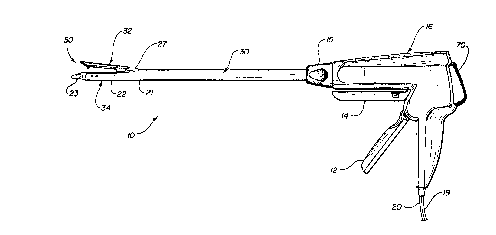
Fig. 1 is a side elevational view of an
endoscopic electrocautery linear stapling
and
cutting instrument of one embodiment of the present
invention;
10Fig. 2 is a side cross sectional view of the
instrument of Fig. 1;
Fig. 3 is a partial cross sectional view of the
distal end of the instrument of Fig. 1 in an open
position;
15Fig. 4 is a partial cross sectional view of the
distal end of the instrument of Fig. 1 in a closed
unfired position;
Fig. 5 is a partial cross sectional view of the
distal end of the instrument of Fig. 1 in a closed,
20fired position;
Fig. 6 is a front cross sectional view of the
distal end of the instrument of Fig. 4 taken along
the line 6-6;
Fig. 7 is a bottom isolated view of the
anvil
25jaw of the instrument of Fig. 1;
Fig. 8 is a top isolated view of a cartridge
of
the instrument of Fig. 1;
Fig. 9 is a side cross sectional view of the
jaw of Fig. 7 along the line 9-9;
30Fig. 10 is a flow chart illustrating a
feedback
system of the present invention;
,,,
;< v.
_ 2128136
- 9 -
Fig. 11 is a bottom isolated view of the anvil of
another embodiment of the present invention;
Fig. 12 is a top view of a cartridge of a circular
cutter of the present invention;
Fig. 13 is a bottom view of the anvil of a circular
cutter of the present invention.
Detailed Description of the Preferred Embodiments
Referring now to Figs. 1-9, there is illustrated a
preferred embodiment of the present invention. An
endoscopic electrocautery linear cutting and stapling
instrument 10 is shown having a body 16 coupled to a shaft
30 with a lumen extending therethrough and an end effector
50 extending from the distal end 21 of the shaft 30. The
shaft 30 is formed of an insulative material and has an
electrically conductive sheath 38 extending through its
lumen. A channel 39 extending through the sheath 38
guides co-axial movement of a driver means 44 within the
channel 39. In this particular embodiment, the driver
means 44 includes a firing trigger 14 associated with the
body 16, coupled to a flexible firing rod 40 coupled to a
driving rod 41, coupled to a block 43. The block 43 is
coupled to a cutting means 11 and a staple driving wedge
13, which the driving means 44 advances by way of the
block 43 into the end effector 50.
The end effector 50 comprises two interfacing jaw
members 32, 34. The end effector 50 is secured by way of
j aw member 3 4 to the channe 1 3 9 . The j aw member 3 2 i s
movably secured to jaw member 34. The body 16 has a
clamping trigger 12 for closing the jaws 32, 34 which
END-110
_2128136
- 10 -
longitudinally advances a close rack 45 coupled to the
proximal end of the sheath 38. The close rack 45 advances
the sheath 38 co-axially through the shaft 30. The sheath
38 advances over a camming surface 27 of jaw 32 to close
the jaws 32 and 34 onto tissue situated between the jaws.
As described in more detail below, the close rack 45 also
acts as a switch to close the circuit which communicates
electrical energy to the end effector 50.
Referring now to Figs. 3-9 an enlargement of the end
effector 50 of the instrument 10 is illustrated. The jaw
members 32 and 34 are shown in an unclamped position in
Fig. 3, in a clamped, unfired position in Fig. 4 and in a
clamped, fired position in Fig. 5. Jaw member 32
comprises an anvil 18, a U-shaped first,pole 52 extending
longitudinally with respect to the jaw 32, and a U-shaped
insulating material 55 surrounding the outside of the
first pole 52. Jaw member 32 has an inner surface 33 which
faces an inner surface 35 of jaw 34. The inner surface 33
includes first pole 52 which comprises two electrically
communicating electrode bars 53, 54 comprised of stainless
steel, extending substantially along the length of the
inner surface 33. The bars 53, 54 are separated by a
knife channel 42 extending longitudinally through the
first pole's center to form its U-shape. The surface of
the bars are formed in flat strips to provide more surface
area contact with tissue. Two series of pockets 36, 37
located on anvil 18, for receiving staple ends, extend
along the inner surface 33, lateral to and outside of bars
53, 54 respectively. The electrode bars 53, 54 and the
insulating material 55 form a ridge 56 extending out
relative to the anvil portion 33a of the inner surface 33
(Fig. 6). The anvil 18 is formed of an electrically non-
END-110
2128136
- 11 -
conductive material. A second pole 51 is located on jaw
34 opposite electrode bars 53, 54.
Jaw member 34 comprises a cartridge channel 22 and a
cartridge 23. The cartridge 23 includes a track 25 for
the wedge 13, knife channel 26 extending longitudinally
through the center of the cartridge 23, a series of
drivers 24 extending into track 25 and staples 100
arranged in two sets of parallel double rows. When tissue
is engaged between the jaws 32, 34, the driver means 44
may be actuated or fired using trigger 14 to advance the
cutting means 11 and wedge 13 through the engaged tissue
to staple and cut the tissue. When the firing mechanism
14 is actuated, the wedge 13 is advanced through the track
25 causing the drivers 24 to displace towards the staples
100, thereby driving the staples 100 through tissue and
into anvil pockets 36, 37.
A gap pin 29 located on the inner surface 33 towards
the tip of the anvil 18 fits into a gap 28 is formed on
the inner surface 35 of the cartridge 23. The gap 28 and
gap pin 29 serve to align the knife channels 42, 26 with
each other, and for the staples 100 to line up with the
pockets 36, 37.
A knob 15 located on the distal end of the body 16
rotates the shaft 30, sheath 38, channel 39 and end
effector 50 which are directly or indirectly coupled to
the knob 15 so that the knob 15 may be used for rotational
placement of the end effector jaws 32,34.
Bipolar energy is supplied to the end effector 50
from an electrosurgical generator 60 through wires 19, 20
END-110
_2128136
- 12 -
extending into the body 16 of the instrument. The
generator 60 is user controlled by way of a footswitch 65.
Wire 19 which provides electrical current to the
first pole energy, is coupled through a wire or other
electrical contact means 61 to electrical contact 62
located on the distal end of close rack 45. Wire 20 which
carries the current of the opposite pole, is coupled
through a wire or other electrical contact means 66 to a
disc contact 67 located at the distal end of the close
rack 45 and electrically isolated from contact 62.
A disc contact 63 , associated with the f first pole 52 ,
located at the distal end of the body 16 is in electrical
communication with a wire or other contact means 64.
Contact means 64 extends through channel 39 to end
effector jaw 32 where it contacts first pole 52. The disc
contact 63 permits the knob 15 to rotate while contact is
maintained between the disc contact 63 and the contact
means 64. The contact means 64 is electrically insulated
from the sheath 38.
When the clamping trigger 12 is actuated, the close
rack 45 moves distally so that the contact 62 associated
with the first pole comes in electrical communication with
the disc contact 63 and the disc contact 67 associated
with the second pole 51 comes in electrical contact with
the electrically conductive sheath 38. The sheath 38
moves over the electrically non-conducting camming surface
27 and is in contact with the electrically conducting
cartridge channel 22. The cartridge channel 22 is in
electrical communication with second pole 51, electrically
opposite of the first pole. Thus the electrical circuit
END-110
_ 2128136
- 13 -
is closed when and only when the clamping trigger 12 is
closed.
In operation, the end effector 50 of the instrument
is located at a tissue site where tissue is to be cut.
The jaw members 32, 34 are opened by pressing a release
button 70 which releases a button spring 71 and permits
the close rack 45 to move proximally. Tissue is then
placed between the interfacing inner surfaces 33, 35
respectively of the jaw members 32, 34. The clamping
trigger 12 is squeezed to cause the sheath 38 to move over
the caroming surface 27 and thereby close the jaws 32, 34
and simultaneously close the electrical circuit as
described above. The gap spacing pin 29 causes the anvil
18 to be held roughly parallel to the cartridge 23. The
electrode bars 53, 54 and the insulating material 55,
which together form the ridge 56, compress the tissue
against the inner surface 35 of jaw member 34 on which
return electrode 51 is contained. A gap of about between
l.5mm and 2.Omm exists between jaw members in the
compression zone. A user then applies RF energy from the
generator 60 using the footswitch 65 or other switch.
Current flows through the compressed tissue between the
second pole 51 and the bars 53, 54 of the first pole 52.
Preferably the bipolar energy source is a low
impedance source providing radio frequency energy from
about 300 kHz to 3 MHZ. Preferably, the current delivered
to the tissue is from 0.1 to 1.5 amps and the voltage is
from 30 to 200 volts RMS.
An audible, visible, tactile, or other feedback
system may be used to indicate when sufficient
cauterization has occurred at which point the RF energy
END-110
_2128136
- 14 -
may be turned off. An example of such a feedback system
is described below. After the RF energy is turned off,
the cutting means 11 is advanced and the staples 100 are
fired using the firing trigger 14. Firing is accomplished
by rotating the firing trigger 14 acting as a lever arm
about pivot 14a. The driver means 44 advances the
cutting means 11 and wedge 13. The cutting means 11 cuts
the tissue in between the bars 53, 54 where the tissue has
been cauterized. Thus, the cut line is lateral to the
coagulation lines formed by the bar electrodes. The wedge
13 simultaneously advances the drivers 24 into the staples
100 causing the staples 100 to fire through tissue and
into the pockets 36, 37 of the anvil 18. Staples 100 are
applied in two longitudinal double rows on each side of
the cutting means 11 as the cutting means cuts the tissue.
Operation of linear staplers are known in the art and
are discussed, for example, in U.S. patent Nos. 4,608,981,
4,633,874, and U.S. Application Serial No. 07/917,636
incorporated herein by reference.
In one embodiment the cartridge provides multifire
stapling capabilities by replacing the double row of
staples with a single row. In the laparoscopic stapling
and cutting devices presently in use, a single shot
replaceable cartridge is used. In order to provide better
hemostasis, this type of stapler was designed to provide
a double row of staples for each parallel row. Because of
the size of the space necessary to contain the double row
of staples, a refireable cartridge with stacked staples
has not been preferred because of the additional space
required for stacking staples. In the multifire stapling
embodiment a single row of staples is used. Using a
single row of staples permits stacking of staples in the
END-110
2128136
- 15 -
space previously occupied by the second row of staples,
providing multifire capabilities. In a further
embodiment, no staples are required and the electrical
current provides the necessary hemostasis.
A preferred embodiment of the present invention
includes a feedback system designed to indicate when a
desired or predetermined degree of coagulation has
occurred. This is particularly useful where the
coagulation zone is not visible to the user. In a
particular embodiment, the feedback system measures
electrical parameters of the system which indicate
coagulation level.
The feedback system may also determine tissue
characteristics at or near a coagulation zone which
indicate degree of coagulation. The electrical impedance
of the tissue to which the electrical energy is applied
may also be used to indicate coagulation. Generally, as
energy is applied to the tissue, the impedance will
initially decrease and then rise as coagulation occurs.
An example of the relationship between electrical tissue
impedance over time and coagulation is described in
Vaellfors, Bertil and Bergdahl, Bjoern "Automatically
controlled Bipolar Electrocoagulation," Neurosurg. Rev. p.
187-190 (1984) incorporated herein by reference. Also as
desiccation occurs impedance increases. Tissue
carbonization and or sticking to instrument as a result of
over application of high voltage may be prevented using a
feedback system based on tissue impedance characteristics.
Other examples of tissue characteristics which may
indicate coagulation include temperature and light
reflectance.
END-110
_2128136
- 16 -
Referring to Fig 10., a flow chart illustrates a
feedback system which is implemented in a preferred
embodiment of the present invention. First, energy is
applied to the tissue. Then the system current and
voltage applied to the tissue is determined. The impedance
value is calculated and stored. Based on a function of
the impedance, for example, which may include the
impedance, the change in impedance, and/or the rate of
change in impedance, it is determined whether desired
(,coagulation has occurred. If coagulation has occurred to
a desired degree, an indication means indicates that the
energy should be turned off. Such an indication means may
include a visible light or an audible sound. The feedback
means may also control the generator and turn the energy
off at a certain impedance level. An alternative
embodiment provides a continuous audible sound in which
the tone varies depending on the impedance level. An
additional feature provides an error indication means for
indicating an error or instrument malfunction when the
impedance is in below a normal minimum and/ or above a
maximum range.
Fig. 11 illustrates an alternative embodiment. The
poles 151, 152 are arranged similar to as in Fig. 6, but
with each pole as a series of electrically connected
electrodes staggered along the length of the knife channel
with insulating material in between staggered electrodes.
Knife channel 142 separates poles 151 and 152 into two
elongated series of electrodes.
Figs. 12 and 13 illustrate a circular cutter of the
present invention with stapling means. Fig. 12
illustrates the stapler cartridge 200 with an interfacing
surface 233. A double row of staple apertures 201 through
END-110
- 17 - ~ ~ 2 a ~ 3 6
which staples are driven into tissue are staggered
about the outer circumference of the surface 232. A
first pole 252 encircles the inner circumference of
the surface 233. A circular cutting knife 211 is
recessed within the cartridge 200 radially inward
from the inner circumference of the surface 233.
Fig. 13 illustrates an anvil 218 having a
second pole 251 electrically opposite of the first
pole 252. An insulator 255 on the cartridge 200
electrically isolates the first pole 252 from the
second pole 251. The anvil 218 includes pockets 237
for receiving staples and a compression ridge 256
for compressing tissue against the first pole 252
and insulator 255 of the cartridge. The circular
cutter is operated similarly to the circular stapler
described in U.S. patent No. 5,104,025 incorporated
herein by reference. Prior to stapling and cutting
however, tissue welding electrical current may be
delivered between the first pole 252 and the second
pole 251 to tissue.
In an alternative embodiment, the circular
cutter may be used without staples. Electrical
current is delivered through the poles to weld and
coagulate tissue, then the knife may be advanced to
cut tissue in a procedure such as an anastomosis.
Several variations of this invention has been
described in connection with specific embodiments
involving endoscopic cutting and stapling.
Naturally, the invention may be used in numerous
applications where hemostatis is desired.
Accordingly, will be understood by those skilled in
the art that various changes and modifications may
be made in the invention without departing from its
scope, which is defined by the following claims and
their equivalents.