Note: Descriptions are shown in the official language in which they were submitted.
WO 93/17322 PCT/GB93/00337
1. 2130343
The ability to measure particles by shape and size is of
importance to many groups of people. The food and chemical industries
are concerned from a quality control point of view; biologists are
interested in characterising cells and monitoring changes in and
differences between cells;' environmental scientists are concerned
with airborne particles and their effect on air quality and health.
This list is by no means exhaustive, it is merely intended to
illustrate the driving force behind the attempts to develop accurate
and reliable measurement instrumentation, and to theoretically
understand the nature of the problem.
There are currently two main optical scattering methods in use
in commercially available particle measurement systems. The first
method attempts to size particles by measuring their static or dynamic
behaviour in fluid. These systems generally measure deposition rate,
acceleration in a jet stream, or Brownian motion. The second method
attempts to size particles by measuring the light scattered from an
illuminated particle or ensemble of particles either at a few specific
angles or over a large solid angular range. Apart from image analysis
systems, none of the commercial instruments is capable of
characterising particles by shape, non-spherical particles being sized
by assigning an equivalent spherical diameter, although this diameter
depends on the measurement method used. What is worse, is that some
instruments are known to become inaccurate when tested with
non-spherical particles of regular shape, so measurements taken with
particles of arbitrary shape have to be treated with some caution.
Instruments which attempt a shape classification are based on
image analysis, which.requires taking an image of a small number of
particles and performing complex image processing. The particle
sample has to be prepared beforehand so that it is in a form suitable
for image processing, i.e. it has to be processed so that individual
particles can be seen with minimal overlapping. Thus there is a
considerable time delay before the results are available. The method
also requires fast computers in order to do the analysis reasonably
quickly. Some of the other instruments also suffer a time delay
WO 93/17322 PGT/GB93/00337
2.
~Z30343
before measurements are available, and whether this is important
depends on the application. It is not necessarily Important for batch
testing powders for example, but it is of potential importance when
monitoring a working environment for asbestos fibres or
micro-organisms. '
Several commercial laser based instruments are available which
will size particles, as disclosed in "Particle Size Analysers Product
Roundup". Powder and Bulk Engineering, Feb 1991; pp 42, and other
research instruments have been built to investigate various aspects of
particle sizing. For example, an instrument has been developed to
size particles using the oscillation in intensity of the scattered
light, as disclosed in "Drop Sizing by Laser Light Scattering
Exploiting Intensity Angular Oscillation in the Mie Regime." by
Ragucci. R., Lavaliere, A. and Massoli, P. Particle and Particle
Systems Characterisation, Vol 7, 1990; pp 22I. Most of the '
instruments analyze an ensemble of particles and, as stated
previously, they assume a spherical particle or particles, and do not
give any indication of non-sphericity.
Research reported in "Light Scattering Instrument to
Discriminate and Size Fibres Part 2: Experimental Sy&tem". Particle
and Particle Systems Characterisation, Vol 6. 1989; pp 144, has been
reported using an instrument designed to discriminate and size fibrous
material. In this research, particles are passed through a laser beam
in single file using a laminar airflow system similar to the design
described below. The forward scattered light is collected by a lens
and passed through a polarizing beamsplitter. The intensity of the
light in two orthogonal polarizations is then recorded using ,
photo-multiplier tubes. Results show that near spherical particles
can be discriminated from fibrous particles by taking the ratio of the
polarized intensities, provided that the particle diameter is above
1.5 microns approximately.
An instrument which has been developed to size particles uses
the laser Doppler velocimetry technique of "Strengths and limitations
of the phase Doppler technique for simultaneous measurements of
particle velocity and size." by Livesley, D.M. Proceedings-SPIE
International Society for Optical Engineering Vol 952, 1988; pp 454,
and this has also shown a capability of discriminating near spherical
CA 02130343 2002-03-11
__ . ' a a e. w w v ve a
we~ww a a w r w a ww w a w
,. ~'~. ~ ~ ~ v w ! a v a v v s d a
w w w s w a w a o www wwee
w w w w s w w a ' s a
w w w w w v w w s a s a w a s v
3.
particles from fibrous ones. This technique is based on refraction of
rays by the particle, so it is limited to particles larger than 5
microns. The instrument uses two coherent laser beams which interfere
with each other, creating a series of fringes in the scattering
volume. The spacing of the fringes depends on the wavelength of the
lasers and the angle between the beams. Three photo-multiplier tubes
are used at different angles of forward scatter which together give an
indication of particle speed, size, and non-sphericity. The speed is
obtained from the time it takes the particle to traverse from one
fringe to the next. The size is obtained from the phase difference in
the signals from two detectors, which is a function of speed and
particle surface convexity, the rate of sweep of the refracted ray as
the particle traverses the fringe being larger for a smaller particle.
The third detector allows a second phase difference to be measured,
and a different in the two measured phases is seen when the particles
are non-spherical.
We have disclosed in "An instrument for the classification of
airborne particles on the basis of size, shape, and count frequency."
Atmospheric Environment, Vol 25A No. 3/4, 1991; pp 645 by Kaye,
P.H., Eyles, N.A., Ludlow, I.K., and Clark, J.M., and in Applications
EP-A-0316171 and EP-A-0316172 an airborne particle classifier (APC)
which has some capability of determining particle shape as well as
size: it is shown in Figure 1 of the accompanying drawings and
described in detail below. The system is capable of collecting
information on a maximum of 10,000 particles per second, and is thus
capable of quasi-real time operation. However, the shape information
is severely limited because of the small number of detectors, and it
is unlikely that it could be used to differentiate unambiguously
between different types of non-spherical particle, e.g. fibres and
platelets. There is also uncertainty in the trajectory and
orientation of particles as they pass through the beam, and it is
difficult to determine and allow for the effect of these on the
scattering with only three detectors.
US-A-4606636 describes an arrangement where a flow stream
carrying particles is carried in a transparent capillary tube along
the axis of a non-divergent quadric reflector. A beam of light
intercepts the tube at the focus of the reflector, non-reflected
AMENDED SHEE?
CA 02130343 2002-03-11
~, . ' ~ v , w v a w v W w
- ~ v w . v w s v a v v w v v v o
v
~ v v s w v v w a s a v
' f
'%'i
r w v w w w w w w Q o w w w
:-, w w w
M w
;i
:2 w w w w w w w w w a
.1:.
v v w v w w w w w w w
w
a
4.
scattered rays are intercepted and reflected scattered rays are
received on a photosensitive cell or optical scanner which feeds a
processing system.
All these prior art systems using quadric reflectors operate on
__5 the assumption that the light beam will impinge on a particle at the
focus of the reflector. In practice, this is not true. In practice
the flow stream will always have a finite thickness and particles
carried thereby will not always cross the focus of the reflector.
This results in variations of ray path which can result in rays
becoming non-monotonic (that is rays scattered at low angles and
reflected cross those scattered at higher angles) before being
recorded. Images from monotonic and non-monotonic rays are completely
different.
Thus no real-time method of shape analysis is yet available, and
little investigative work has been done on non-spherical particles.
According to the present invention apparatus for the analysis of
individual particle characteristics from an aerosol or other
suspension of particles includes: a scattering chamber including an
optical system having an ellipsoidal reflector with an orifice therein
leading to a rear chamber;
a monochromatic light source adapted to transmit a collimated
beam of light along the main axis of the reflector;
means for directing a gas stream of finite thickness containing
particles through the beam of light at substantially a main focus of
the reflector;
the optical system being arranged to collect the scattered light
from a solid angle of at least 3~ around the region where the
particle stream crosses the light beam;
a detector having a two dimensional array of a multitude of
sensors arranged to form an image from light scattered from a particle
in the stream of particles and collected by the optical system;
and a data processor, the data processor being adapted to
compare parameters of the image with parameters stored in a memory to
determine the nature of the particle;
characterised in that the recorder is associated with an imaging
screen positioned behind a secondary focus of the reflector such that,
whatever part of the finite thickness of the gas stream occupied by
,r,~;~EhiDED 5~'P"~
CA 02130343 2002-03-11
' ' ~ s w w v o w o v w a
,,.._.. ' ewrww wwww wwws wwo
_ s w
~ ~ ' w s v. v
~~~-'.:a,~ w w w w a ~ ~ a w w w n w w a w o w w
..w " v w w v a w w
w w swww wa ww aww w, a
5.
the particle when it passes through the beam of light, rays of light
scattered from the particle and imaged thereon by the optical system
are monotonically ordered with respect to the angle of scattering of
the rays from the particle so that the image recorded on the imaging
screen is substantially independent of the position of the particle in
the gas stream.
The detector is preferably a charged couple device (ccd) video
recorder.
The imaging screen may be an image intensifier or the image
screen of the camera.
The apparatus might advantageously include trigger means, which
might have sensor means for sensing scattered light not collected by
the optical system, and thereby determining the presence of a particle
in the beam of light, and for triggering the detector to store the
image associated with the particle. The sensor means might comprise a
photomultiplier tube onto which are focussed scattered rays which pass
through the orifice in the optical system to the rear chamber.
In instruments including a photomultiplier provision might be
made for changing the positions of photomultiplier and detector, or
even replacing the photomultiplier with another detector.
The trigger means might also include a controller having an
input from the detector means and an output to the detector, and might
be adapted to act only with a single particle in the beam.
The parameters compared might relate to entire images, to parts
of images, or to resolving diffraction or interference maxima and
minima.
According to another aspect of the invention a method of
analysing individual particle characteristics from an aerosol or other
suspension of particles includes the steps of directing a gas stream
of finite thickness containing particles through a scattering chamber;
the scattering chamber including an optical system having an
ellipsoidal reflector with an orifice therein leading to a rear
chamber;
a monochromatic light source adapted to transmit a collimated
beam of light along the main axis of the reflector;
means for directing the gas stream through the beam of light at
substantially the main focus of the reflector;
AMENDED SHEET
CA 02130343 2002-03-11
- w w f w s
1 . w . w o v s v v v w s v v
w w 1 1 1 1 w
s. s w s w s sew
a y w eels
yu~~-' v w v w
'~' s ewww ew se ewe a
we s
6.
the optical system being arranged to collect the scattered light
from a solid angle of at least 3?f around the region where the
particle stream crosses the light beam;
the scattering chamber being associated with a ccd video
recorder having a two dimensional array of a multitude of sensors
arranged to form an image from light scattered from a particle in the
stream of particles and collected by the optical system;
the video recorder passing information to a data processor, the
data processor being adapted to compare parameters of the image with
parameters stored in a memory to determine the nature of the particle;
characterised in that the recorder is associated with an imaging
screen positioned behind the secondary focus of the reflector such
that, whatever part of the finite thickness of the gas stream occupied
by the particle when it passes through the beam of light, rays of
light scattered from the particle and imaged thereon by the optical
system are monotonically ordered with respect to the angle of
scattering of the rays from the particle so that the image recorded on
the imaging screen is substantially independent of the position of the
particle in the gas stream.
Preferred embodiments of the invention are intended to be capable
of classifying particles into one of five broad shape classifications:
spheres, droplets, fibres, platelets and "chunks" (i.e. particles of
comparable size in all three dimensions), and also of differentiating
between particles of differing aspect ratio.
In this specification, the term "scattering profile" is intended
to mean the three-dimensional scattered light intensity distribution
about the particle. The scattering profile is unique for particles of
given shape, orientation, and dielectric structure for a given
wavelength of illumination.
In order that the invention may be better understood, an example
will now be described in detail with reference to the accompanying
drawings, in which:
Figure la, to which reference has already been made, is an
elevation of a conventional particle analysis instrument,
Figure 1b is a corresponding plan view;
Figure 2 is a sectional view showing a scattering chamber
according to the present invention.
p,M~ND~~ S~l~r'~
i
CA 02130343 2002-03-11
, . ' ~ v v a v a v v a w v o a
'
'
_ : o o s o 0 00 o a o
i . ae.eo
~ ~ ~ ~ v a n w v v v a o
v
,z w v w v a v v a w v v v w
w w
v a
w w w a s v v s v
o eooo ew sw oeo ee a
Figure 3 is a diagrammatic representation of the system of the
present invention:
Figure 4 is a sectional view through the sample inlet assembly,
Figures 5 to 8 are ray trace diagrams illustrating the passage
of light in the scattering chamber;
Figure 9 shows the collected image of light scattered from a 1
micron polystyrene sphere between 30° and 140°;
Figure 10 shows the collected image of light scattered from a
water droplet between 30° and 140°; and
Figure 11 shows the collected image of light scattered from an
arbitrary airborne particle between 5° and 300.
The prior art device illustrated in Figures la and 1b, as
described in EP A 0316172, has a laser 10, for example a HeNe laser,
projecting a beam 11 through a beam expander 12, iris diaphragm 13 and
cylindrical lens 14 at right angles into a scattering chamber 15. In
the scattering chamber 15 the beam 11 is reflected by a mirror 16 to
pass along the axis of a first ellipsoidal reflector 17, positioned in
the main chamber 15, through an orifice 18, along the axis of a second
ellipsoidal reflector 19 in a rear chamber 20 and through a beam dump
21. At the rear of the scattering chamber 15 is a collimating lens 22
and a collection system comprising three photo-multiplyer tubes 23.
At the focal point of the second ellipsoidal reflector 19 in the rear
chamber 20 is a radiation collector 24 leading to a photomultiplier
lens 25. A sample inlet tube 26 and sample outlet tube 27 are
positioned in the scattering chamber 15 such that a sample of gas can
be passed through the focal point of the first ellipsoidal reflector
17.
In use a sample is drawn by a pump 28 through the tubes 26, 27,
such that particles therein are illuminated by the beam 11. Light
deflected by the particles through a substantial angle are reflected
by the first ellipsoidal reflector 17 and pass through the collimating
lens 22 to the photomultipliers 23. Light deflected at lower angles
pass through the orifice 18 into the back chamber 20 where they are
reflected by the second ellipsoidal reflector 19 onto the radiation
collector 24 and thence to the photomultiplier 25. Non- reflected
light is dumped by the beam dump 21.
The outputs of the photo-multiplier tubes are digitized and
AMENDED SHEET
i
CA 02130343 2002-03-11
~ ~ . ~ w w w s w a w a w ~ ~
,~ . w. w w v ~ w v v w a a a
s w w v a v w v w v w v v
4 w w w w w ~ w w w www wwww
w w w w v w w w s w
a w wwww ~~ ww www W w
stored using dedicated electronics and analysed using a computer, for
example by comparison with data or with known shapes as described in
EP-A-0316171.
Due to the considerable promise shown with the instrument
described with reference to Fig. 1, the new instrument shown in Figs.
2, 3 and 8 retains the same ellipsoidal reflector light collection
system and the same type of particle delivery system. As the present
invention relies for its operation on specific positioning of certain
items and of calibration it will be described with reference to an
actual experimental instrument.
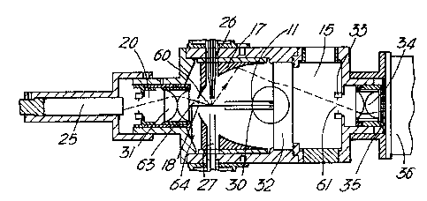
In an instrument according to the present invention (Figure 2) a
laser beam 11 delivered by a laser system similar to that shown in
Figures 1 is contained initially within a shroud 30 and directed along
the axis of an ellipsoidal reflector 17 in a scattering chamber 15,
through an orifice 18 and then onto a beam dump in the form of a
silvered prism 63 glued to a lens of a forward scatter lens assembly
31 and directing light onto a matt black surface 64. The lens
assembly 31 is adapted to focus scattered light onto a photomultiplier
tube 25 in a rear chamber 20. The laser was a Lasermax model
LAS-200-670-10 diode module with integral power supply and collimating
optics. This laser has a power output of 10 mW at a wavelength of 670
nm. The output is plane polarized and operates in the TEMoo mode,
with a cross section of 4 mm by 1 mm. The module was mounted in the
housing by set screws which enabled it to be aligned with the chamber.
The beam passes through a quarter wave plate to render it circularly
polarized, through an iris diaphragm and then through a cylindrical
lens before entering the scattering chamber 15. The beam 11 at a main
focus 60 of the ellipsoidal reflector 17 was thus approximately
elliptical with dimensions 3 mm wide and 120 microns deep. The laser
housing could be moved inside the mounting to allow for cylindrical
lens focal length tolerances. The ellipsoidal reflector 17 was chosen
for its collection capabilities, had the main focus 60 and a secondary
focus 61 separated by 98.6mm, and had a quoted solid angle collection
of 84~L of the sphere surrounding the main focus 60 (3.3i( of the 4i'~
solid angle). A sample inlet tube 26 and sample outlet tube 27
connected to a pump, for example a vane type pump whose output can be
adjusted, are arranged to supply a sample stream across the light beam
A~1~N~~~ 5~~~
CA 02130343 2002-03-11
o ~ ~ ~ s s ~ ~ a a ~ ~
~-. , ~ ~ , ~ ~ ~ ~ ~ ~ ~ ~ ~ ~ ~ ~ ~
~ ~ ~ ~ a s ~ ~ ~ ~ ~ ~ ~
~ ~ ~ v a v ~ ~ w ~ 1 ~ v m a ~ ~
.~f:~ ~ ~ ~~~ ~~~ ~1
~ ~ ~ 1 ~ 1 ~ A ~ ~ 9 ~ ~ ~
9.
11 at the main focus of the ellipsoidal reflector 17. The optical
arrangement is such that light scattered by a particle in a sample and
being reflected by the ellipsoidal reflector 17, which encompasses a
solid angle of at least 3 ?f , passes through an optical window 32 and
an iris 33 positioned at the secondary focus of the reflector 17. The
optical window 32 increases the distance between the main and
secondary foci of the reflector 17 and the iris 33 reduces the amount
of background scatter. From the iris 33 reflected light passes
through a pair of plano-convex lenses 34 to an image intensifier 35
positioned adjacent a video camera 36. The camera was a typical
charged couple device (ccd) video camera with a 110880 (385 by 288)
element array. The position of the image intensifier (or any
alternative imaging screen) in the system is critical, as will be
discussed below with reference to Figures 5 to 8 of the accompanying
drawings.
The recording, analysis and viewing arrangements of the
invention may best be seen from Figure 3. A computer 37 contains,
inter alia, a frame grabber 38 which has an input from the camera 36
and a camera controller 39 which has an input from the photomultiplier
25 and an output to the camera 36. The frame grabber 38 was a
commercial board with a 256 by 256 pixel array, which was available
with commercial software for post processing of images. The computer
has the normal storage and calculation facilities, and outputs to an
image monitor 40 and to a computer monitor 41.
The sample inlet assembly (Figure 4) including the sample inlet
tube 26 has the tube 26 leading from a sheath air chamber 50 to which
can be supplied, through a sheath air inlet 51, filtered air.
Co-axial with the inlet tube 26 is a sample air tube 52 leading from a
sample chamber 53 to lie within the inlet tube 26 where it terminates
adjacent a narrowing 54 in the tube 26. The exact positioning of the
termination relative to the narrowing 54 can be adjusted by an
adjustment screw 55, and the extension of the sample inlet tube into
the scattering chamber 15 can be adjusted by the adjustment screw 55.
In use during calibration clean filtered sheath air is delivered
via the air inlet 51 and sheath air chamber 50 to the sample inlet
tube 26, and sample air, containing particles of a known type is
delivered to the sample air tube 52 from the sample chamber 53.
PMDNDED SNEE'~
CA 02130343 2002-03-11
' o v w ow ww w w o
..~ ww, ww w w w a w w ow w w w
o w w w w a w w w w w s w
w w w w a w w v s s www owww
a w w w a w w w w w
a w wwww as ww aww ww s
10.
Operation of the pump 28 draws sample air, surrounded by a sheath of
clean filtered air, out of the sample inlet tube 26, across the laser
beam 1l, and into the sample outlet tube 27. The sample air is drawn
through the chamber at (2.5-6)1/m creating a columnar, laminated
airflow with a diameter of lmm across the gap between the tubes.
Particles thus pass in single file through the laser beam 11 within
0.5 mm of the main focus 60 of the ellipsoidal reflector. The laser
beam 11 impinges on any particle in the sample air at substantially
the main focus 60 of the ellipsoidal reflector 17 and as a result
light is scattered. Scattered light impinging on the ellipsoidal
reflector 17 is reflected through the optical window 32, iris 33 and
lenses 34 onto the image intensifier 35. Scattered light not
impinging on the reflector 17 passes through the orifice 18 and
forward scatter lens 31 where it is focussed onto the photomultiplier
tube 25. Light not scattered is dumped.
The photomultiplier tube 25 passes a signal to the camera
controller 39, which monitors the signal. When a predetermined level
is reached a signal is sent to the camera 36 causing the camera to
image the pattern on the image intensifier 35 and pass this image to
the frame grabber 38 whence it is stored in the computer 37. The
image, which is distinctive of the type of particle and (if relevant)
of its alignment, may be viewed on the image monitor 40.
It will be realised that in practice the sample air stream will
have a finite thickness. Particles will not therefore always cross
the main focus 60 of the reflector 17 when the laser beam 11 impinges
thereon. This is illustrated in Figures 5 to 8. In Figure 5 is shown
a ray trace of the complete optical system for the particular
ellipsoidal reflector 17, optical window 32, iris 33 and lenses 34.
The dimensions are in mm measured from an origin at the secondary
focus of the reflector 17 and the traces correspond to scattering
angles 30° to 140° in 5° steps.
Figure 6 shows a detail of the ray trace of Figure 5, in the
region of the image intensifier 35 which is shown at a position +44 mm
from the origin. It will be seen that at this position the rays are
monotonic (that is rays scattered at low angles and reflected do not
cross those scattered at higher angles).
Figures 7 and 8 show respectively traces of rays for particles
es~~~.~NC'~ED SHEEN
CA 02130343 2002-03-11
~ o v o v o m ~ ~ o ~ ~ A
. - _..3 - ~ 4 ~ ~ ~ ~ 4 ~ ~ t ~ ~ D ~ ~ ~ ~
6 ~ ~ ~ 0 ~ ~ 1 ~
O ~ ~ A A ~ 1 ~ ~ ~ ~ ~ 1 0 A 11
.,,;i",~' a ~ ~ 1 w ~ ~ ~ ~ s
w ~ w ~ ~ ~ ~ n ~ ~ s a ~ ~ w ~
11.
crossing the beam 11 0.5mm forward (toward the vertex of the reflector
17) and 0.5mm rearward of the main focus 60 of the reflector 17, and
it will be seen that shortly to the left and right of the 44mm
position the rays become non- monotonic, whilst being monotonic at the
44mm position. Whilst the rays remain monotonic the images presented
by a given particle remain substantially identical (other than some
degree of distortion) whatever the crossing position of the particle
relative to the main focus 60 and can therefore be successfully
compared with a memory of that image. However once the rays become
non-monotonic the image is completely changed and will not be
comparable. It is therefore essential that the image intensifier 35,
or other imaging screen, is placed in a position where rays will be
monotonic whatever the position a particle crosses the laser beam 11.
The optimum position of the image intensifier 35 can be determined by
trial and error by passing a stream of particles through the
instrument whilst moving the image intensifier 35 to determine the
position where complete duplication of images is lost. Alternatively
a manouvrable sample inlet tube 26, supplying a very narrow sample
jet may be used to test the effects of particles passing at known
distances from the main focus 60. It will be realised that using very
narrow sample jets such that all particles will cross the laser beam
11 substantially exactly at the main focus 60 of the reflector 17 will
provide an instrument that will be too slow acting to be of practical
value. For the instrument described here as calibrated for commercial
use a sample jet width of about lmm would be typical.
It will be realised that, in practice, at times two or more
particles will cross the laser beam 11 so closely together that
scattered light from both will be imaged unless precautions are taken.
This can be avoided by sensing the time during which the camera
controller 39 is receiving an intake from the photomultiplier unit 25
and aborting the triggering of a camera action if this time is too
great (indicating the presence of more than one particle).
When the position of the image intensifier 35 and the timing of
the camera controller have been set the instrument can be calibrated
by passing a series of particles of known size, shape and constitution
therethrough and storing the images or other parameters (such as
diffraction or interference maxima or minima, or details of portions
r~MENDED SHEk
I
CA 02130343 2002-03-11
' 2 a ~ ~ v o a a o ~ ~ w
< ca ~~ ~ a ~ a ~ ~ 0a ~ ~ c
. ' a ~ a ~ a~ a ~ o ~ ~ a ~
n ~ a a a w ~ ~ w a ~ ~ ~ m o ~ ~
~','':-'.'a a w v v a ~ v a a w
a ~ wa~~ wa ~~ a w ~~ ~
12.
of the image) in a library for future comparison. Once one instrument
has been calibrated it can, of course be expected that instruments of
similar construction will provide similar results, and the calibration
may prove to be suitable for such similarly constructed instruments.
When storing images to hard disc the image capture rate is
limited by factors such as the speed limitations of the disc, and for
the system described above might be approximately two images per
second.
With the instrument described above, the light scattered between
28° and 141° from a particle was collected by the ellipsoidal
reflector 1~, (i.e. the scattered light was collected from a solid
angle of at least 3 ~ around the region where the particle stream
crosses the beam 11). The reflector 1'j was mounted in a cylindrical
holder which could be moved to position it correctly. This was
necessary because although the optical and mechanical dimensions of
the reflector are specified, no relationship between them is given.
The light scattered between 5° and 28° from a particle passed
to the
pair of plano-convex lenses 31.
Compared with the performance of the instrument of Figure 1 the
present invention gives greatly increased spatial resolution. By use
of a charge coupled device (ccd) video camera coupled with an image
intensifier four advantages are gained:
1. The resolution is increased by replacing three detectors
with the 110880 (385 by 288) elements of the ccd array.
2. The light input to charge output conversion efficiency is
virtually constant across the array because it is manufactured in one
piece of silicon.
3. The light gain of the image intensifier is also virtually
constant over its aperture due to its construction.
4. Use of a video camera means that images can be captured on a
standard computer frame grabber board, and therefore can be processed
and stored faster than with a still camera using photographic film.
The image intensifier is required in this example of the
invention because of the small number of photons scattered from a
particle during its transit through the laser beam. The time of
transit of a particle through the beam is approximately (2-5)
microseconds depending on flow rate, during which time typically
~:.,~.avN~~=~ ~'~~~
CA 02130343 2002-03-11
~ ' , ' ~ ' Q ~ w g o w o w o
- ~ ~ ~ ~ ~ ~ ~ ~ ~ ~ ~ A ~ ~ ~ !
- ! ~ ~ ~ 69 1 ~ ~ ~ ~ 0
~ ~ ~ ~ ~ ~ ~ ~ * ~ 1 ~ ~ P 4 ~
f;i:'iy) 9 ~ 1 ~ 4 ~ ~ A ~ 1
.;;f'~' w ~ ~ ~ ~ ~ o o ~ ~ a t ~ 1 ~ ~
13.
several thousand photons are scattered depending on particle size,
when using a focused lOmW helium-neon laser as in the instrument. The
number of photons scattered depends approximately on the fourth power
of the particle size in the size range of interest (approximately 1-10
microns), and hence varies considerably. The camera 36 has an
asynchronous trigger facility which allows it to be used in a similar
manner to a still camera. This is necessary so that the scattered
light can be captured during the time that a particle is in the beam
11.
The maximum number of images that the camera 36 can output is 25
per second, i.e. it can record data on 25 particles per second, as
opposed to the data rate of 10,000 particles per second of the
original instrument. This is not a disadvantage however, since the
instrument in this example is used for basic research into scattering
profiles from particles of different shape. The high spatial
resolution offered by the camera 36 allows the determination of an
optimal detector configuration for use in real time instruments with
more rapid particle handling rates, also embodying the invention.
Such an optimised detector configuration may well have fewer elements,
for example 32 by 32 than a camera CCD array so as to allow a higher
particle analysis rate. The configuration need not be symmetrical,
and may be optimised to suit analysis of a specific particle shape.
Examples would include a custom multi-element photodiode array
arranged geometrically for optimal shape characterisation.
In one particle size range of particular interest (1-10
microns), a significant proportion of the total scattered light is
scattered in the angular range up to 30°. This light passes through
the hole 18 in the ellipsoidal reflector 1'7, and is thus not collected
by the camera 36. Light scattered in this angular range may be of
importance in determining particle shape and the instrument is
preferably designed so that this angular range could be investigated
if necessary. -
The light passing through the orifice 18 is normally collected
by a photo-multiplier tube 25 which generates a pulse to trigger the
camera 36, and when the scattering chamber 15 is designed to allow the
camera 36 and photo-multiplier tube 25 housing to be interchanged the
arrangement allows the camera 36 to record scattering in either the
,:~.~r~E~ SHEEN
CA 02130343 2002-03-11
~ _ ~ . . , o ~ w n o ~ w R y p
.. ~ ~ 9 ~ ~ ~ 1 ~ ~ P ~ ~ Q a ~ ~ O
a i ~ v so ~ ~ a v 1 o v
,'e~~ p ~ ~ a o ~ ~ ~ o ~ ~~~ eow~
~~~'<:1~ 2 1 ~~a ~~R ~w
v ~ ~ ~ ~ w o a w ~ m v w ~ ~ ~
i4.
range 5°-30° or the range 30°-140°.
Previous results recorded from fibres have shown that there
could be differences in orientation between particles as they traverse
the gap between the inlet and outlet tubes 26, 27. To investigate
this more thoroughly the distance between the inlet 26 and outlet 27
tubes was made adjustable, so that the distance between the ends of
the tubes and the laser beam could be varied. The depth of the
particle delivery tube 52 inside the concentric clean air tube 26 was
made adjustable so that the aerodynamic focusing effects of this
system could be investigated.
In the original instrument the clean air filter was an integral
part of the instrument, which meant that only total airflow could be
monitored. In the new example of Fig. 2 it was a requirement that
the clean air flow rate could be monitored separately as well as the
total, so that the ratio between particle laden air and clean air
could be monitored and adjusted.
The camera controller 39 will usually be a purpose-built board
acting in a system with a micro-computer which enables the grabbing
and storing of frames to be controlled from a keyboard. The computer
37 performs any post processing of the images which may be necessary.
The optical system was designed with the help of a dedicated ray
tracing computer program package, so that the effects of particles
passing through the scattering volume at different positions could be
determined. This was necessary because the ellipsoidal reflector 17
causes the images to be a non-linear representation of the scattering
angle, and this in turn is a function of particle position. The
design parameters were
1. The total solid angle collected should be as large as
possible.
2. The system should be designed to cope with changes in
particle position within the scattering volume.
3. The diameter of the projected image on to the camera
faceplate should equal to the camera aperture.
The first criterion is best satisfied by collecting the light
with an ellipsoidal or parabolic reflector. Criteria 2 and 3 were
found by modelling to be satisfied by the condenser lens 34
arrangement. Several different configurations of lenses were modelled
~:i ~'wDSD SN'E~ '!
CA 02130343 2002-03-11
~ ' . . . C ~ ~~ fee ~~ 9 ~~ a
a~ ~~ o ~ ~ ~ ~ ~ s~ ~ ~ a
. ~,-_..~ ~ c. 1 ~ ~ a a v ~ ~ ~ r ~ a
1 a ~ . a o v ~ ~
0 ~ ~ ~ ~ ~ ~ ~
S ~ 4 i~ Q ~ ~ a ~ 0
1 9 a ~ ~ n ? 1 ~ O ~ 1 ~ ~ P
i5.
on the ray tracing software before arriving at the one shown in Figure
as a suitable solution.
With reference to the frame grabber board 38 this preferably
contains in its software an expert system for classifying spatial
5 distributions of the captured video frame diffraction maxima
corresponding to known particles of different shapes and sizes, and
for then identifying those characteristics in test particles. The
analysis may include recording features of the spatial arrangement of
the image maxima for each of several particles for whom the relevant
characteristics are known, and then using those features to set the
predetermined criteria for analysis of test particles.
The camera controller 39 is newly designed to control the camera
36 triggers and frame grabber 3$ and has four main functions as
follows:
1. Trigger generation. The board uses the output of the
photo-multiplier tube to generate two trigger pulses, one for the
image intensifier 35 and the other for the video camera 3~. The image
intensifier 35 is turned on for the duration of time that the particle
is in the scattering volume and thus acts as an electronic shutter.
The camera trigger pulse is of 3 ms duration and starts after the
trigger is removed from the image intensifier. This timing
arrangement was found to improve the quality of the images by reducing
w
ccd overspill between elements (smearing). The persistence of the
image intensifier phosphor ensures that no data are lost. The trigger
pulses can be disabled from the keyboard.
2. Noise floor setting. The level below which the camera 36
will not be triggered can be set from the keyboard. This prevents the
camera being falsely triggered by noise from the photo-multiplier
tube.
3. Time of flight checking. The board contains a counter which
records the length of time that a particle is in the laser beam to a
resolution of 125 ns. This can be read from the keyboard or by
programs and is used to discriminate between genuine single particles
and either particles floating into the beam outside the scattering
volume, or two or more particles following each other through the
beam. It is not possible to detect if two or more particles pass
through the beam alongside each other, unless this is evident from the
AMENDED SHEET
CA 02130343 2002-03-11
~ ~ ~~ ~~ ~~ ~ ~~ ~
' ~ ~ ' ~ ~ w ~ ~ ~ , ~ ~ w ~ ~ ~
~ ~ ~ ~ ~ ~ a ~ ~ ~ ~ ~ ~
,CI ~ ~ ~ ~ ~ R A ~ ~ C ~~~ 11~1
~ 1 ~ ~ ~ P ~ 11 ~ ~
~ ~ ~ ~ ~ ~ ~ ~ A ~ ~ ~ 1 ~ ~ ~
1~.
seen, which is consistent with the theory of scattering from spheres.
There is a good qualitative agreement between the image and
theoretical predictions. In particular, it can be seen that the
minimum which occurs at an angle of approximately ~0° in the
horizontal plane disappears in the vertical plane, which agrees with
theoretical predictions. The scatter angles at which the maxima and
minima occur are also in good agreement.
Simulations with 2.95 micron and 4.3 micron diameter spheres
have also been carried out with similar correlation, although the high
periodicity of the fringes and the non-linear nature of the
experimental data make quantitative comparison at this stage more
complex.
Figure 10 shows a typical result from a water droplet generated
using a water spray, with scattering between 30° and 140°. Since
the
spray generates a wide droplet size distribution, the diameters of the
individual droplets creating the scattering profiles are not known.
Simulations of scattering profiles from 2 micron and 2.5 micron
diameter spherical water droplets have been carried out and these show
that relatively more light is scattered in the 90° to 140°
angular
range than is the case with polystyrene spheres. This can also be
observed in the experimental data.
With the camera 36 positioned to capture small scattering angle
data from 5° to 30°, the laser 10 was fitted with a 22x
transmission
broadband neutral density filter on its output to reduce the beam
intensity 11, because without this, the amount of scattered light is
such that the ccd array is saturated, and the image is normally
completely white for droplets of the size range generated.
Figure 11 shows an image taken when the instrument is operated
with the inlet open to the atmosphere in the laboratory.
To summarise, the instrument possesses, inter alia, the
capability of resolving diffraction maxima and minima with particles
in the approximate size range (1-10) microns. The lower limit is
governed by the number of scattered photons, which varies
approximately in proportion to the fourth power of the diameter of the
particle for particles of this size, and therefore changes by several
orders of magnitude in the size range of interest. The lower limit
could be reduced by using a more powerful laser but this would cause
AMENDED SHEET
i
CA 02130343 2002-03-11
o ~ ~~ ~~ e~ ~ ~~ ~
~ ' ' ~ a o ~ s a ~ o 0 0 ~ " ~ ~ ~ ~
0 0 ~ ~ ~ ~ o ~ ~ o o ~ o
!,~ a a ~ ~ o a o ~ ~ a ~~~ ~eoe
::'-:~' ~ ~ s w w w ~ ~
.. . ~
a ~ o ~ ~ ~ w ~ o ~ ~ ~ ~ ~ ~ ~
18.
camera saturation problems with larger particles. The upper limit is
caused by the number and closeness of the scattering maxima and minima
and could be increased by using a camera with a higher resolution ccd
array and redesigned instrument optics.
The nature of the ellipsoidal reflector 17 causes scattering
angles below approximately 70° to be compressed into a relatively
small area on the ccd array, and therefore detailed examination of
this region could be facilitated by redesigning the lens system to
expand this region, at the expense of losing scattering information at
IO larger angles. The instrument would readily accommodate such changes.
With the use of a reflector 17 to collect light over a large
solid angle, in combination with a lens system 34 which produces an
image at the camera imaging plane largely insensitive to the
particle's position within a column, and without any intermediate
collecting lenses (for which it would be necessary to provide a
transformation to correct for positional sensitivity introduced by
such lenses), the instrument successfully images light scattered from
individual particles even at small angles, without significant
distortion.
The instrument still of course has some sensitivity to changes
in particle position in the scattering volume, especially at small
scattering angles. While this is a disadvantage when calculating the
angles where diffraction maxima and minima occur, it is possible to
take this into account by observing the size of the central shadow
region. There will, however, always be some uncertainty in the
calculations, especially at small angles.
The apparatus can also be exploited to determine the orientation
of particles as they traverse the airflow between the inlet and outlet
tubes 26. 27. The two tubes can be moved in relation to the laser
beam 11, which effectively enables the scattering volume to be moved
to different positions of the airflow across the gap. The distance
between the tubes can also be changed so that airflows of different
lengths can be investigated, and an investigation of the aligning
effects of an electrostatic field can be carried out by putting a
voltage on the outlet tube, which is electrically isolated from the
rest of the instrument.
Although it is not possible to trace one particle across the
t~'l,~IEO SH
i
CA 02130343 2002-03-11
. . w w w w
' 1' ' '
, w o ,
: s
f.
ww
1 v
, w w w
w
w o w w w o
w ww wsw
s w s s w s a w s
w w swww ww ww ww ww
19.
complete airflow, it is possible to determine whether there is any
preferred orientation of particles at any particular position in the
airflow, and if so, whether this orientation changes as the particle
moves with the flow.
From an investigation of the aerodynamics it should be possible
to design a particle delivery system which ensures an almost constant
orientation. This is seen to be a requirement in a practical
instrument capable of shape discrimination since the number of
detectors could be much less than on the research instrument of Figure
2, and changes in orientation would almost certainly produce an
incorrect shape result.
The apparatus could also be used to investigate the deformation
of liquid droplets as they traverse the airflow. If any deformation
can be detected, it may be possible to differentiate between solid
spheres and droplets.
Droplets of known size would be generated using an aerosol
generator, and experiments performed with liquids of different
viscosities recording scattering profiles as droplets traverse the gap
between inlet and outlet tubes.
One could then determine if there is any elongation or
relaxation of droplets due to shear forces in the airflow across the
gap, which would cause a non-spherical scattering profile. From the
scattering profiles already obtained from water droplets, it is
expected that any non-spherical scattering profile due to deformation
will be small, and detection will almost certainly require computer
analysis. Appropriate algorithms would detect deformations once the
scattering profiles have been obtained.
Theoretical modelling of the aerodynamics of the particle
delivery tube system can also be used, and compared with the
experimental results. It should be possible then to determine an
optimum delivery system so that droplets can be discriminated from
solid spheres and non-spherical particles.
Although the apparatus has been used so far with aerosols in air
or other gas, the apparatus could be used to detect characteristics of
particles supported in a fluid or liquid medium, such as a colloidal
suspension, with minor modifications to the chamber and optics. The
detailed design of the optical and electrical components could also of
AMENDED SHED
CA 02130343 2002-03-11
~ ' , . ' , ' o o a a a o 0 0 o s a o
. . ~ ' ~ ~o 00 0 o a a a o 00 0 0 0
P A
a a a o ~ ~ y o s o 0 0 0 0 0
,.°~ a ~ w w v v s a w a o
v a v v a v v a v v v ~ v w v
20.
course differ; for example, ccd arrays even as low as 32 x 32 pixels
would be feasible, and the array could be of any regular or irregular
shape.
It will be realised that, whilst the above described instrument
has been described as having a camera 36 positioned usually to receive
the greater percentage of scattered radiation and using the
photomultiplier tube 25 as part of the camera trigger device, with the
alternative of reversing these positions, it would in fact be possible
to position cameras in both these positions, and to use the
information from both. This would need some form of triggering device
which might, for example, be a photomultiplier tube 25 to which some
portion of the scattered light is diverted.
It will also be realised that the image intensifier 34 may not
always be necessary, and that the imaging screen on which the image
falls may, for example, be that of the camera 36.
;'~~ ~~;0~0 ~~~~