Note: Descriptions are shown in the official language in which they were submitted.
213~8~
--1--
1552 (203-1180)
ENDOSCOPE ATTACHMENT FOR CHANGING ANGLE OF VIEW
BACKGROrJND OF THE INVENTION
l.Field of the Invention
lo The invention relates to accessories for endoscopes
and, more particularly, to endoscope sheaths which change an
angle of view of an endoscope.
2. Description of the Related Art
Endoscopes have long been used in surgery to view
internal portions of a patient's body from a narrow incision
in the body exterior or through a naturally occurring hollow
viscus. Endoscopes used for this purpose are long, slender
instruments having a shaft which is either rigid or
flexible, depending upon the procedure being performed. In
general, endoscopes include an objective lens positioned
adjacent a dista:L end, and an image transmission system
which may include a fiber optic bundle, relay rods or
lenses, or a solicl state sensor to transmit the image to the
viewer. Endoscopes also are usually equipped with an
illumination system, such as a fiber optic bundle, which
illuminates the area being imaged.
Generally, a camera adapter is provided at the proximal
end of the endoscope to permit the image to be displayed on
a monitor for viewing by the entire surgical team. It is
also known to provide a fluid and/or gas conduit which
permits the surgeon to clean the distal-most imaging lens
and/or clear the region in front of the distal-most lens for
2 1 3 ~
-2
optical viewing. See, for example, U.S. Patent Nos.
4,667,656; 4,770,163; and 4,838,246.
Most endoscopes used for medical procedures have a
fixed forward viewing angle. Different areas of the body
can be imaged by changing the position of the endoscope or,
in the case of flexible endoscopes, by bending the distal
tip. In these endoscopes, the objective lens is disposed
perpendicular to the longitudinal axis of the instrument
such that the area directly in front of the instrument is
viewed by the user. Other configurations for endoscopes
include side-viewing, and oblique angle of view endoscopes.
In certain procedures, in addition to forward viewing
endoscopes, it is desirable to have the capability for
changing the angle of view during different stages of the
procedure. For example, when examining the lining of a body
cavity, e.g., esophagus, intestinal walls, it is
advantageous to employ a side-viewing or oblique angle of
view endoscope. Presently, the ability to view at different
angles can only be accomplished by maintaining a variety of
expensive high quality reusable side-viewing endoscopes.
It is known in the art to provide attachments which
change the angle of view of conventional reusable
endoscopes. Typical devices of this type are disclosed in
U.S. Patent Nos. 4,747,661 and 4,787,370. In general, such
attachments fit adjacent only the distal end of the
endoscope and include complex optical elements which
increase the cost of the endoscopes and reduce their
efficacy for single-use applications. Additionally, many of
the known attachments require special adaptive elements on
the distal end of the endoscope in order to properly secure
the attachment. The attachments do not cover substantially
the elongated endoscopic portion of the endoscope. As a
result, it is difficult to clean and re-sterilize the
`~ i'`Z; ~ ~ ~r ~ Z ~
213~ ~8~
-3
expensive and delicate optics of the endoscope prior to
reuse.
Accordingly, it would be advantageous to provide a
device which may be readily inserted over or connected to an
endoscope and which functions in changing the angle of view
of the scope without the need for complex attachments. It
would also be advantageous if such device extends
substantially the length of the elongated endoscopic portion
of the endoscope to protect the endoscope from
contamination. It would be a further benefit if the device
incorporated an integral illumination and/or irrigation
system which provides properly aimed illumina~ion of the
area to be viewed and irrigation of the viewing lens surface
of the endoscopic sheath and/or the surgical field. It would
also be desirable to provide a coupler between a light
source and the illumination system which improves the
accumulated light by increasing the packing fraction of the
fibers at the coupler interface. It would be further
desirous if the device could be employed for single use
applications.
SUMMARY OF THE INVENTION
Generally stated, the present invention is directed to
an endoscopic sheath for protecting and/or changing an angle
of view of an endoscope. A distal portion of the sheath
houses structure for changing the angle of view of an
endoscope. In a preferred embodiment, this structure
includes a prism. The distal portion of the sheath may
additionally house structure for changing the angle of
illumination of the illumination optics portion of an
endoscope. This may be a prism, a mirror, a curved light
guide, or structure for angling at least one optical fiber.
~oth the structure for changing the angle of view of an
endoscope and the optical member for changing the angle of
2~7 ~
-4
illumination of an endoscope are positioned within the
sheath distal portion to align with the respective imaging
or illumination elements of the endoscope.
BRIEF DESCRIPTION OF THE DRAWINGS
Preferred embodiments of the invention are described
hereinbelow with reference to the drawings wherein:
Fig. 1 is a perspective view of an endoscope and an
endoscope sheath in accordance with one embodiment of the
present invention;
Fig. 2 is a side plan view of the endoscope sheath of
Fig. 1 with an illumination light guide, and further with
the end~scope sheath axially aligned with an endoscope;
Fig. 3 is a partial perspective view with portions cut
away of the endoscope sheath partially mounted to the distal
end of an endoscope;
Fig. 4 is a side view in partial cross-section of the
distal end of an endoscope sheath in accordance with a first
embodiment illustrating the prism for changing the angle of
view, as well as an illumination system incorporated into
the sheath, and an endoscope disposed in the sheath;
Fig. 5 is a side plan view of a sheath in acc~ordance
with the present invention having an integral light guide
and coupler extending proximally therefrom;
Fig. 6 is an enlarged side view of the coupler in
accordance with a preferred embodiment of the present
invention;
Fig. 6A is an enlarged side view of an alternative
coupler in accordance with the present invention;
Fig. 7 is a cross-sectional view taken along line 7-7
of Fig. 6 showing the glass rods disposed in the coupler;
Fig. 8 is a cross-sectional view taken along line 8-8
of Fig. 6 showing the arrangement of fibers in the light
guide;
2 ~ 3 ~
" -5
Fig. g is a side view in partial cross-section of the
distal end of an endoscope sheath in accordance with a
second em~iodiment illustrating a prism for changing the
angle of view, with the illumination system of the endoscope
sheath cooperating with the illumination system of the
endoscope disposed in the sheath;
Fig. 10 is a side plan view in partial cross-section of
an endoscope sheath in accordance with a third embodiment
illustrating a prism for changing the angles of view and
illumination, and having an endoscope positioned therein;
Fig. 11 is an exploded perspective view with parts
separat d of a fourth embodiment of the endoscope sheath of
the present invention incorporating an elongated sheath
portion and a prism mount having a 30~ prism for altering
the angle of view;
Fig. 12 is a side schematic view in partial cross-
section of the distal end of the endoscope sheath
illustrating ray path and orientation through the prism of
the endoscope sheath;
Figs. 13A and 13B are side and frontal views
respectively of the 30 deflection prism of Fig. 12;
Fig. 14 is a perspective view of the prism mount of
Figs. 11;
Fig. 15 is a side view of the prism mount of Fig. 14;
Fig. 16 is al cross-sectional view taken along the lines
16-16 of Fig. 15;
Fig. 17 is a side cross-sectional view of the elongated
sheath portion and prism mount of the endoscope sheath of
Fig. 11 illustrating the illumination system and fluid
system incorporated in the sheath, and showing an endoscope
disposed in the sheath;
Fig. 18 is a side view of an alternative embodiment of
the elongated sheath of Fig. 11 incorporating an elongated
2 ~ ~ ~ Q~
.:
. .
sheath portion and a prism mount having a 60D prism for
altering the angle of view;
Fig. 19 is a perspectiv~ view of the proximal end
portion of the elongated sheath of Figs. 11 and 18
illustrating alignment inserts incorporated therein to
properly align the elongated sheath when positioned over the
endoscope;
Fig. 20A is a side plan view of the prism mount of Fig.
18 illustrating the positioning of the optical fiber of the
elongated sheath portion with respect to the prism mount;
Fig. 2OB is an enlarged side plan view showing the one-
piece prism mount structure and the 60 prism;
Fig. 21 is a perspective view of the prism mount of
Fig. 18;
Fig. 22 is an exploded perspective view of a kit
incorporating two endoscope sheaths of the present invention
and packaged with an endoscope in a molded-type blister
pack;
Fig. 23 is a perspective view of another alternative
embodiment of the endoscope sheath of Fig. 1 including a
flexible sheath portion with a distal tip connected thereto
for housing a prism; and
Fig. 24 is an exploded perspective view of a kit
incorporating the endoscope sheath of Fig. 23 and packaged
with an endoscope in a molded type blister pack.
DE~AII,ED DESCRIPTION OF THE PREFERRED EMBODIMENTS
Referring now to the drawings, in which like reference
numerals identify similar or identical elements throughout
the several views, Fig. 1 illustrates an endoscope sheath in
accordance with the principles of the present invention.
Endoscope sheath 10 is configured and dimensioned to be
positioned over a conventional forward-viewing endoscope
1000 to change the angle of view of the endoscope.
~31~
- -7
Referring now to Fig. 1, in conjunction with Fig. 3,
endoscope 1000 may be any known conventional endoscope and
may be eithar rigid or flexible. The preferred endoscope to
be used with endoscope sheath 10 of the present invention
includes endoscope housing 1002 and an elongated endoscopic
portion 1004 extending distally of ~he housing 1002.
Examples of endoscopes which can readily be utilized with
elongated sheath 10 of the present invention are disclosed
in U.S. Patent Nos. 3,089,484; 3,257,902; 4,784,118;
4,964,710 and 5,188,092, the contents of each being
incorporated herein by reference. As used herein, "distal"
refers to the portion of the endoscope or endoscope sheath
furthest from the operator, i.e., in the direction of the
endoscopic portion from housing 1002, while "proximal"
refers to the portion closest to the operator.
A function of endoscopic portion 1004 is to transfer
illuminating light from endoscope housing lOOZ to the distal
end of the endoscopic portion 1004 to provide illuminating
light to the operative site. In an exemplary configuration,
endoscopic portion 1004 includes an outer covering 1006 and
an annular array of fiber optic elements 1008 extending
between proximal illuminating coupling port 1010 of
endoscope housing 1002 and the distal end of endoscopic
portion 1004 to convey light to the distal end of the
endoscope. Preferably, the fiber optic elements 1008 are
positioned adjacent the inner wall of the outer covering in
an annular configuration as shown in Fig. 3. Any known
illumination source may be connected via a light guide 200
(Fig. 2) to coupling port 1010 to provide the illuminating
light for the fiber optics 1008. Such illumination sources
include, for example, the Lumatec model Superlite light
source, halogen lamps, Argon or He-Ne-lasers, tungsten
filament incandescent lamps, etc.
2~31~
--8--
Endoscopic portion 1004 incorporates an image
transferring system which may include a plurality of fiber
optic elements 1012 (Fig. 4) to transfer an image formed at
an image plane to eyepiece 1014 of the endoscope (Fig. 1)
for viewing. Alternatively, a series of optical lens
components may be used instead of fiber optics 1012 to
transfer the image to the viewer. Known relay optical
systems include those shown and described in U.s. Patent
Nos. 3,089,484; 3,257,902 and the aforemen~ioned 4,964,710.
It is also envisioned that a video system including a
monitor may be operatively connected to housing 1002, such
as by coupling to eyepiece 1014, to provide a video image of
the body tissue being viewed. While the preferred
embodiments of the present invention will be described in
conjunction with an endoscope having a central image
transferring system and an annular array of illumination
fibers, it will be understood by those skilled in the art
that the endoscope sheath could be readily adapted for use
with other endoscope configurations such as non-concentric
imaging and illumination optics.
Referring now to Figs. 1 and 2, in conjunction with
Figs. 3 and 4, the novel endoscope sheath 10 of the present
invention will be described in detail. Endoscope sheath 10
includes a housing portion 12 and an elongated sheath
portion 14 extending distally from the housing portion 12.
Housing portion 12 includes a proximal illuminating inlet
connector 16 for reception of a light guide or tube 200
(Fig. 2) which is connected to a light souxce such as those
described above with respect to the endoscope.
Endoscope sheath 10 is preferably of sufficient length
to extend over substantially the entire endoscopic portion
1004 of endoscope 1000 to protect and isolate of the
endoscope during surgical procedures and prevent direct
contact of endoscopic portion 1004 with the body and body
21314~
g
fluids. Thus, the user may conveniently switch to and from
different angles of view without needing to keep multiple
clean endoscopes on hand. Elongated sheath 14 is preferably
formed of a sufficiently rigid material such as a
biocompatible plastic, stainless steel or the like to be
placed over a rigid endoscope. Where the sheath is used
with a rigid endoscope the sheath itself need not have
considerable rigidity in and to itself since support is
provided by the rigid endoscope. The inner diameter of
elongated sheath 14 preferably approximates the outer
diameter of endoscopic portion 1004 to form a friction fit
between the sheath and the endoscope to maintain endoscope
sheath 10 on endoscope 1000 during the surgical procedure.
In the alternative, the inner portion of elongated sheath 14
of endoscope sheath 10 may be provided with a compressible
material having a first non-compressed internal diameter
slightly smaller than the outer diameter of the endoscopic
portion 1004. Insertion of the endoscopic portion 1004
initially expands the compressible material of elongated
sheath 14, which then returns to its original diameter to
frictionally engage endoscopic portion 1004. Other means to
maintain endoscope 1000 within endoscope sheath 10 can be
utilized such as clamps, screws, a bayonet fastener,
collets, etc. See, for example, U.S. Patent 5,217,441.
Referring now to Figs. 2-4, endoscope sheath 10 may
also be provided with an illumination system which
preferably includes a fiber optic bundle 18 which extends
from the inlet connector 16 of housing 12 through a channel
20 (Fig. 4) formed in the wall of elongated sheath 14 to the
distal end portion 22 of the elongated sheath 14. Optical
fiber bundle 18 may be fabricated from any suitable optical
material including glass and optical plastics. A preferred
material for optical fiber 18 is polymethylmethacrylate
(PMMA) having a relatively large numerical aperture to
~31~0
--10--
provide a field of illumination equal to or greater than the
angled field of view. The integral illumination system of
endoscope sheath lo ena~les the surgeon to alter the
illumination angle to illuminate the new, angied area to be
viewed independent of the illumination system of the
endoscope. Further details of the illumination system are
discussed below. As will be appreciated, where the sheath
contains a separate illumination system, the endoscope used
therewith need not contain a second illumination system.
Thus, it is contemplated that the sheath could be used with
an endoscope having only an imaging optical system without
an illumination optical system.
Referring to Fig. 4, the distal end portion 22 of
elongated sheath portion 14 houses the optical elements used
to change the angle of view of the endoscope. In general,
these optical elements include an optical member 24 which
cooperates with imaging portion 1012 of the endoscope 1000
to change the angle of view of the imaging portion. In this
embodiment, optical member 24 comprises a 30 deflection
prism used to change the direction of view of endoscope 1000
from forward-viewing to viewing at an oblique angle, i.e.,
at a 30 angle relative to the longitudinal axis of
endoscope 1000. In a preferred embodiment, prism 24 is a
hybrid prism, i.e., one which principally reflects light,
but, also refracts the light so as to change the angle of
view. In the alternative and depending on the particular
application, prism 24 may be a full reflection prism or a
full refraction prism. A full refraction prism is
preferably used when it is desirable to change the angle of
view less than about 30 relative to the endoscopic axis
since such refraction prisms may introduce undesirable
aberrations, e.g., chromatic aberration, when used as the
sole means for changing the angle of view. Reflecting
surface 25 of prism 24 may be a metalized mirror having an
2 ~
aluminum base which is coated with at least one layer of
silicon dioxide and possibly one or more coats of black
paint to protect the metalized layer.
The optical components of distal end portion 22 of
endoscope sheath 10 also includes a concave lens 26 which is
positioned between prism 24 and the area to be viewed. Lens
26 assists in directing the incident rays through optical
member 24 and acts as a window, sealing the prism from the
external environment. A convex lens 28 is positioned
between optical prism member 24 and objective lens lQ16
disposed at the distal end of the endoscope image
transferring system 1012. Concave lens 26, prism 24 and
convex lines 28 couple the image into the endoscope which
maintaining the field of view, albeit at an altered angle.
Preferably, the optical components are arranged in an afocal
system thus avoiding the need for refocusing of the
instrument. Although depicted in Fig. 4 as two separatP
elements, optical member 24 and convex lens 28 may
alternatively be formed as a single element, e.g., as a
prism having a proximal surface portion configured as a
convex lens. The optical components of endoscope sheath 10
may be fabricated from suitable optical materials such as
glass or optical plastics.
Fiber bundle 18 of the illumination system of endoscope
sheath 10 is curved downwardly at its distal end towards the
center axis of elongated sheath 12 to orient the tips of the
illumination fibers to provide uniform illumination to the
angled ~ield of view. Such orientation of fiber bundle 1~
alters the angle of illumination from a position at
substantially 0 relative to the longitudinal axis of the
endoscope 1000 to an angle of about 30 relative to the axis
to correspond to the angle of view provided by prism 24.
Accordingly, the area to be viewed will be directly
illuminated by the illumination system. Further, it is
2 ~
-12-
preferable to provide for an opaque barrier, such as barrier
sheath portion 30 to separate the curved distal end of fiber
bundle from the optical components 24, 26. Barrier portion
reduces veiling glare by disposing the illumination
outlet distal to and offset from the imaging optics.
Referring now to Figs. 5-8, there is illustrated an
alternative embodiment of the endoscope sheath of the
present invention. Endoscope sheath 31 includes housing
portion 33 and an elongated sheath portion 35 extending
distally from the housing portion 33. Elongated sheath 35
is dimensioned to extend over substantially the entire
endoscopic portion of an endoscope of the type described in
connection with the embodiment of Fig. l.
A light guide 37 is integrally formed with endoscope
sheath 31 through housing portion 33 and includes a fiber
optic light guide tube 39 and light source coupler 41
attached to one end portion of the guide tube 39. Light
guide tube 39 includes an optical fiber bundle 43 (Figs. 6
and 8) having a plurality of individual optical fibers 43a
which extend through the length of the tube and into
proximal housing portion 33 of endoscope sheath 31 where the
fibers extend to the distal end portion 45 of elongated
sheath 35.
The optical Eibers 43a of fiber bundle 43 are arranged
in a clover leaf configuration as shown in Fig. 8 within
light tube 39. The optical fibers 43a are arranged in a
side by side configuration (similar to the configuration of
optical fibers 18 in Fig. 3) and through a channel formed in
the wall of elongated sheath 35 to the distal end portion 45
of endoscope sheath 31. Preferably, optical bundle 43
includes 5 optical fibers however any number of fibers may
be used depending on the application and design
spe~ifications. The fibers preferably are fabricated from
polymethyl methacrylate (PMMU,) and have a fluorinated
2~3~go
-13-
polymer cladding. The diameter of each fiber is about
1.5mm.
Referring particularly to Figs. 6 and 7, in conjunction
with Fig. 5, light guide coupler 41 includes coupler housing
47 which is fabricated from a metallic material such as
aluminum. Coupler housing 47 includes a plurality of
longitudinal bores 49 extending completely therethrough
which are formed by a drilling or boring operation.
Alternatively, the coupler with a bore, could be extruded
from any suitable heat resistant material, such as, metal
ceramic or heat resistant plastic. Longitudinal bores 49
house a plurality of cylindrically shaped glass rods 51
which extend from light source inlet end portion 41a of
coupler 41 to a position identified by reference numeral 53
intermediate the inlet end 41a and light tube coupler end
41b of the coupler. The remaining portion of coupler
housing 47 defined between position 53 (Fig. 6) and light
tube coupler end 41b accommodates the ends of optical fibers
which extend beyond light tube 39. In particular, the size
and number of fibers in the fiber optic bundle. A single
optical fiber 43a is positioned within each longitudinal
bore 49 of coupler housing 47 such that each single fiber
43a is in face to face contacting relation ~ith a single
glass rod 51 at position 53. Optical bundle 43 may be
secured within coupler 41 by conventional means.
Specifically a heat resistant adhesive or friction fit is
preferred.
Fig. 6A shows an alternative preferred coupling 47a
configured substantially similar to coupling 47 of Fig. 6
with the exception of inner bore 119~ In this embodiment,
inner bore 119 is substantially circular in cross-section
and dimensioned to receive a pair of glass rod mounting
sleeves 111 and a fiber optic mounting sleeve 113 therein.
Preferably, both the glass rod mounting sleeve 111 and fiber
2~3~ ~8~
-14-
optic mounting sleeve 113 are fabricated from an engineering
metal or plastic and are spaced apart so as to inhibit the
conduction of heat longitudinally through coupling 47a. The
fibers 43a of the fiber optic bundle 43 extend proximally of
fiber optic mounting sleeve 113 and are positioned in
abutment with glass rods 49. This configuration establishes
air gaps 117 between sleeves 111 and 113 which inhibit heat
conduction to fibers 43a.
The face tc face arrangement of glass rods 51 and
optical fibers 43a within the longitudinal bores of coupler
housing 47 increases the packing fraction of the fibers 43a
and glass rods 51, which thereby results in a high
transmittance of light from the glass rods to the optical
fibers with minimal light loss. For example, in a seven-
fiber fiber optic bundle system the relative output of a
system with this arrangement of fibers and glass rods was at
least double that of conventional fiber optic systems having
a high number of fibers and better than conventional liquid
systems.
Glass rods 51 are preferably fabricated from a heat
resistant material such as clad glass rods, i.e., glass rods
having a (glass) cladding with a lower index of refraction
than that of the core glass material. Such clad glass rods
are available from Electro Fiber opticS. Preferably, the
diameter of each glass rod 51 is identical to the diameter
of each individual optic fiber 43a, i.e., about 1.5mm.
Also, in the preferred embodiment, 5 glass rods 51 are
provided to supply light to the five individual optical
fibers 439 of optical bundle 43.
Referring now to Fig. 9, there is illustrated an
alternative embodiment of the endoscope sheath of the
present invention. Endoscope sheath 40 is similar in most
respects to endoscope sheath 10 of Figs. 1-4 and includes an
elongated sheath pcrtion 42 extending from a housing portion
A, ' ~ R`~Ra A`R ~ `',a~ R~/i' Fl ~ ~
2 ~ 3 ~
-15-
(not shown) and having incorporated therein an integral
illumination system in the form of a fiber optic bundle 44
which extends through a channel 46 formed in sheath portion
42. In this embodiment, the distal end portion 48 of
endoscope sheath 40 employs a 60 deflection prism element
50 to alter the angle of view 60 relative to the axis of
the endoscope. In addition, at the extreme distal end
portion 48 of the elongated sheath 40 at a position adjacent
the distal end of fiber optic bundle 44 is a prism element
52. Prism element 52 changes the angle of illumination from
substantially 0 relative to the endoscope axis to 60
relative to the endoscope axis to correspond to the 60
angle of view. Thus, the oblique area to be viewed is
directly illuminated by the illumination system. A concave
lens 53 may be disposed adjacent prism 52 and functions in
diverging the light rays passing through prism 52 to
increase the area of the body cavity illuminated by fiber
optic bundle 44. An opa~ue barrier portion 54 is present
between the distal light emitting end of fiber bundle 44 and
prism 50 to reduce veiling glare. Distal end portion 48 also
includes a concave lens 56 and convex lens S8 which function
in a similar manner to their corresponding components
described in connection with the embodiments of Figs. 1-4.
Fig. 10 illustrates an alternate embodiment of the
endoscope sheath of the present invention. Endoscope sheath
includes an elongated sheath portion 62 which is
positioned over endoscope 1100 having illumination system
1102 in the form of fiber optic bundles. Endoscope sheath
60 does not have an independent illumination system as the
prior two embodiments. Endoscope sheath 60 includes an
elongated sheath portion 62 and a distal end portion 64
connected to the sheath portion 62~ Distal end portion S4
includes an optical element 66 which changes both the angle
of view of the imaging system 1104 of the endoscope 1100 and
2 ~ s a
-16-
for changi~g the angle of illumination of the illumination
portion 1102 of the endoscope. Element 66 is a prism which
aligns with both the imaging system 1104 and the
illumination portion 1102 of endoscope 1100 and can
direct/reflect light for illumination and imaging at
substantially the same angle to the longitudinal axis.
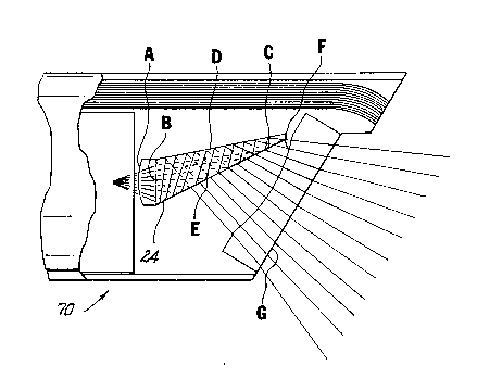
Referring now to Figs. 11, 12, 13A, 13B and 14-17,
there is illustrated another alternative embodiment of the
present invention. Endoscope sheath 70 includes an elongated
sheath member 72 and a prism mount 74 which is adapted to be
mounted to the distal end portion of elongated sheath member
72. Elongated sheath 72 is to be positioned over the
endoscopic portion 1002 of the endoscope 1000 and includes
an illumination system in the form of fiber optic hundle 76
and a fluid and/or gas conduit 78 which extends along the
length of the sheath member 72. As illustrated in Fig. 17,
fiber optic bundle 76 and conduit 78 extend through channels
80, 82 respectively formed in a wall of sheath member 72. An
illumination inlet connector 84 at the proximal end of
sheath member 72 receives an illumination guide tube 500
extending from a light source to supply the fiber optic
bundle 76 with light. An inlet port 86 is also formed on the
proximal end portion of the elongated sheath 72 and receives
a fluid supply tube 600. The fluid mechanism includes an
outlet port or nozzle 88 which directs over and/or is in
front of the distalmost lens surface 102 of prism mount 74
to clean the lens surface so as to remove body fluids which
may accumulate thereon during the surgical procedure and/or
to clear the region in front of the lens to enhance viewing.
The novel prism mount 74 is adapted to be positioned
within and attached to the distal end portion of the sheath
member 72. Prism mount 74 preferably defines an outer
diameter which is substantially equal to or slightly less
than the inner diameter of the distal end of sheath member
-17- 2~ 3~ ~i8~
72 to form a frictional fit between the two components to
thereby mount the prism mount 7 4 to the sheath member .
Adhesives and/or sealing compounds may be used to s~cure the
prism mount to the sheath member and to seal the distal end
of the sheath. Prism mount 74 includes a 30 hybrid prism
90 for changing the angle of view approximately 30 relative
to the axis of the endoscope. Fig. 12 illustrates the ray
path and orientation through the optical elements of prism
mount 74. Th~ geometrical characteristics of the optical
components of prism mount 74 are defined bv lens surfaces A-
F as shown in Fig. 12. The on-axis geometrical and optical
parameters of the optical components of optical sheath 70
are recorded in Table 1 below. Table 1 is a follows:
TABLE 1
Surface Radius Thickness Medium Index
A 0.488 0.035 Bk7 1.52
B Plano N/A F2 1.62
C Plano N/A F2 1.62
D Plano N/A F2 1.62
20 E Plano .081 AIR 1.00
F 0.371 .087 lEpoxy 1.56
G Plano
*dimensions are in inches
1 Emerson & Cuming Stycast 1267 Epoxy
Figs. 13A and 13B illustrate the specific
dimensions and angles of a preferred 30 hybrid prism in
accordance with the invention. Table 2 below outlines these
dimensions in detail. Table 2 is as follows:
TABLE 2
30 Surface or Angle Dimension Angle (degreesl
i 0.117 N/A
ii 0.304 N/A
iii 0.2S3 N/A
i ' " ' "` ' ', ` - ' ' ' .' ' '.' ' '.'.. ' . ' ' ' ~' .`. -; - . , .. ,; .. . .... . . .. . ..
2 ~ 3 ~
18-
iv 0.240 N/A
v 0.011 N/A
vi 0.387 N/A
a N/A 10846'
B N/A 1912'
* dimensions are in inches
Prism 90 is preferably inserted within a
correspondingly dimensioned opening 92 (Figs. 14-16) formed
in the wall of prism mount 74 and secured within the prism
mount by snap fit or other friction fit, adhesives or the
like. The upper portion of prism mount 74 defines a
recessed region 94 which is dimensioned to accommodate the
optical fibers 76 extending from elongated sheath member 72.
Prism mount 74 also includes angled channel 96 adjacent
recessed region 94 which accommodates the extreme distal end
of the optical fibers 76. Channel 96 bends the fibers
towards the longitudinal axis of endoscopic portion 1004 to
alter the illumination angle in a manner similar to that
described in connection with the embodiments of Figs. 1-4,
i.e., channel 96 redirects optical fibers 76 such that the
light rays are directed from prism mount 74 at a 30 angle
relative to the axis of the endoscope. The barrier portion
98 (Fig. 17) defined between channel 96 and the optical
components of pri.sm mount 74 minimizes veiling glare in a
manner similar to that described above. Prism mount 74 may
also contain a channel 100 which receives the distal end of
fluid conduit 78. Channel 100 terminates in nozzle 88 which
directs fluid over and/or in front of lens surface 102 to
clean the lens surface and/or clear the region in front of
the lens for optimal viewing. The remaining optical
components of prism mount 74 include concave lens 104
positioned at the distal end of prism mount 74 and having
lens surface 102, and convex lens 106 disposed at the
proximal end surface of prism 88. Lens 102 and lens 106
2 ~
--19--
function in a similar manner to their corresponding lenses
of the embodiment of FigsO 1-4. Sealants may be llsed to
seal the distal end of the sheath, such as at fibers 76 and
conduit 78.
Referring now to Figs. 18-21, there is illustrated
another alternative embodiment of the present invention.
Endoscope sheath 110 is similar to the endoscope sheath
described in connection with the embodiment of Figs. 13-17
and includes an elongated sheath 112 having an fiber-optic
bundle illumination system 114. Elongated sheath 112
includes aligning insert members 116 (Fig. 19) disposed
within the interior of sheath 112. Insert members 116 may
extend along the length of elongated sheath 112 and are
dimensioned to reduce the effective inner diameter of
elongated sheath 112 to approximate the outer diameter of
endoscopic portion 1004. Insert members 116 may also serve
to form a friction fit between the sheath 110 and the
endoscope portion 1004. Aligning inserts 116 also define a
channel 118 to accommodate the fiber optic bundle 114. See
Fig. 19. Similar inserts are shown in Fig. 11. It is
contemplated that the elongated sheath and alignment inserts
could be integrally molded together as one piece to redu~e
cost and facilitate assembly. It is also contemplated that
the plurality of illumination fibers in the sheath may be
replaced by a light transmissive solid molded plastic light
guide conforming to the shape of the illumination channel in
the sheath, or a liquid-containing light guide similarly
conforming to the light channel.
Prism mount 120 includes a 60 prism 122 which is
mounted within the prism mount 120 for changing the angle of
view 60~ relative to the longitudinal axis of the endoscope.
Prism 122 includes wing portions 123 (Fig. 21) disposed on
opposing side walls thereof which engage flanges 125 of
prism mount 120. Because the distal tip of the prism is
2 ~
-20-
captured in notch-forming tabs on the mount the notch-
forming tabs, wing portions and flanges securely hold prism
122 to the mount. Prism mount 120 also includes a light
guide 124 which may be integrally formed with prism mount
120 and disposed adjacent the distal end of fiber optic
bundle 114. Light guide 124 alters the angle of
illumination 60 relative to the axis of the endoscope
without requiring bending of the fiber optic bundle 114.
Light guide 124 preferably has a total internal reflection
surface Y for directing illumination from the fibers toward
the angled field of view. Prism mount 120 also includes an
opaque barrier 126 (Fig. 20A) positioned between light guide
124 and prism 122. The barrier 126 comprises a reflective
or opaque layer such as foil which fits into channel 127
between prism 122 and light guide 124 and extends proximally
to the overlap mirrored surface of prism 122. Barrier 126
minimizes veiling glare and leakage of light between prism
122 and light guide 124.
As best shown in Fig. 20B, the remaining optical
components of prism mount 120 include convex lens surface
128 which is formed on the proximal surface of prism 122 and
lens 130 molded into the mount. Lenses 128, 130 function in
a similar manner to their corresponding lens components of
the embodiment of Figs. 1-4. Advantageously, prism mount 120
may be integrally molded of plastic or glass to include lens
130 and light guide 124 obviating the need for separate
formation and mounting of these components. Further, as
previously mentioned, the prism mount system advantageously
includes wing portions 123 on the prism and flanges 125 on
the mount for engaging and retaining prism 122, thereby
facilitating assembly of prism to the mount in snap-fit
relation. Thus, where prism mount 120 has integrally formed
therein the concave lens 130, the device may be assembled by
simple snap-fitting prism 122 onto mount 120, fitting mount
2131~
-21-
120 to sheath 112 with fibers 114 abutting total integral
reflection light guide 124, and mounting the tip to the
sheath. Prism mount 120 can engage sheath 112 by friction
or other fit with or without adhesives or sealants, as
required.
Referring to Fig. 20B, the geometrical
characteristics of ~he optical components mounted in prism
mount 120 are defined by lens surfaces H - N. The on-axis
geometrical and optical parameters of the optical components
are recorded in Table 3 below. Table 3 is as follows:
TABLE 3
Surface Radius Thickness Medium Index
H 0.4671 N/A Polystyrene 1.59
I Plano N/A Polystyrene 1.59
15 J Plano N/A Polystyrene 1.59
K Plano 0.0793 Air 1.00
L 0.4904 0.0440 Polystyrene 1.59
M Plano N/A
Fig. 20B also illustrates the specific dimensions
and angles of the optical components of prism mount 120.
Table 4 outlines these dimensions in detail. Table 4 is as
follows:
TABLE 4
Surface or Anqle Dimension Anqle
x 0.2145 N/A
xi 0.3516 N/A
q N/A 99.5u
a N/A 28.5
Referring now to Fig. 22, there is illustrated a
kit 201 incorporating the previously described endoscope
sheaths of the present invention. The kit 201 may include
an endoscope 1000 and one or more of the endoscopic sheaths
S each having a different angle of view. For example,
sheaths with a 30 prism and/or a 60 prism can be packaged
2:~31~Q
-22-
with an endoscope in a single kit so the user can select
(and interchange) the sheath and place it over the endoscope
prior to insertion into the body to achieve the desired
angle of view in the surgical procedure. It is also
envisioned that a 0 sheath could be included to protect the
endoscope and maintain the cleanliness of the scope during
non-oblique imaging. Thereafter, when a change of the angle
of view is required to perform the surgery, a sheath
incorporating a 30 or a 60 prism may be interchanged with
lo the 0 sheath. A further advantage of a 0 sheath is that
an improved illumination system having a higher transmission
efficiency may be used in place of or to supplement the
illumination system of the endoscope. Yet a further
advantage is that ar endoscope not having a fluid and/or gas
conduit for cleaning the distal-most imaging lens and/or
clearing the region in front of the distal-most lens can be
provided with such a conduit by using a sheath having such a
conduit.The package used may include a molded plastic cover
or lid 202 and a base 204 which is secured to the lid along
respective peripheral portions thereof. Alternatively, it is
contemplated that a kit may be provided containing a
plurality of endoscope sheaths without an endoscope. Such a
kit may be useful, for example, with reusable endoscopes.
Referring now to Fig. 23, there is illustrated
another alternative embodiment of the endoscope sheath of
the present invention. Endoscope sheath 150 includes
elastic sheath member 152 which is fabricated from a
flexible material such as latex rubber and is attached to
prism mount 154 in any known manner such as by welding,
adhesives or the like. The elastic sheath 152 may be pulled
onto endoscopic portion 1004 and extend proximally to
enclose substantially all of the elongated endoscopic
portion of the endoscope. Elastic sheath 152 is preferably
dimensioned to form a snug fit about the endoscopic portion
2~3~a
-23-
1002. Prism mount 154 includes a prism to change the angle
of view as previously described. This embodiment of
endoscopic sheath may have particular application with
flexible endoscopes. Fig. 24 illustrates the endoscope
sheath 150 of Fig. 23 packaged along with a conventional
endoscope 1000 as part of a kit having a 30D endoscope
sheath and 60 endoscope sheath.
While the invention has been particularly shown
and described with reference to the preferred embodiments,
it will be understood by those skilled in the art that
various modifications and changes in form and detail may be
made without departing from the scope and spirit of the
invention. For example, it is conceivable that other prisms
such as 45 prism or right angle prism may be readily
employed. Additionally, plural prism systems, and rotatable
prisms which change the angle of view from forward to side
viewing, e.g., roof prisms can also be utilized. Such
rotatable prisms may be controlled proximally by the user.
Accordingly, modifications such as those suggested above,
but not limited thereto, are to be considered within the
scope of the invention.