Note: Descriptions are shown in the official language in which they were submitted.
~ 3~3~ JJM--79
~: :
WOIJND IMPI~NT MATERIALS ~"
The present invention relates to novel bioabsorbable
materials for use as or in wound implants, and to methods of ,
preparation of those materials.
Healing of cavity wounds depends on the production by
the wound of substantial quantities o~ matrix materials and
granulation tissue as natural ~iller, and the de~
keratinization and migration of cells at the periphery of
the wound across the moist surface o~ the neoangiogenic
10 matrix. Currently, such wounds are treated with dressings -~
designed to maintain a moi~t environment and to prevsnt
fluid loss, infection, adherence and trauma. Additionailly,
alginates and hydrocolloids have been used to absorb excess
exudate and contribute to granulation induction. These
materials have the obvious disadvantage that they are not
designed to be 'absorbed' by the wound and therefore must be
removed from the cavity, usually with irrigation and
disruption of wound reparation.
An effective alternative to alginates and
hydrocolloids would be similar materials constructed from
absorbable biomaterials with a determined pharmacological
fate that could be left in situ throughou~ and after wound
healing. Hitherto, the materials suggested for this purpose
have included bioabsorbable sponge~ formed by freeze-drying
solutions or suspensions of bioabsorbable pol~ners.
Advantageously, these bioabsorbable polymers are
natural biopolymers such as ~ollagen, fibrin, fibronectin
or hyaluronic acid. Such materials are not only highly
biocompatible and biodegradable, but they can also assist
30 wound healing by promoting the proliferation of fibroblasts, ~ ;
and by promoting angiQgenesis.
For example, US-A-4970298 (Frederick H. Silver et alJ
describes a biodegradable collagen matrix suitable for use
as a wound implant. The matrix is ~ormed by freeze drying
a dispersion containing collag~n, crosslinking the collagen
via two crosslinking steps and freez~-drying the crosslinked
matrix. The matrix may also contain hyaluronic acid and
fibronectin.
' `
~ 7``~ o)~
23~8
. JJM--79
W090~00060 (Collagen Corporation) describes collagen
implants that are formed by flash freezing and then freeze-
drying a suspension of cvllagen fibrils without chemical
cross-linking. The implants have a bulk density of 0.01 to i-
0.3 g/cm3 and a pore population in which at least about 80%
of the pores have an av~rage pore size of 35 to 250 ~m.
This wound healing matrix also serves as an effective
s~stained de7ivery system for bioactive agents. ~ .j
EP-A~0274898 (Ethicon Inc.) describes an absorbable
implant material hav~ing an open cell, foam-like structure
and formed from resorbable pGlyasters, such as poly-p~ -
dioxanone, other polyhydroxycarboxylic acids, polylactides
or polyglycolides. The open-cell plastic matrix is
reinforced with one or more reinforcing elements of a
textile nature formed from a resorbable plastic and embedded
in the matrix. The open-cell plastic matrix is made by
freeze-drying a solution or suspension of the plastic
material in a non-aqueous solvent. The pore size of the
open-cell plastic matrix is from 10 to 200 ~m.
JP-A-03023864 (Gunze KK) clescribes a wound implant
material comprising a collagen sponge matrix reinforced with
fibres of poly-L-lactic acid. The collagen spong~ matrix is
formed by freeze drying a solution of porcine
atherocollagen. :~
The above bioabsorbable sponge implants are formed by
freeze-drying solutions or suspensions of a bioabsorbable
material in a solvent. However, it is generally difficult ~`~
to control the pore size and overall density of sponge
materials made in this way. Normal freeze-drying procedures
30 result in sponges having large pores and low density. Such :~
sponges are weak, and tend to be resorbed too quickly to be
suitable in practice for use as wound implants. The
physical weakness of the sponges has been addressed by
embedding bioabsorbable reinforring fibres in the sponge
matrix, but the reinforcing fibres cannot prevent the rapid
braakdown and resorption of the sponge matrix in situ.
The rate of resorption o~ the freeze-dried sponges has
ty~pically been reduced by chemical cross-linking of the
i~ ~ 3 2 3 6 8 JJM-79
. ,
~ 3
polymer making up the sponge. For example, the collagen in
a collagen sponge can be cross linked with carbodiimide or
glutaraldehyde to make it insoluble and to reduce the rate
of breakdown of the collagen by collagenase present at the
wound site. This ch~mical cross-linking by its very nature
makes the collagen less biocompatible and less wound-
friendly. Moreover, even with cross linking, it is
difficult to obtain a controlled and optimised rate of
cellular invasion and resorption of the implant.
Some control over the pore size and density of freeze-
dried sponges can be achieved by varying parameters such as
the concentration of the starting solution or suspension and
the rate of freezing. Smaller pore sizes can be ob ained by
"flash-~reezing" the solution or suspension, since this
results in the formation of smaller ice crystals in the
froz~n solution. However, even flash-freezing followed by
freeze drying results in a sponge of quite low bulk density,
with highly disperse pore sizes typically in the range of 35
to 250 ~m.
Accordinyly, it is an object of the present invention
to provide a bioabsorbable wound implant material that has
high strength and controlled porosity.
The present invention provides a wound implant
material comprising a plurality of bioabsorbable
microspheres bound together by a bioabsorbable matrix. The
term "bioabsorbable microspheres" refers to substantially
spherical particles of one or more bioabsorbable materials.
Preferably, the degree of non-sphericality of the particles,
as defined by the average ratio o~ the largest diameter to
the smallest diameter of each particle, is less than 2.0,
more preferably less than 1.5 and most preferably less than
1.2. A ratio of 1.0 would correspond to perfectly spherical
particles. The microspheres may be solid or hollow, or may
comprise microcapsules encapsulating a solid, liquid or gel
comprising a pharmacologically active substance, a
biopolymer or a growth factor. The microspheres need not be
of uniform size, but prefarably at least 90% of the
microspheres have diameters- between 50 ~m and 1500 ~m.
~1 3~3~8
-: - JJM--7 9
: 4
More preferably, at laast 90% of the microspheres have
diametars between 200 ~m and 1000 ~m. Most prefer~bly, at
least 90% of the microspheres have diameters between 500 ~m ~ -
and 800 ~m.
The bioabsorbable matrix may be a solid or a semi-
solid such as an aqueous gel of a biopolymer. Pre~erably,
the matrix is a bioabsorbable colid obtained by air drying
or freeze-drying a gel solution or suspension of a
bioabsorbable polymer in a solvent. The bioabsorbable
matrix may comprise the same material as the microspheres,
or may comprise oth~r materials.
It can thus be seen that the wound implant materials
according to the present invention are aggregates of solid
microspheres bound together by the bioabsorbable matrix
material. Preferably, the materials c~ntain at least 30% by
volume of 1:he microspheres. More preferably, the materials
contain at least 40% by volume, and most preferably at least
50~ by volume of the microspheres. It will be appreciated
that, based on closest packing of spheres, the materials may
20 contain up to 72% by volume of ~micro~pheres of identical ~ -
size, and a still higher fraction by volume if the
microspheres are size disperse.
The porosity of the materials according to the present
invention may b~ controlled both hy varying the size of the
microspheres and by varying the volume fraction of the
microspheres in the material. Average pore æizes in the
range 50 ~m-250 ~m have been described as optimal for tissue
ingrowth.
The pre~erred material for the bioabsorbable matrix
is collagen in solid, gel or sponge form. The volume of the
bioabsorbable matrix is not more than 70% of the total
volume of the material according to the present invention.
Preferably, the bioabsorbable matrix does not occupy the
whole o~ the interstitial space between the microspheres,
but instead is concentrated in the region of contact between
microspheres, where it functions as a glue to hold the
microspheres together. Pre~erably, the bioabsorbable matrix
materials do not comprise more-than 20% by volume and/or 20%
~ ' ,~, '
~ ~L3~3~ JJM-7~
by weight of the materials according to the present
invention, and more preferably ~hey do not comprise more
than 10% by volume and/or ~0% by weight of the materials.
Preferably~ the microspheres and/or the matrix
comprise one or more ~ioabsorbable polym~rs independently
selected from the group consisting of polymers or copolymers
of lactic acid and/or glycolic acid~ collagen, cross-linked
collagen, hyaluronic acid, cross-linked hyaluronic acid, an
alginate or a cellulose derivative. Preferably, the
microspheres or the matrix, or both, additionally contain
pharmaceutically active compounds such as fibronectin, a
cytokine, a growth factor, an antiseptic, an antibiotic, a
steroid or an analgesic.
The wound implant materials according to the present
invention may be reinforced by including fi~res or a mesh of
a suitable bioabsorbable polymer such as
polylactic/polyglycolic acid or oxidised regenerated
cellulose.
It will also be appreciated that single pieces of the
materials according to the present invention can be made
with more than one porosity. For example, a layered
structure could be made by builcling up layers containing
microspheres of different sizes, thereby giving dif~erent
porosities in dif~erent layers of the materialO
The wound implant materials according to the present
invention c~n be cut into any suitable shape for use as or
in a wound implant.
The present invention also ~ncompasses a method of
making a wound implant material as described above,
comprising the steps of preparing bioabsorbable
microspheres; dispersing the bioabsorbable microspheres in
a solution or suspension of a bioabsorbable material in a
solvent; and removing the solvent by evaporation.
Preferably, the solvent is removed by freeze drying.
The mlcrospheres may b~ made by any of the methods
known in the art O These methods are reviewed, for example,
by R.C. Oppenheim in Polymeric Particles and Microspheres
~uiot and Couvreur, editors, Chapter I, pp 1~25 (CRC Prass,
. ,-.`. :,, :.,, .' .'., . ~ - . ... ..
~132368 JJM-79
1986). The most commonly used method comprises di~persing
a water-insoluble bioabsorbable polymer in a nonaqueous,
volatile solvent, followed by mixing the solvent with water
and an emulslfier, emulsifying the mixture and then
5 evaporating the solvent under reduced pressure. Cross- -
linking agents and/or pharm~ceutically active compounds may
be included in the emulsio~. ~2thods o making
bioabsorbable microspheres are also describ~d in US-A-
3092553, EP-A 0119076, EP-A-0351296, W091/06286 and
W091/15193. The as-prepared microsphere~ are qenerally ~ize
disperse~ having diameters in the range 0.01 ~m to 1500 ~m.
It is generally found that larger microspheres suitable for
the practice of the present inventiQn are obtained from
water-in-oil emulsion by cross-linking and evaporation.
Smaller microspheres are obtained from oil-in-water
emulsions.
Large biopolymer microspheres suitable for the
practice of the present invention may also be obtained by
the extrusion of a laminar ~low of an aqueous dispersion of
the biopolymer. The laminar flow is then broken up by
vibrations into droplets, which :Eall into a cross-linking
bath to form the cross linked microspheres.
Specific techniques for forming biopolymer
microspheres in the size range o~ interest f3r the present
25invention are described in detail in EP-A-0381543 and
W092/02254. Biopolymer microspheres suitable for the
practice of the present invention may be obtained from
Bioetica, 32 Rue Saint~Jean-de-Dieu, 69007 Lyon, France~ :
under the Trade Mark "Type A Collaspheres".
30Preferred size ranges can be isolated by filtration:~
or c~ntri~ugation.
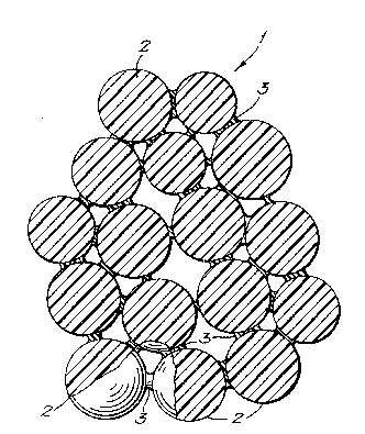
An embodiment of the present invention will now b~ :~
described further~ by way of example, with reference to the
accompanying drawing, which shows a schematic cross-section
35 through a material according to the present invention. ~;
E m~le 1
A cross-linked ester of hyaluronic acid prepared as
described in EP-A-026511~ ~Fidia SpA) is dissolved in a
JJM--79
~13236~
; 7
volatile organic solvent and fibrous collagen is added to
the resulting solution. The solution is emulsified in water
using gelatin as the emulsifier. The organic solvent is
removed under reduced pressure at room temperature to leave
a suspansion of hyaluronic acid ester/collagen microspheres
dispersed in the water. Microspheres in the size
range 600 ~m-~OO~m are isolated by filtration, dried, and
mixed into a 7% collagen/water gel. The mixture is then
fr2ez~-dried and cut into 5 cm x 5 cm x 0.5 cm doses. The
density o~ the material is 50 mg/cm3, of which 3 mg/cm3 is
the collagen matrix and 47 mg/cm3 is the microspheres.
The reticulation of the resulting implant material is
assessed by electron microscopy. This shows con~istent pore
sizes of between 50 and 250 ~m.
A cross-section through resulting implant material is
shown schematically in Figure 1. Referring to the Figure,
thQ implant material 1 comprises microspheres 2 stuck
together by the collagen matrix 3. The matrix 3 does not
fill the whole of the interstitial space between the
microspheres, but leaves the pores between the microspheres
substantially open.
Example 2
A wound implant material i~ prepared as in Example 1,
with addition of hyaluronic acid at a concentration of 0.1
to 2 mg/cm3 based on the weight of the dry finished
material, to the collagen/water gel. The resulting material
benefits rom the chemotactic effect of hyaluronic acid
assisting cellular ingrowth.
The materials prepared as above have a more consistent
pore size than conventional bioabsorbable sponge implants.
This allows more precise control of cellular ingrowth and
rate of resorption in situ. The bulk density of the
materials according to the present invention (10-100 mg/cm3)
may be made higher than that of conventional freeze-dried
sponges depending on the application, resulting in a
stronger and more slowly absorbed implant. Furthermore, the
rate of absorption of the microspheres can be tailored
within a wide range. Thi~ allows, for example, the
~13 2 3 6 8 JJM--79
`i"' 8
preparation of implants that are absorbed more slowly than
a conventional freeze-dried collagen sponge.
The above examples are intended for the purpose of
illustration only. Many other embodiments falling within
the scope of the accompanying claims will be apparent to the
skilled reader.
~ -'. '. ,'
', :"