Note: Descriptions are shown in the official language in which they were submitted.
2~.4~~~
METHOD AND APPARATUS FOR THE ALIGNMENT OF
A FEMORAL KNEE PROSTHESIS
The present invention relates generally to method and
apparatus for establishing the correct alignment and orientation
for a femoral knee prosthesis during total knee arthroplasty
surgery and pertains, more specifically, to determining the correct
position and orientation of cutting guides with respect to a
patient's femur so that the femur can be cut to fit the femoral
knee prosthesis and the femoral knee prosthesis will be implanted
in an anatomically correct orientation.
During knee resurfacing arthroplasty, commonly called knee
replacement surgery, the distal surfaces of the femur are cut away
and replaced with a metal cap to simulate the bearing surfaces of
the femur. The proximal surface of the tibial is modified in a
similar way, to provide a metal-backed plastic bearing surface.
The metal femoral component of the new prosthetic joint transfers
the weight of the patient to the tibial component such that the
joint can support the patient's weight and provide a near-normal
motion of the knee joint.
Several studies have indicated that the long term survival of
a prosthetic knee joint is dependant on how accurately the
components of the knee joint are implanted with respect to the
weight bearing axis of the patient's leg. In a correctly
functioning knee, the weight bearing axis passes through the center
of the head of the femur, the center of the knee and the center of
the ankle joint. This weight bearing axis typically is located by
1
2~.~~Q8~
analyzing an X-ray image of the patient's leg, taken while the
patient is standing.
The X-ray image is used to locate the center of the head of
the femur and to calculate the position of the head relative to
selected landmarks on the femur. The selected landmarks are then
found on the patient's femur during surgery and the calculations
used to estimate the actual position of-the femoral head. These
two pieces of information are used to determine the correct
alignment of the weight bearing axis for the femur, commonly
referred to as the mechanical axis of the femur. To completely
define the correct position for the femoral component of the knee
prosthesis, the correct relationship between the center of the
femoral head and the knee joint and the rotation of the knee joint
about the mechanical axis must be established. This information is
determined from landmarks on the distal portion of the femur. The
correct alignment for the tibial component of the knee prosthesis
is determined by finding the center of the ankle joint and relating
its position to landmarks on the tibia. This point and the center
of the proximal tibial plateau are used to define the weight
bearing axis, or mechanical axis, of the tibia. The correct
relationship between the ankle joint and the knee joint and the
rotation of the knee joint about the mechanical axis are determined
by reference to the distal portion of the femur and landmarks on
. the tibial plateau.
Various mechanical alignment instruments are used to assist
the surgeon in making cuts on the distal femur and proximal tibia
2
which will allow the femoral and tibial components of the
prosthetic knee implant to be attached to the femur and tibia.
These mechanical alignment instruments permit the surgeon to fix
cutting guides in place with respect to the selected landmarks on
the bones so that the cuts will be correctly oriented with respect
to the mechanical axes determined from the X-ray image.
There are two general types of alignment instruments in common
use. These are intramedullary and extramedullary alignment
systems. Intramedullary alignment systems use the inside of the
femur or tibia, the medullary canal, as one of the selected
landmarks for establishing alignment. Extramedullary alignment
systems use only the external surfaces of the body to establish
alignment.
A typical extramedullary alignment system requires the surgeon
to visually align a slender rod with the center of the knee and the
center of the femoral head for alignment of the femoral component,
then align a similar rod with the center of the ankle and the
center of the tibial plateau for alignment of the tibial component.
The centers of the femoral head and ankle are found by palpation or
are established with an intraoperative X-ray. If correctly placed,
the rods will lie parallel to, and offset from the mechanical axes.
Once aligned, the rods are used as a guide to fix the location of
the cutting guides with respect to the femur and the tibia so that
the cuts can be performed.
A typical intramedullary alignment system requires the surgeon
to insert rods into the medullary canal of the femur and of the
3
tibia. If properly placed, these rods should lie on the axis of
the bones. In the case of the tibia, the mechanical axis is very
close to the axis of the bone. In the case of the femur, the axis
of the bone is quite different from the mechanical axis due to the
offset nature of the hip joint, and this difference must be
measured from the pre-operative X-ray and used to correct the
alignment of the femoral cutting guides.
Both intramedullary and extramedullary approaches to alignment
have numerous inherent drawbacks and sources of error.
Extramedullary alignment depends on accurate visual estimation of
the alignment of the extramedullary rods. Location of the femoral
head by palpation is difficult and error-prone, particularly with
obese patients. Use of intraoperative X-rays improves the result
somewhat, but is time consuming and exposes the patient and
operating room personnel to radiation. X-rays also are subject to
distortion and require visual interpretation and estimation to
analyze correctly, as X-rays offer only one planar view in two
dimensions.
Intramedullary alignment approaches provide only sightly
better results, in that the knee joint alignment is still
determined by estimating the difference between the bone axis and
the mechanical axis from a potentially distorted X-ray image. In
addition, intramedullary rods must be introduced very carefully,
not only to make sure they align correctly with the medullary
canal, but also to make sure that the insertion of the rods does
4
not create an embolism, which could seriously injure or even kill
the patient.
An ideal alignment system finds the mechanical axis of the
patient's leg directly, without the need for preoperative or
intraoperative X-rays, estimation, calculation, location of hidden
or obscured landmarks, or surgical intervention outside of that
required for access to the knee joint surfaces. The ideal
alignment system depends only on the accepted definition that the
mechanical axis passes through the center of the head of the femur,
the center of the knee joint and the center of the ankle, in order
to locate the mechanical axis.
The present invention provides method and apparatus for
locating the mechanical axis of a patient's femur by directly
locating the center of rotation of the head of the femur. As such,
the present invention attains several objects and advantages, some
of which are summarized as follows: Enables accurate location of
the direction of the mechanical axis of the femur interoperatively,
without invading the medullary canal and without the necessity for
surgical intervention beyond that already required for access to
the knee being replaced; provides a relatively simple procedure
capable of being performed quickly just prior to preparing the
femur for distal cuts; attains a high degree of accuracy with
minimal procedural steps and apparatus; enables a direct
. determination of the direction of the mechanical axis of the femur
without reliance upon visual estimation or interpretation;
provides apparatus capable of long-term reliable performance.
5
CA 02142083 2004-08-26
n A
The above aspects and advantages, as well as further aspects and
advantages, are attained by the present invention which may be
described briefly as method and apparatus for determining the
direction of the mechanical axis of a femur of a patient in relation
to the corresponding knee of the patient, the method comprising: the
step of and means for placing the knee of the patient in an
equilibrium position wherein external forces on the knee are balanced
and the knee remains essentially stationary at the equilibrium
position; the step of and means for applying a force to the femur at
a predetermined location relative to the mechanical axis of the
femur; the step of and means for directing the applied force in a
direction such that the knee is undeflected from the equilibrium
position while the force is applied to the femur in said direction
and the step of and means for employing said direction of the applied
force to indicate the direction of the mechanical axis of the femur.
The invention will be understood more fully, while still further
aspects and advantages will become apparent, in the following
detailed description of preferred embodiments of the invention
illustrated in the accompanying drawing, in which:
FIG. 1 is a schematic representation of the alignment method and
system of the present invention;
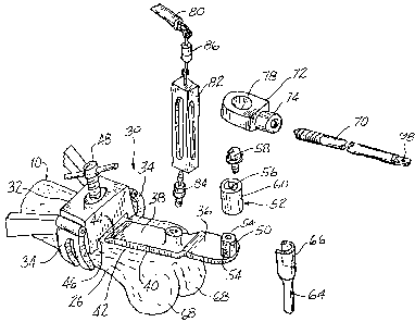
FIG. 2 is an exploded pictorial perspective view, partially
schematic, of the alignment system of the present invention at the
distal end of a femur;
6
~~~~o~~
FIG. 3 is a pictorial perspective view, similar to FIG. 2, but
only partially exploded;
FIGS. 4 and 5 are enlarged fragmentary side elevational views,
partially in cross-section, of a portion of the alignment system
illustrating the method of the present invention;
FIG. 6 is an enlarged fragmentary side elevational view
similar to FIGS. 4 and 5, but showing the location of guides at the
distal end of the femur; and
FIG. 7 is a side elevational view similar to FIG. 6, with an
alternate guide.
Referring now to the drawing, and especially to FIG. 1
thereof, the femur of a supine patient is illustrated schematically
at 10 and is seen to include a femoral head 12 and a distal end 14
at the knee K of the patient. The femur 10 is constrained for
rotation about the femoral head 12 and the mechanical axis 16 of
the femur 10 passes through the center of rotation 18 of the
femoral head 12 and the center 20 of the knee K of the patient. It
has been suggested that the location of the mechanical axis 16 can
be determined by freely suspending the leg of the patient to permit
free rotation of the femoral head 12 and then applying a tensile
force at the center 20 of the knee to rotate the femur 10 until the
mechanical axis 16 is aligned with the direction of the tensile
force. Then, the direction of the tensile force serves as an
. indication of the location of the center of rotation 18 and the
direction of the mechanical axis 16 relative to the center 20 of
the knee, thereby locating the mechanical axis 16 and enabling that
7
. 2~.~2f~~~
location to be used for the proper placement of cutting guides at
the knee.
In practice, however, where a patient is supine on an
operating table, the patient s leg cannot be fully freely suspended
since the lower leg or the foot of the patient must remain on the
operating table. As a result, a tensile force applied to the knee,
as suggested above, must overcome external forces over and above
the force necessary merely to rotate a freely suspended femur 10,
thereby tending to introduce some deviation in the direction of the
applied tensile force from the direction of the mechanical axis 16.
The present invention eliminates the effect of external forces in
the determination of the direction of the mechanical axis 16 by
eliminating the requirement for rotating the femur 10 in response
to an applied tensile force and relying, rather, on the fact that
the femur 10 will not rotate when a force is applied to the femur
in a direction aligned with the mechanical axis 16 so as to pass
through the center of rotation 18. Accordingly, in the method and
apparatus of the present invention, the leg of the patient is
partially suspended, at the knee K, so as to balance external
forces at the knee and locate the knee at an equilibrium, or
suspended, position. A force, illustrated in the form of a tensile
force F, is applied to the distal femur 26, at the knee K, at a
predetermined location relative to the mechanical axis 16. Force
F is moved so as to be applied in directions parallel to the
coronal plane, as illustrated in phantom as well as in full lines
in FIG. 1, and any deviations in the location of the knee K from
8
214~~~
the suspended position, that is, any movements of the knee K within
the coronal plane to either side of the suspended position while
force F is applied to the knee, are observed until force F is
oriented in a direction wherein the knee is undeflected from the
suspended position and remains stationary at the suspended
position. The direction of force F which produces no deflection of
the knee from the suspended position, as illustrated in full lines
in FIG. 1, is aligned parallel with the mechanical axis 16 and thus
determines the direction of mechanical axis 16 in the coronal plane
relative to the knee of the patient. The direction of the
mechanical axis 16 in the sagittal plane is determined in a
conventional manner, as will be explained in greater detail below.
Once the direction of the mechanical axis 16 is fully determined,
that direction is employed as a reference for the proper location
of cutting guides used in the preparation of the distal femur 26
for the reception of a femoral knee prosthesis, as will now be
described.
Turning now to FIGS. 2 and 3, distal femur 26 is shown being
prepared for the determination of the direction and location of the
mechanical axis of the femur 10 and the subsequent implant of a
femoral knee prosthesis (not shown). Apparatus constructed in
accordance with the present invention is illustrated generally at
and is seen to include securing means shown in the form of a
femoral clamp 32 having clamping jaws 34 which grip the femur 10 to
25 secure the femoral clamp 32 upon the exposed femur 10. An anterior
reference member in the form of a bearing holder 36 includes an
9
. . .
anterior reference bar 38 having an anterior reference surface 40
which is seated against the anterior cortex 42 of distal femur 26
when the anterior reference bar 38 is engaged with the femoral
clamp 32, as seen in FIG. 3. Thus, anterior reference bar 38
includes a ramp 44 providing a wedge-shaped proximal end for
facilitating insertion of the anterior reference bar 38 into a
complementary channel 46 in the femoral clamp 32 and assuring
direct contact between the anterior reference surface 40 and the
anterior cortex 42. Femoral clamp 32 includes a clamping screw 48
which is tightened to clamp the anterior reference bar 38 in place,
as seen in FIG. 3. Once clamped in place, with anterior reference
surface 40 in intimate, fixed contact with anterior cortex 42,
anterior reference bar 38 will be aligned with the sagittal
component of the mechanical axis of femur 10.
A stud 50 is affixed at the distal end of the bearing holder
36 and projects in an anterior direction, normal to the coronal
plane, to receive a bearing 52 placed over the stud 50 and secured
to the stud 50 against rotation on the stud 50. To that end, stud
50 includes opposite flats 54 and bearing 52 includes a central
opening 56 having a complementary configuration for securing the
bearing 52 on the stud 50. A retainer screw 58 is affixed to the
stud 50 to hold the bearing 52 in place on the stud 50 so that the
bearing 52 provides a cylindrical bearing surface 60 extending in
the anterior direction along an axis 62 normal to the coronal
plane. An intercondylar post 64 includes a clip 66 which is
snapped over the bearing 52 to secure the intercondylar post 64 to
the bearing holder 36 with the intercondylar post 64 depending from
the bearing holder 36, normal to the coronal plane, in the
posterior direction.
Prior to clamping the bearing holder 36 in place, as seen in
FIG. 3, bearing 52 is secured on stud 50 and intercondylar post 64
is clipped to bearing 52. Then, the proximal end of the anterior
reference bar 38 is engaged with the femoral clamp 32, with the
intercondylar post 64 assisting in the proper positioning of the
bearing holder 36, by virtue of the placement of the intercondylar
post 64 between the condyles 68 of the distal femur 26 and
perpendicular to the coronal plane. Once the bearing holder 36 is
clamped in place, as seen in FIG. 3, the intercondylar post 64 is
removed from the bearing 52, and the cylindrical bearing surface 60
of the bearing 52 is exposed, extending along axis 62 normal to the
coronal plane and intersecting the mechanical axis 16, as
illustrated at 69 in FIG. 4. As seen in FIG. 4, as well as in FIG.
3, an alignment member in the form of an elongate alignment rod 70
is coupled with a collar 72 by means of a threaded coupling 74 and
includes a pointed tip 76 which initially is recessed with respect
to a bore 78 in the collar 72. Bore 78 is complementary to the
cylindrical bearing surface 60 of bearing 52 so that alignment rod
70 can be coupled with bearing 52 by slipping collar 72 over
bearing 52, with collar 72 journaled for rotation on bearing 52, to
enable pivotal movement of the alignment rod 70 about axis 62.
The leg of the patient is partially suspended by connecting
the bearing holder 36 to a support arm 80 located above the femur
11
10, as seen in FIGS. 3 and 4. A vertical alignment and suspension
device, shown somewhat schematically at 82, is connected between
the bearing holder 36 and the support arm 80, as by suspension
couplings 84 and 86. Support arm 80 is a part of a positioning
system which may be manipulated. by the surgeon to swing the support
arm 80 directly over the femur 10 so as to facilitate attachment of
the vertical alignment and suspension device 82 at couplings 84 and
86, and suspension of the patient's leg. Then the patient's leg is
elevated until the weight of the leg is substantially supported by
the support arm 80. Once the patient's leg is suspended, with the
weight of the leg largely supported by the support arm 80, the
position of the support arm 80 is fixed and the patient's knee K is
placed in the suspended position illustrated in FIG. 4, in which
suspended position the vertical alignment and suspension device 82
indicates that the line of suspension 90 is truly vertical with
respect to gravity. In this equilibrium position of the knee, all
external forces on the knee are balanced, and the knee remains
essentially stationary. One positioning system currently available
for use in positioning support arm 80 is known as the ENDEX
endoscopy positioning system sold by Andronic Devices Ltd. of
Richmond, B.C., Canada. Vertical alignment and suspension device
82 may be in the form of a simple mechanical plumb bob arrangement
which provides a visual indication of plumb, that is, vertical
. alignment along the line of suspension 90, or may be in the form of
an electronic plumb indicator.
12
CA 02142083 2004-08-26
r
As best seen in FIGS. 4 and 5, a powered surgical drill 92
subsequently is coupled to the distal end of the alignment rod 70,
through a force indicator 94, by means of a coupling arrangement
shown in the form of a hook 96, affixed to the surgical drill 92 for
rotation by the surgical drill 92 and passed through an eye 98 at the
distal end of alignment rod 70. The surgeon then pulls upon the
surgical drill 92, in the direction illustrated, to apply a force
along the alignment rod 70, which force is transmitted to the bearing
52 and the bearing holder 36 and observes the force indicator 94 to
gage the amount of force exerted. Preferably, a tensile force of at
least about ten pounds is applied to alignment rod 70 to establish
force F. Force F thus is applied to the femur 10 at the
predetermined location established by the location and orientation
of bearing 52 by means of the surgical drill 92 coupled to the knee
K through the alignment rod 70, the force indicator 94, the hook 96
and the eye 98 and pulled upon by the surgeon to establish the
tensile force. As force F is applied to the alignment rod 70, the
angular direction of the force F is changed by the surgeon, in
directions parallel to the coronal plane, by angular pivotal movement
of the alignment rod 70 about axis 62, with collar 72 journaled on
bearing surface 60 of bearing 52 serving as means for directing the
applied force F to align force F so that the knee K is maintained
stationary at the suspended position and is undeflected from the
suspended position, as observed by indications provided by the
vertical alignment and suspension device 82, while force F is applied
to the femur 10 at the knee R.
13
CA 02142083 2004-08-26
w
Upon reaching the angular position of alignment rod 70 where the
knee K remains undeflected from the suspension position while force
F is applied to the knee K, the alignment rod 70 is locked in place
by actuating the powered surgical drill 92 to rotate alignment rod
70 about the longitudinal axis of the alignment rod 70, as indicated
by the arrow in FIG. 5. Such rotation of the alignment rod 70
advances the pointed tip 76 of the alignment rod 70, by means of the
threaded coupling 74, to embed the pointed tip 76 in the bearing 52,
as seen in FIG. 5 and secure the angular position of the alignment
rod 70 relative to the fixed bearing 52, the pointed tip 76 of the
alignment rod 70 and the bearing 52 thus serving as means for
employing the direction of the applied force F to indicate the
direction of the mechanical axis of the femur 10. The coupling
arrangement provided by the hook 96 and eye 98 assures that both the
force along alignment rod 70 required to establish force F and the
torque required to rotate alignment rod 70 to lock the alignment rod
70 in place are applied without a moment which would tend to displace
the alignment rod 70 from the proper angular position. Bearing 52
preferably is constructed of a synthetic polymeric material having
sufficient lubricity to facilitate the necessary angular movements
of the alignment rod 70, as described above, while enabling a fixed
connection through the use of pointed tip 76. Once used, the bearing
52 is discarded and replaced by a new bearing 52; hence, the material
of the bearing 52 should render the bearing 52 economically
expendable.
With the alignment rod 70 affixed on the bearing 52, as
l, 4
CA 02142083 2004-08-26
described above, the direction in which the alignment rod 70 extends
is parallel with the mechanical axis 16 of femur 10 and the direction
of the mechanical axis 16 is determined. Further, since alignment
rod 70 is parallel with the mechanical axis 16, alignment rod 70 now
is available for use in locating cutting guides for making the cuts
necessary to prepare the distal femur 26 for the reception of the
femoral knee prosthesis to be implanted. Turning now to FIG. 6, the
surgical drill 92 and the force indicator 94 are removed from the
alignment rod 70, the vertical alignment and suspension device 82 is
uncoupled from the bearing holder 36 and the support arm 80 and the
support arm 80 is affixed directly to the bearing holder 36 so that
the femur 10 is held in place, essentially rigidly, by the support
arm 80.
The alignment rod 70 now is available to receive a distal
femoral condyle locator 100 which is slipped over the distal end of
the alignment rod 70 and translated along the alignment rod 70 until
the femoral condyle locator 100 engages the distal end of the femur
10. The femoral condyle locator 100 includes a sleeve 102 for
sliding along the alignment rod 70 and a locator surface 104 which
is maintained perpendicular to alignment rod 70 by the engagement of
the sleeve 102 with the alignment rod 70. Once in place, as
illustrated in FIG. 6, femoral condyle locator 100 is secured in
place by a set screw 106. A femoral drill guide 110 then is mounted
upon the femoral condyle locator 100 by engaging pins 112 through the
femoral drill guide 110 and into corresponding holes 114 in the
femoral condyle locator 100 to lock the femoral drill guide 110 in
CA 02142083 2004-08-26
place. Femoral drill guide 110 includes a plurality of drill
alignment holes 118, any matched pair of which may be selected by the
surgeon for drilling corresponding locator holes 120 in the femur 10.
Thus, locator holes 120 are placed in appropriate position relative
to the mechanical axis 16 of the femur 10 for the reception of
standard cutting guides for the resection of the distal femur 26.
Apparatus 30 is removed from distal femur 26 by removing the femoral
drill guide 110 from the femoral condyle locator 100, then removing
the femoral condyle locator 100 from the alignment rod 70, then
uncoupling the alignment rod 70 from the bearing 52, uncoupling the
support arm 80 from the bearing holder 36, loosening the clamping
screw 48 to detach the bearing holder 36 from the femoral clamp 32
and then removing the femoral clamp 32 from the femur 10. Locator
holes 120 are then available for use in connection with conventional
cutting guides.
In an alternate arrangement illustrated in FIG. 7, rather than
locating the femoral drill guide 110 on the femoral condyle locator
100, a distal femoral resection guide 130 is located on the femoral
condyle locator 100, as by pins 132 extending through the femoral
resection guide 130 to enter a corresponding selected set of holes
114 in the femoral condyle locator 100. The distal femoral resection
guide 130 then is locked to the alignment rod 70, by virtue of pins
132 engaged with holes 114 in the femoral condyle locator 100 which
is secured in place by set screw 106. Slots 134 are provided in the
distal femoral resection guide 130 in position to guide a cutting
instrument, such as a saw, for executing distal femoral cuts 136.
16
CA 02142083 2004-08-26
. r
Apparatus 30 then is removed from the femur 10, as described above
and resection of the distal femur 26 is completed in a conventional
manner, utilizing the distal femoral surfaces 138 established by
femoral cuts 136.
It will be seen that the present invention attains the several
objects and advantages summarized above, namely: Enables accurate
location of the direction of the mechanical axis of the femur
interoperatively, without invading the medullary canal and without
the necessity for surgical intervention beyond that already required
for access to the knee being replaced; provides a relatively simple
procedure capable of being performed quickly just prior to preparing
the femur for distal cuts; attains a high degree of accuracy with
minimal procedural steps and apparatus; enables a direct
determination of the direction of the mechanical axis of the femur
without reliance upon visual estimation or interpretation; provides
apparatus capable of long-term reliable performance.
It is to be understood that the above detailed description of
preferred embodiments of the invention are provided by way of example
only. Various details of design, construction and procedure may be
modified without departing from the true spirit and scope of the
invention, as set forth in the appended claims.