Note: Descriptions are shown in the official language in which they were submitted.
~ 0 94/0856~ PCT/US93/09878
2I~ 6I15
INTERDIGITATION-FUSION LIPOSOMES AND GELS
Related U.S. A~lication Data
This application is a continuation-in-part of
application Serial No. 07/961,277, filed October 14,1992,
which is a continuation-in-part of application Serial No.
07/664,576, filed March 5, 1991, now abandoned, which is a
continuation-in-part of application Serial No. 07/464,528,
filed January 12, 1990, now abandoned.
Field of the Invention
10This invention relates to interdigitation-fusion (IF)
liposomes and gels. These liposomes and gels capture high
solute to lipid ratios. The term solute encompasses
bioactive agents, including contrast agents. This invention
also relates to the discovery of interdigitation of lipids
to produce IF gels and liposomes, and the further discovery
that such interdigitation to form liposomes according to the
present invention is size dependent.
The present invention relates to a method for
producing IF liposomes and gels. In the method of the
present invention, liposomes formed by sonication, extrusion
or alternative size reduction processes such as
homogenization to the appropriate size are fused in the
presence of a suitable inducer. This process produces a
composition of the present invention in gel form. The gel
itself can be employed, for example, for delivery of
bioactive agents, or can be used to form IF liposomes, which
in turn possess very high internal volumes and encapsulate
large amounts of solute.
WO 94/08565 PCT/US93/098 ~
2146~
To produce IF liposomes from the gels, the gels are
incubated at a temperature usually but not necessarily above
the transition temperature (Tm) of the lipid used, such that
liposomes are formed. The temperature required by the
methods of the invention is that temperature for any given
mixture of lipid, solute and inducer that induces a change
in the material properties of the mixture thereby producing
the IF liposomes of the invention. The liposomes formed
from IF gels are IF liposomes. Preferably, but not
necessarily, the inducer is also removed during the
incubation step. The result is a composition comprising
liposomes cont~;n'ng high solute to lipid ratios.
Back~round of the Invention
The therapeutic properties of many drugs may be
dramatically improved by the ~m;nistration in a liposomally
encapsulated form [See, for example P.N. Shek and R.F.
Barber, Mod. Med. ~n~, 41, 314-382, (1986)]. In certain
cases, for example, in the administration of amphotericin B
and doxorubicin [Lopez-Berestein, et al., J. Infect. Dis.,
20 151, 704-710, (1985) and Rahman, et al., Cancer Res., 40,
1532 (1980)] toxicity is reduced while efficacy is
maintained or even increased. The benefit obtained from
liposomally encapsulated agents may be fortuitous, and
likely results from the altered pharmacokinetics and
biodistribution of the entrapped drug. [Ostro, et al.,
Amer. J. Hosp. Pharm., Vol. 46 Aug 1989]
A num.ber of methods are presently available for
"charging" liposomes with bioactive agents. For example, in
a liposome-drug delivery system, a bioactive agent such as a
drug may be entrapped in the liposome and then administered
to the patient to be treated. See, for example, Rahman et
al., U.S. Patent No. 3,993,754i Sears, U.S. Patent No.
~ 0 94/08565 PCT/US93/09878
21~1gli~
4,145,410; Papahadjopoulos, et al., U.S. Patent No.
4,235,871; Lenk et al., U.S Patent No. 4,522,803; and
Fountain et al., U.S. Patent No. 4,588,578. In addition to
this basic method for entrapping a bioactive agent, if the
bioactive agent is lipophilic, it may associate with the
lipid bilayer. However, many of the pharmaceutical
formulations produced utilizing the traditional methods
suffer from the disadvantage of low drug to lipid ratio,
scaleup problems and the use of toxic solvents.
In addition to the above-described methods, numerous
bioactive agents have been shown to accumulate in liposomes
in response to an imposed proton or ionic gradient known as
"remote loading" [See, for example Mayer, et al., Biochim.
Biophys. Acta, 857, 123, (1986), Mayer, et al.,
Biochemistry, 27, 2053, (1988) and M.B. Bally, et al., Chem.
Phys. Lipids, 47, 97, (1988)]. This loading technique
allows independent variation of any of the liposomal
parameters. Much higher drug to lipid ratios can be
achieved in comparison to conventional techniques [Mayer, et
al. Chem. Phys. Lipids, 40, 333 (1986)]. The procedures and
materials for remote loading are disclosed in Bally et al.,
U.S. Patent No. 5,077,056, issued December 31, 1991, and
Mayer et al., U.S. Serial No. 07/636,015, filed January 4,
1991. The relevant portions of this patent and patent
25 application related to remote loading are incorporated
herein by reference.
Liposomes are completely closed lipid bilayer
membranes which contain entrapped aqueous volume. Liposomes
P may be unilamellar (single bilayer membrane) or
30 multilamellar (onion-like structures characterized by
multiple bilayer membranes, each separated from the next by
an aqueous layer). The bilayer is composed of two lipid
monolayers having a hydrophobic "tail" region and a
W094/08565 PCT/US93/098 ~
21~6'~.~S ,,
hydrophilic "head" region. In the membrane bilayer, the
hydrophobic (nonpolar) "tails" of the lipid monolayers
orient toward the center of the bilayer, whereas the
hydrophilic (polar) "heads" orient toward the aqueous phase.
The basic structure of liposomes may be made by a variety of
techniques known in the art.
One class of liposomes that may be used in the
practice of the invention are those characterized as having
substantially equal lamellar solute distribution. This
class of liposomes is denominated as stable plurilamellar
vesicles (SPLV) as defined in U.S. Patent No. 4,522,803 to
Lenk, et al., monophasic vesicles as described in U.S.
Patent No. 4,588,578 to Fountain, et al., and frozen and
thawed multilamellar vesicles (FAT MLV) wherein the vesicles
are exposed to at least one freeze and thaw cycle; this
procedure is described in Bally et al., U.S. Patent No.
4,975,282, issued December 4, 1990, entitled "Multilamellar
Liposomes Having Improved Trapping Efficiencies" and
incorporated herein by reference.
Liposomal encapsulation could potentially provide
numerous beneficial effects for a wide variety of
pharmaceutical agents and a high drug to lipid ratio should
prove important in realizing the potential of liposomally
encapsulated agents. The use of liposomes to administer
drugs has raised problems with regard both to drug
encapsulation and drug release during therapy. For example,
there is a continuing need to increase drug to lipid ratios
so as to m; n i mi ze the lipid load presented to the patient.
Interdigitation of lipids is a phenomenon which has
been recently explored in considerable detail by James L.
Slater and Ching-Hsien Huang in Progress Lipid Res., 27,
325-359, 1988. In general, the art describes the
~ 0 94/08565 PCT/US93/09878
214~1i5
interdigitation of various lipid species resultiny from
either the presence of various inducers and/or acyl chain
length asymmetry (See Figures lA and lB). There has been no
report in the literature, however, of the size dependency
for fusing liposomes during interdigitation to produce the
IF gels and liposomes of the present invention.
Obiects of the Present Invention
It is an object of the present invention to provide
interdigitation-fusion gels and liposomes, which may be used
for delivering solute in a number of applications, including
therapeutic applications.
It is another object of the present invention to
provide an interdigitation-fusion gel which contains
saturated lipid and may additionally contain non-saturated
lipids and effective concentrations of bioactive agents for
formulation into compositions for topical or oral
administration to a mammal, including humans.
It is a further object of the present invention to
provide a method for producing interdigitation-fusion
liposomes and gels which accumulate high concentrations of
bioactive agents.
It is an additional object of the present invention to
provide therapeutic methods for treating ~nimAls, especially
mammals, including hllm~n~ with interdigitation-fusion
liposomes and gels having a high solute to lipid ratio.
It is yet another object of the present invention to
provide ph~rm~ceutical compositions based upon the
interdigitation-fusion gels and liposomes of the present
invention.
WO 94/08565 PCT/US93/098 ~
21~611~ -
It is still another object of the present invention to
provide methods for making the interdigitation gels and IF
liposomes of the present invention.
It is another object of the present invention to
provide a novel method for trapping bioactive agents.
These and other objects of the present invention may
be readily understood from the detailed description of the
invention which is set forth herein.
S'~mm~ rv of the Invention
The present invention relates to interdigitation-
fusion (IF) liposomes and gels which can contain solute.
These liposomes and gels capture high solute to lipid
ratios, including bioactive agent. These IF gels and
liposomes find use in a number of applications, including
cosmetic, pharmaceutical and agricultural applications. In
combination with bioactive agents, these liposomes and gels
may be administered topically or systemically to plants and
AnlmAlS, especially mAmm~15, including hl~mAn~. In addition
to the above applications, the IF gels and liposomes of the
present invention are also useful in combination with resin
technology, in particular, paint technology.
The compositions of the present invention comprise a
sized liposome, preferably about 0.4 microns in diameter or
less, more preferably about 0.05 microns in diameter or
less, and most preferably about 0.025 microns in diameter,
in com.~bination with a solute, for example a bioactive agent
and an amount of an interdigitation inducer effective to
fuse the liposomes to produce an IF gel. The initial
liposomes may alternatively be FAT M~Vs. IF liposomes may
be produced from the IF gels of the present invention.
~ 0 94/08565 PCT/US93/09878
214611~
Preferably, in the IF liposomes and gels of the present
invention, a saturated lipid, for example,
dipalmitoylphosphatidylcholine (DPPC),
dimyristoylphosphatidylcholine (DMPC),
di-0-hexadecylphosphatidylcholine and
distearoylphosphatidylcholine, as well as lipids in which
the unsaturated carbon-carbon double bonds in the acyl side
Ch~ ' n.~ of the lipid are in the trans configuration, such as
transdielaidoylphosphatidylcholine and
dipalmitelaidoylphosphatidylcholines, or mixed fatty acid
lipids such as palmitoyloleoylphosphatidylcholine (POPC) or
l-stearoyl-2-oleoyl phosphatidylcholine (SOPC), as well as
other unsaturated lipids, may also be used.
In certain embodiments, unsaturated lipids can be
employed in combination with the saturated lipids of the
invention. It is generally preferred that when DOPC is the
unsaturated lipid employed, it be used with the saturated
lipid DPPC in no more than a proportion of 50 mole percent
unsaturated lipid.
The interdigitation-fusion gel that is produced when
sized liposomes are fused, preferably in the presence of an
inducer, which may or may not contain a bioactive agent, may
be used without further modification, or alternatively, the
gel may be further modified, for example, by usually but not
necessarily heating to a temperature above the lipid
transition temperature (Tm) but in any case incubating the
mixture to a temperature which will induce a change in the
material properties of the mixture and thus induce the
formation of IF liposomes from the IF gels of the invention,
and further possibly removing the interdigitation inducer
contained therein, to produce IF liposomes.
WO 94/08565 ~ 1 ~ 6 1 1 S PCT/US93/098
The IF liposomes of the present invention may be used
to capture surprisingly high solute to lipid ratios,
including bioactive agents. These IF liposomes may be used
without further modification or they may be further sized to
produce liposomes of varying and/or homogeneous size using
techniques and methodologies readily available in the art,
and which will be reviewed hereinbelow.
In the present invention, the preferred
interdigitation inducer is a short-chain alcohol, such as
those having 1 to 4 carbon atoms, preferably ethanol because
of the ease with which it can be removed to produce IF
liposomes from the IF gels of the present invention.
However, any inducer that produces a fused IF gel from sized
liposomes may be used in embodiments of the present
invention.
Exemplary inducers for use in the present invention
include, for example, polyols such as glycerol, ethylene
glycol, short-chain alcohols such as methanol, ethanol,
propanol, isopropanol and n-butanol and anesthetics such as
chlorpromazine, tetracaine, phenylethanol, benzyl alcohol
and phenylbutanol, and others including polymixin, myelin
basic protein, choline, acetylcholine, Tris buffer and
chaotropic salts such as, for example, thiocyanate.
Ethanol, however, is preferred because of its ease of
removal and ph~rm~ceutical compatibility. The amount of
inducer used in the present invention comprises an amount
effective for producing interdigitation-fusion gels from
sized liposomes. It is to be noted that in certain
embodiments of the present invention the saturated lipid
used to make IF gels and liposomes of the present invention
may be a self-inducer, i.e., the saturated lipid will
produce IF gels and liposomes of the present invention
without the need to add an inducer. In particular, the use
~ 094/08565 PCT/US93/09878
214611~
of di-0-hexadecylphosphatidylcholine (DHPC) in this aspect
of the present invention as exemplary, is noted.
Alternatively or additionally, as noted herein, a solute
which may be a bioactive agent may itself also be an
inducer.
In another embodiment of the present invention,
hydrostatic pressure may be used as the interdigitation
fusion inducer. In such cases, small liposomes are caused
to form IF gels by the application of hydrostatic pressure.
In particular embodiments of this aspect of the present
invention, small liposomes comprising DPPC or DSPC can be
induced by pressure to interdigitate, thus forming
intermediary gels that form liposomes upon heating of the
lipid above its phase transition temperature.
As a further aspect of this embodiment of the present
invention, high hydrostatic pressures may be used to kill
bacteria and to sterilize liposomal preparations. Thus,
hydrostatic pressure can be used not only to induce
interdigitation fusion, and but also to sterilize the
resultant liposome preparations.
The IF liposomes and gels of the present invention may
contain concentrations of virtually any solute, including
bioactive agents such as vitamins, hormonal agents,
antimetabolites, antimicrobial agents, antifungal agents,
local anaesthetics, bronchodilators, beta-adrenergic
blockers, antihypertensive agents, antidepressants,
anticonvulsants, antihistamines, antimalarial agents,
analgesics, antibiotics, immunogens, immunomodulators,
antigens, nutrients, proteins, peptides, nucleosides, oligo
and polynucleotides, ribonucleic acid (RNA~ and
deoxyribonucleic acid (DNA) and analogs of RNA and DNA,
antineoplastic agents, antihistaminic agents,
WO 94/08565 PCT/US93/098 ~
21~iiS
neuropharmacologic agents including sedatives and hypnotics,
steroidal and nonsteroidal antiinflammatory agents, diuretic >
agents, antiarrhythmic agents and vascular dilating agents,
among others, including radiographic contrast agents,
5 nuclear magnetic resonance (NMR) contrast agents and
antiviral agents. Additional bioactive agents for use in
the present invention include nutrients such as proteins,
fatty acids and carbohydrates. In certain preferred
embodiments of the present invention, radiocontrast agents,
10 NMR contrast agents, peptides and certain antibiotics, for
example, cephalosporins are utilized in the present
invention. Exemplary radiocontrast agents for use in the
present invention include, for example, iohexol, iopamidol,
iotroxic acid, ioxoglate, iotrolan, ioversol, iothalamate,
15 iodimide, iodipamide, iopentol, iodixanol, metrizamide,
mixtures thereof and their pharmaceutically acceptable
salts. Bioactive agents can be naturally occurring,
synthetic, or semi-synthetic antimicrobial agents.
Exemplary antimicrobial agents are aminoglycosides such as:
20 gentamicin, amikacin and tobramycin. In preferred
compositions of the present invention, the IF gels and/or
liposomes contain high concentrations of the bioactive
agent. In general, the bioactive agent/lipid weight ratio
of the IF gels and liposomes of the present invention are as
high as from about 1:10 to about 15:1. Of course, these
weight ratios are exemplary only and in certain cases it may
be necessary to provide drug and lipid in weight ratios
above or below these ratios.
In certain embodiments of the present invention the
solute or bioactive agent can also function as the inducer.
In such cases, it is not necessary to remove the
inducer/bioactive agent as in other embodiments of the
present invention.
-- 10 --
~ 0 94/08565 PCT/US93/09878
214~11S
Suitable bioactive agents for use in the present
invention include any agent which exhibits biological
activity when administered topically or systemically in the
liposomes or gels of the present invention. Numerous
bioactive agents may be included with the IF gels of the
present invention, preferably for topical or oral delivery.
The same agents may be included in the IF liposomes of the
present invention for topical ~ml ni stration.
The present invention also relates to a method for
producing IF liposomes and gels cont~' ning a bioactive agent
of varying concentration. In the method of the present
invention, sized liposomes, preferably about 0.4 microns in
diameter or less, and more preferably about 0.05 microns in
diameter or less and most preferably about 0.025 microns in
diameter, formed by sonication, extrusion, homogenization or
an alternative process, or alternatively, FAT MLVs, are
fused in the presence of ethanol or other suitable inducer.
Depending upon the interaction of the bioactive agent with
the lipids used in the IF gels and liposomes of the present
invention, the agent may be added before or after the
inducer is added. The addition of inducer results in the IF
gel of the present invention. The gel including bioactive
agent can be administered to a patient, or alternatively,
converted to IF liposomes of the present invention.
To produce IF liposomes of the present invention, the
IF gels are exposed to a temperature usually but not
necessarily above the transition temperature of the lipid
used and, additionally, the inducer may be removed. The
temperature re~uired by the methods of the invention is that
temperature, for any given mixture of lipid, solute or
inducer that produces a change in the material properties of
the mixture thereby forming the IF gels and IF liposomes of
-- 11 --
W O 94/08565 ., P~r/US93/098 ~
~ 1 4~
the invention. The result is a composition comprising IF
liposomes containing high concentrations of bioactive agent.
The IF liposomes produced by this method may vary in
size generally from about 100 microns to about 0.025
microns, more preferably about 20 ~m to about 0.025 um, and
may contain high concentrations of bioactive agent. These
IF liposomes may be further size reduced using any of the
techniques available in the art.
In a further aspect of this invention, a second
material may be incorporated into interdigitation-fusion
vesicles by post-gel incorporation, as described in detail
in Example 29, below. The second material may be a second
lipid, or may be some other material which would otherwise
interfere with the interdigitation fusion process.
Brief Descri~tion of the Drawinqs
Fiaure lA is a schematic representation of the
different acyl chain arrangements possible in bilayers. A
represents a noninterdigitated bilayer comprising a
phospholipid cont~;n;n~ a symmetrical, saturated
phospholipid C(16):C(16)phosphatidylcholine. B represents a
partially interdigitated bilayer comprising an asymmetrical
saturated phospholipid C(16):C(lO)phosphatidylcholine. C
represents a mixed interdigitated bilayer comprising
C(16):C(lO)phosphatidylcholine. D represents a fully
interdigitated bilayer comprising
C(16):C(16)phosphatidylcholine in combination with an
effective amount of an inducer.
Fiaure lB is a schematic representation showing the
effect of temperature and an inducer (ethanol) on the
interdigitation of a saturated species of phospholipid.
- 12 -
O 94/08565 21 ~ 6~ PCT/US93/09878
Fiaure 2 represents interdigitation of
Dipalmitoylphosphatidylcholine (DPPC) liposomes as a
function of the initial size of the liposomes and the
concentration of ethanol. Interdigitation is determined by
diphenylhexatriene (DPH) fluorescence intensity. DPH
fluoresces maximally when incorporated into the lipid
bilayer. Interdigitation results in the reorientation of
DPH with a concomitant decrease in fluorescence. Fo = DPH
fluorescence in the absence of ethanol. F = DPH
fluorescence in the presence of ethanol. Excitation = 351
nm. Emission was detected between 380 and 580 nm and
quantitated by weighing.
Fiqure 3 represents lipid mixing of DPPC liposomes as
a function of size and ethanol concentration as judged by
resonance energy transfer (RET) between NBD-PE and
rhodamine-PE incorporated together in a marker population of
liposomes.
Fiaure 4a graphically represents the 14C sucrose
encapsulation percentage as a function of ethanol
concentration. The internal volume of DPPC IF liposomes as
a function of increased ethanol concentration is shown in
Fiqure 4b, solid circles representing internal volume
determined by 14C sucrose encapsulationi open circles refer
to the CAT 1 EPR measurement. Fiqure 4c represents the
percentage of DPPC recovered as a result of failure of IF
liposomes to form at 1.0 M ethanol concentrations, with the
result being the SWs r~m~;n-ng did not successfully
centrifuge.
Fi~ure 5 shows internal volumes of IF liposomes formed
from various lipids using 14C sucrose, TEMPONE EPR and CAT 1
EPR methods (solid, diagonal, and shaded bars,
respectively).
WO 94/08565 PCT/US93/098 ~
?. t ~
Fiaure 6 shows the internal volume of DPPC IF
liposomes as a function of initial size of liposomes prior
to the addition of ethanol. Internal volumes of these
liposomes were calculated by 14C sucrose encapsulation as
well as CAT 1 EPR and TEMPONE EPR methods (solid, diagonal
and shaded bars, respectively).
Fiaures 7a and 7b graphically represent the
incorporation of DPPG into DPPG-DPPC IF liposomes. The
internal volumes of the IF liposomes are shown in Fiaure 7a
as a function of mole fraction of DPPG (volumes measured by
14C encapsulation, open circles; volumes measured by the
broadening agent TEMPONE EPR technique, closed circles).
Fiaure 7b shows the percent recovery of Pi (closed circles)
and 14C labeled sucrose (open circles) as a function of
DPPG.
Fiaures 8a and 8b graphically represent the internal
volume and encapsulation as a function of initial DPPC
concentration, wherein Fiaure 8a shows that the
encapsulation percentage of sucrose increases with the
initial DPPC lipid concentration. Fiaure 8b shows the
internal volume of the DPPC IF liposomes measured by both
the 14C sucrose method (closed circles) and the EPR method
(open circles).
Fiaure 9a and 9b are Malvern particle size
distributions of IF liposomes at (A) 10 mg/ml and (B) 20
mg/ml lipid.
Fiaures 10a and 10b graphically represent the effects
of cholesterol on formation of DPPC IF liposomes. Fiaure
10a shows the "final" cholesterol concentration of the IF
liposomes (open circles) and the final percentage of 14C
sucrose encapsulated (open squares) as a function of initial
- 14 -
~ 0 94/08565 PCT/US93/09878
2~ S',
cholesterol percent of the liposomes prior to addition of
ethanol. Fioure 10b shows the decrease in internal volume
of the DPPC-cholesterol IF liposomes as a function of
cholesterol content (14C sucrose encapsulation, CAT 1 EPR
and TEMPONE EPR methodsi open circles, open triangles and
closed circles respectively).
Fiaures lla and llb graphically represent the effects
of dioleoylphosphatidylcholine (DOPC), (unsaturated lipid)
on formation of IF liposomes. Fiaure lla shows the internal
volume of IF liposomes containing varying amounts of DOPC by
14C sucrose encapsulation and TEMPONE EPR methods (open
squares and closed s~uares, respectively). Fiqure llb shows
the lipid recovery following the formation of IF liposomes
cont~;n'ng varying amounts of DOPC.
Fiaure 12 is a histogram demonstrating the effect of
lipid incubation time above and below the DPPC Tm (5
minutes, 30 minutes, 60 minutes and 120 minutes), and
incubation procedure (room temperature "RT", or 50C), on
the resulting internal volume of the IF liposomes.
Throughout this specification, the term "RM temp" means
25C, unless otherwise specified.
Fiaure 13 is a graph showing the effect of pressure
and temperature on formation of PIF vesicles composed of
DPPC. Small DPPC liposomes were placed in Teflon~
polytetrafluoroethylene sample holders and the indicated
pressures were applied for 15 minutes at the indicated
temperatures.
Fiaure 14 is a graph showing the effect of pressure
and temperature on formation of PIF vesicles composed of
DSPC. Small DSPC liposomes were placed in Teflon~ sample
. .
W O 94/08565 PC~r/US93/098 * ~6115
holders and the indicated pressures were applied for 15
minutes at the indicated temperatures.
Fiaure 15 is a graph showing the effect of pressure
sterilization on bacteria at 40C.
Fiaure 16 is a yraph showing the effect of pressure
sterilization on bacteria at 50C.
Fiaure 17 is a graph showing the effect of pressure
sterilization on bacteria at 60C.
Fiaure 18 is a graph showing the effect of the mole
fraction of DPPG on the internal volume of large DPPC/DPPG
interdigitation-fusion vesicles (IFVs).
Fiqure 19 is a graph showing the effect of the
addition of DOPC or cholesterol on the internal volume of
large DPPC interdigitation-~usion vesicles (IFVs).
Fi~ure 20 is a graph showing the effect of post-gel
incorporation of DMPC S W s on the internal volume of large
DPPC IFVs.
Fi~ure 21 is a graph showing the effect of temperature
on the membrane fluidity of various DMPC, DPPC, and
DPPC/DMPC IFVs.
Fiaure 22 is a graph showing the effect of post-gel
incorporation of cholesterol on the internal volume of large
DPPC IFVs.
Fiaure 23 is a graph showing the effect of post-gel
incorporation of DOPC S W s on the internal volume of large
DPPC IFVs.
- 16 -
~ 0 94/08~65 PCT/US93/09878
21~115
Fiaure 24 shows phase contrast photomicrographs o the
ethanol-induced DPPC interdigitated sheets and DPPC IFVs.
A. Photomicrographs (400X) of ethanol-induced DPPC
interdigitated sheets formed at 20 mg/ml (3.0M ethanol, 150
mM NaCl, 10 mM Tris-HCl, pH 7.2, room temperature). The
sheet suspension was then disrupted by a five-fold dilution
with 3.OM ethanol NaCl/Tris buffer. B. Photomicrograph
(400X) of 20 mg/ml DPPC IFVs in 3.OM ethanol NaCl/Tris
buffer at room temperature. The IFVs were formed from
ethanol/DPPC interdigitated sheets by raising the sample
temperature above the Tm of the DPPC. The sacle bars in
both micrographs indicate 10 microns.
Fiaure 25 Freeze-fracture electron micrographs of
ethanol-induced DPPC interdigitated sheets and DPPC IFVs.
A. Ethanol-induced DPPC interdigitated sheets (2.5M
ethanol, 150 mM NaCl, 10 mM Tris/HCl, pH 7.2, room
temperature). B. DPPC IFVs formed by incubation of the
interdigitated sheets above the Tm of DPPC. The scale bars
indicate 1 micron.
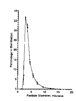
Fiaure 26 shows the size distribution of DPPC IFVs.
The normalized diameter distributions for three DPPC IFV
samples were measured at room temperaturte using a Malvern
3600ER laser diffraction particle sizer. The distributions
are based upon particle number and were calculated by that
instrument. Average values and stAn~Ard deviations for the
three samples are shown. The number averaged diameter for
the samples was 3.54+/-0.12 microns. The IFVs were formed
from DPPC SWs at 20 mg/ml using 4.OM ethanol.
r
Fiaure 27 shows the effect of interdigitation-fusion
paramters on the internal volume of DPPC IFVs. A. Internal
volume as a function of ethanol concentration. For ethanol
concentrations greater than or equal to 2.OM, the DPPC IFVs
O 94/08565 2 ~ 4 6 1 ~ ~ PCT/US93/098
a
were formed by direct addition of ethanol to DPPC S W s at 20
mg/ml. At 1.5M or less, ethanol was prediluted before
addition to avoid mixing artifacts. Error bars indicate
st~n~rd deviations. B. Internal volumes of DPPC IFVs
formed from DPPC L W ETS of various diameters are shown. The
L W ETS were prepared by extruding previously formed MLVs
through two polycarbonate filters. Diameters were
determined by quasi-electric light scattering. C. Internal
volumes of DPPC IFVs formed at different DPPC S W
concentrations. Interdigitation-fusion was induced with
4.OM ethanol.
Fiaure 28 shows the internal volumes for IFVs made
from various saturated acyl chain phosphatidylcholines.
Average internal volumes are shown for DMPC, DPPC, DHPC,
DSPC and DAPC IFVs which were prepared from S W s at 20 mg/ml
using 4.OM ethanol to induce interdigitation-fusion. Error
bars indicate the stndard deviations.
Fi~ure 29 Incorporation of cholesterol into DPPC IFVS
is shown. Final internal volumes of DPPC/Cholesterol IFVs
are compared for two methods of cholesterol incorporation
into IFVs. DPPC. Filled circles indicate DPPC/Chol IFVs
formed directly from DPPC/Chol S W s of varying mole percent
cholesterol. With this method, the internal volumes of the
product IFVs rapidly decreased with increased cholesterol
content. Filled squares indicate internal volumes of
DPPC/Chol IFVs formed when cholesterol was added in the form
of 1:1 DPPC/Chol S W s in 4.0M ethanol after ethanol-induced
DPPC interdigitated sheets were formed. Significantly
higher internal volumes were produced at each mole percent
cholesterol used. Differential sc~nn;ng calorimetry was
used to demonstrate that cholesterol was incorporated
directly into bilayers. At 35 mole percent cholesterol, the
- 18 -
~ 0 94/08565 PC~r/US93/09878
2l~6ll5
DPPC gel-to-liquid crystalline phase transition was
eliminated in DPPC/Cholesterol IFVs formed by either method.
Detailed Descri~tion of the Invention
For purposes of clarity, throughout the discussion of
the present invention, the following definitions will be
used:
"Interdigitation" and "interdigitated" are used
throughout the specification to describe a lipid bilayer in
which the acyl chain region of one lipid in a bilayer
interpenetrates into the other layer of the lipid bilayer.
The term interdigitation shall include full interdigitation,
mixed interdigitation and partial interdigitation. Full
interdigitated liposomes include interdigitated liposomes in
which the acyl ~h~;n~ of the lipid interpenetrate fully or
partially across the width of the lipid bilayer as in Figure
lA(D). Mixed interdigitated liposomes include
interdigitated liposomes in which certain acyl ChA in.~ Of
unsymmetrical phospholipids, generally the longer acyl
20 ~h~;n.~, extend completely across the bilayer span, whereas
the shorter ~h~; n.~ meet end to end in the bilayer midplane
as in Figure lA(C). Another example of mixed
interdigitation liposomes includes liposomes in which
regions of the liposome are either fully or partially
interdigitated and may co-exist with regions that are not
interdigitated. Partially interdigitated liposomes include
interdigitated liposomes in which the acyl rh~; n.~ of
unsymmetrical phospholipids pair such that the longer acyl
chain of one bilayer pairs with the shorter acyl chain of
the other bilayer as in Figure lA(B).
-- 19 --
WO 94/08565 PCT/US93/09 ~
.. . .
ai~6ll~
"Inducer" is used throughout the specification to
describe molecules, including amphipathic molecules of
limited size which localize at the lipid bilayer aqueous
phase interface region of a liposome and produce an
interdigitation-fusion gel which may also be a liquid, of
the present invention. The term also contemplates lipids
and/or solutes such as bioactive agents that may act as
"self-inducers". Hydrostatic pressure may also be an
inducer.
"Pressure induced fusion" ("PIF") is interdigitation
induced by a sufficient application of hydrostatic pressure
applied to sized liposomes to form an interdigitation fusion
gel. Liposomes formed from this gel are referred to herein
as PIF liposomes, or simply PIFs.
"Interdigitation-fusion gel" (IF gel) is used
throughout the specification to describe the product that
results when an inducer is combined in sufficient quantity
to fuse sized liposomes. The resulting sheets of lipid are
fused gels for purposes of the present invention and may
include products of varying viscosity including liquids,
gels and in certain cases, even very viscous products
approaching the solid state.
~ Interdigitation-fusion liposome" (IF liposome) is
used throughout the specification to describe the liposome
that results from IF gels which are generally but are not
necessarily raised above the lipid transition temperature
("Tm"), but in any case are incubated at a temperature to
produce IF liposomes. The inducer may additionally but not
necessarily be removed from the interdigitation-fusion gel.
In certain embodiments in which the liposome contains self-
inducing lipid or the solute is an inducer, the inducer is
not removed. The IF liposomes may contain large
- 20 -
~ 0 94/08565 PC~r/US93/09878
2~ s
concentrations of solute such as bioactive agents in
bioactive agent:lipid ratios of about 1:10 to 15:1.
"Solute" is used throughout the specification to
describe any chemical, including buffers and solvents that
may be entrapped by the IF gels and liposomes of the present
invention. Solutes may include buffers, salts, toxins,
microbes and bacteria, pesticides, insecticides, herbicides,
fungicides, emulsifying agents, cosmetics, unicellular
organisms and a large number of chemical agents, especially
including bioactive agents.
"Bioactive agent" is used throughout the specification
to describe any agent such as chemical agents which exhibits
biological activity when A~m;nistered to living organisms,
including plants, AnimAl5 such as mAmmAls, and especially
including hllmAn~. Bioactive agents include drugs and
nutrients, among others as described hereinabove and
following.
"Saturated lipid" is used throughout the specification
to describe a lipid that may be used to produce the
interdigitation-fusion gels and liposomes of the present
invention. The term saturated lipid includes, but is not
limited to, lipids having symmetrical and/or asymmetrical
acyl side rhAin~ which are saturated, i.e., contain no
double bonds, lipids having unsaturated side chains in which
the unsaturated carbon-carbon double bonds are oriented in
the trans configuration and certain lipids having
unsaturated side chAinc in which the unsaturated carbon-
carbon double bonds are oriented in the cis configuration,
or mixed fatty acid lipids such as for example SOPC and
POPC.
WO 94/08565 PCT/US93/09 ~
~1~6115.
"Interdigitation-fused lipid containing composition"
is used to describe the IF gels and liposomes of the present
invention.
The present invention relates to lipid contA;n;ng
compositions comprising a sized liposome, preferably about
0.4 microns or less to about 0.05 microns or less and more
preferably about 0.025 microns in diameter or less, and an
amount of an inducer effective to fuse the liposomes in
combination with a solute. The initial liposomes may
alternatively be FAT MLVs. In certain embodiments the
solute is a bioactive agent. The compositions of the present
invention may advantageously include bioactive agent at high
concentrations, for example at bioactive agent:lipid ratios
of about 1:10 to 15:1.
In the present invention, the sized liposomes which
give rise to IF gels and liposomes of the present invention
are preferably formed from zwitterionic, cationic, and
anionic lipids and phospholipids comprising fatty acyl
chA;n~, having 12 to 35 carbon atoms, also including therein
saturated (disaturated and partially saturated) and
unsaturated and polar or apolar lipids and phospholipids.
For example, the saturated lipids of the invention
include but are not limited to for example,
dimyristoylphosphatidylcholine,
distearoylphosphatidylcholine,
dipalmitoylphosphatidylcholine,
dimyristoylphosphatidylserine,
dipalmitoylphosphatidylserine, distearoylphosphatidylserine,
dimyristoylphosphatidylethanolamine,
dipalmitoylphosphatidylethanolamine,
distearoylphosphatidylethanolamine, dimyristoylphosphatidic
acid, distearoylphosphatidic acid, dipalmitoylphosphatidic
~ 0 94/08565 PCT/US93/09878
21~6115
acid, dimyristoylphosphatidylinositol,
distearoylphosphatidylinositol,
dipalmitoylphosphatidylinositol, hydrogenated soy
phosphatidylcholine, hydrogenated soy lecithin,
dipalmitoylphosphatidylglycerol,
di-0-hexadecylphosphatidylcholine,
dipalmitoylphosphatidylglycerol,
distearoylphosphatidylglycerol,
dimyristoylphosphatidylglycerol, among others.
Other saturated lipids include but are not limited to
the saturated lipids having symmetrical and/or asymmetrical
acyl side ~h~;n~ which are saturated, i.e., contain no
double bonds, lipids having unsaturated side ~h~; n.~ in which
the unsaturated carbon-carbon double bonds are oriented in
the trans configuration and certain lipids having
unsaturated side ChA; n~ in which the unsaturated carbon-
carbon double bonds are oriented in the cis configuration,
or mixed fatty acid lipids such as for example SOPC and
POPC .
Other lipids for inclusion with the saturated
symmetrical lipid include other liposome forming lipids
including, for example synthetic or natural phospholipids
including mixed chain compositions, for example,
phosphatidylcholines (PC), phosphatidylethanolAm; n ~s ( PE),
phosphatidylserines (PS), phosphatidylglycerols (PG),
phosphatidic acids (PA), phosphatidylinositols (PI),
sphingomyelins (SPM) and cardiolipins, among others, either
alone or in combination.
In addition to the above lipids, additional lipids
including various lysolipids, for example, n-octadecyl-2-
methylphosphatidylcholine, n-octadecylphosphatidylcholine,
1-laurylpropanediol-3-phosphocholine, erythro-N-
- 23 -
W O 94/08565 2 I ~ G 1 ~ ~ PC~r/US93/098 ~
lignoceroylsphingophosphatidylcholine, cholesterol, and
water soluble derivatives thereof such as for example
cholesterol hemisuccinate and alpha tocopherols and water
soluble derivatives thereof such as tocopherol
hemisuccinate, and gangliosides, glycolipids, and
glycosphingolipids which may also be included in
compositions of the present invention. One of ordinary
skill in the art will recognize that the amount and type of
lipid which may be included in compositions of the present
invention may be varied within the teachings of the present
application to produce the compositions according to the
present invention.
In the compositions of the present invention, the
sized liposomes cont~-n;ng significant quantities of at
least one saturated lipid are interdigitated-fused with
addition of an inducer. The sized liposomes of the present
invention generally cont~;n;ng a saturated phospholipid will
undergo full, partial or mixed interdigitation in
combination with an effective amount of the interdigitation
inducer. While not being limited by way of theory, it is
believed that the inducer may function to displace some of
the headgroup-associated water molecules and in general,
causes an increase in the headgroup surface area. It is
preferred that the lipids chosen should undergo full
interdigitation in the presence of the inducer; however, it
is to be recognized that lipids which provide less than
complete interdigitation, i.e., either mixed or partial
interdigitation are also contemplated and are within the
scope of the present invention. Exemplary interdigitation
inducers for use in the present invention include, for
example, short chain alcohols including methanol, ethanol,
propanol, isopropanol and n-butanol, polyols such as
glycerol and ethylene glycol, anaesthetics such as
chlorpromazine, tetracaine, phenylethanol, benzyl alcohol
- 24 -
-
-
~O 94/08565 2I ~ ~11 S PCT/US93/09878
and phenylbutanol, among others, buffers such as Tris and
chaotropic salts such as thiocyanate SCN-, as well as others
referred to hereinabove.
In certain cases, the saturated lipid used to form the
IF gels and liposomes of the present invention are self-
inducers, i.e., these lipids will interdigitate and form IF
gels and liposomes by mixing the lipid in the presence of
solute at varying temperatures without the need to add a
chemical inducer (for example, DHPC). In addition, in
certain cases extremely high pressure may be used to produce
interdigltation without the need to include an inducer.
In accordance with one aspect of the present
invention, interdigitation may be induced by the application
of hydrostatic pressure to a population of sized liposomes.
The period of time and level at which the pressure is
applied to the liposomes must be effective to cause the
interdigitation fusion to occur. Although no specific
m;n;~n~ pressure is required, a preferred pressure level for
liposomes comprised of saturated lipids is at least about
20,000 psi, preferably at least about 40,000 psi. The
pressure should be applied for a period of time sufficient
to cause interdigitation fusion, preferably about one minute
to about one hour.
Good results were obtained using liposomes comprised
of either dipalmitoylphosphatidylcholine (DPPC) or
distearoylphosphatidylcholine (DSPC). In one example, small
unilamellar vesicles (SWs) comprising DPPC or DSPC were
fused to form larger liposomes by the application of
pressure of at least 20,000 psi, and preferably at least
40,000 psi for a period of about 15 minutes. During these
tests, at least a partial fusion of the small liposomes was
noted at the first observation after 5 minutes. This
WO 94/08565 PCT/US93/098~r
process is discussed in more detail in Example 27, below.
Although DPPC and DSPC were successfully interdigitated by
pressure induced fusion (PIF), the PIF process was found not
to induce interdigitation on small liposomes of
palmitoyloleoylphosphatidylcholine (POPC), which has
asymmetrical acyl side rh~; n.~ .
As a further aspect of this embodiment of the present
invention, high hydrostatic pressures may be used to kill
bacteria and to sterilize liposomal preparations. Thus,
hydrostatic pressure can be used not only to induce
interdigitation fusion, and but also to sterilize liposome
preparations. This sterilizing process can be applied as
part of an interdigitation fusion process, or can be used to
sterilize liposomes formed by other processes, such as, for
example, the methods described in U.S. Patent Nos.
4,522,803, 4,588,578 and 4,975,282, discussed above.
Hydrostatic pressures have long been known to have
effects on cellular processes. Currently, high pressure and
moderate temperature is used for the pasteurization and
sterilization of certain food products. Such methods
generally employ the application of pressures as high as
75,000 psi and moderate temperatures. Bacteria, yeasts and
viruses may all be inactivated by the application of high
pressure. The inactivation process, given a fixed pressure,
is known to vary as a function of temperature, chemical
composition of the medium, and time.
In accordance with the present invention, high
hydrostatic pressures and moderate temperatures are used to
kill microbes, including bacteria such as Bacillus subtilis
in Example 28 below, and thereby to sterilize liposomal
preparations. Bacillus subtilis was chosen for use in this
example because it is considered one of the most difficult
- 26 -
~ 094/08565 PCT/US93/09878
2116115
microbes to kill in sterilization processes. In Example 28,
the rate of inactivation for several temperatures at a
number of pressures is described. As can be seen from the
results presented in Figures 15, 16 and 17, the rate of
sterilization is a function of the temperature and pressure
at which the sterilization is performed.
In general, in performing the sterilization process of
the present invention on a selected liposomal composition in
the shortest time, one would determine a suitable
lo temperature and pressure effective for composition. For the
most rapid sterilization, one would use a combination of the
highest temperature and pressure which can be tolerated by
the liposomal composition. The data presented in Figures
15, 16 and 17 for Example 28 provide an example of the
relationship between temperature and pressure for such
sterilizing processes.
A pressure of about 10,000 psi is considered to be a
m;n;mllm level for effecting sterilization in accordance with
the present invention. Preferably, a m;n;ml~m of about
40,000 psi pressure is used to carry out the sterilization
more rapidly, the actual time re~uired being dependent on
the temperature under which the process is performed. The
pressure should be kept below a level which can have a
deleterious effect on the particular liposome composition
being sterilized, either by damaging the liposome structures
or by affecting any of the components of the composition.
In like manner the maximum temperature should also be below
that which causes deleterious effects to the liposome
composition, such as structural or chemical changes to the
liposomes or to any of the components of the composition.
In general, the compositions of the present invention
include an amount of an inducer effective for fusing the
- 27 -
-
WO 94/08~6~ PCT/US93/098 ~ 2 1 ~
sized liposomes. The amount and type of inducer utilized to
produce liposome fusion will vary as a function of the type
of liposome utilized. In general, however, the amount of
inducer used comprises about 1.0% to about 50% of the total
weight of solution which includes a combination of the sized
liposomes, inducer and solute. One of ordinary skill in the
art will recognize to readily vary the concentration of the
inducer within the teachings of the art and the present
application to produce interdigitation gels and liposomes of
the present invention.
While not being limited by way of theory, it is
believed that sized liposomes fuse into lipid sheets (gels)
at certain concentrations of inducer in order to relieve
bilayer strain imposed by a small radius of curvature (See,
for example, Figure 3). The resulting interdigitation-
fusion gel that is produced may capture a high concentration
of solute. This includes encapsulating substances which
otherwise cannot be entrapped in high solute lipid ratios in
liposomes. According to the method of the present
invention, when the IF gels are exposed to temperatures
usually but not necessarily above their L beta I-L alpha
transition temperature ("Tm"), but in any case at a
temperature which changes the material properties of the
mixture such that IF liposomes are formed, and the inducer
is preferably (but not necessarily) removed, liposomes of
high captured volume result. These liposomes may vary in
size as a function of the solute, liposome and inducer
utilized, but generally, will range in size from about 100
~m and more preferably about 20 microns, to about 0.025
microns.
While not being limited by way of theory, it is
believed that interdigitation, which renders the lipid
bilayer less susceptible to perturbation during liposome
~ O 94/08565 2 1 4 ~ ~ ~ 5 ~ PC~r/US93/09878
formation, can be utilized to capture substances which
normally interact with membranes and are difficult to
entrap. For example, interdigltation-fusion liposomes of
the present invention have been used to entrap high
concentrations of aminoglycosides which are very difficult
to entrap in high concentrations because of their tendency
to interact with membranes. IF liposomes have been shown to
entrap gentamicin at a drug/lipid ratio of about 1:2 (w:w)
whereas typically modified small plurilamellar liposomes
(SPLVs) entrap gentamicin at a drug/lipid ratio of about
1: 10 (w:w) .
The production of interdigitation-fusion gels and
liposomes of the present invention involves the initial
formation of sized liposomes about 0.4 microns in diameter
or less, more preferably about 0.05 microns or less and most
preferably no greater than about 0.025 microns.
Alternatively, FAT MLVs can be used, and in some cases,
larger liposomes can be used. Any of the methods available
in the art for producing sized liposomes may be utilized
including the methods described in greater detail
hereinbelow. Typically, liposomes can initially be prepared
by vacuum drying a solution of lipid in organic solvent, for
example, chloroform, to a thin film in a round bottom or
other suitable flask or vessel, followed by hydration of the
lipid film with an aqueous solvent such as for example,
aqueous buffer or saline solution. Alternatively, liposomes
can be formed from admixture of dry lipid powder and aqueous
solvent, preferably for example saline solution or aqueous
buffer.
The liposomes are then sized according to any methods
known in the art such as sonication, extrusion or
homogenization, and further described hereinbelow. After
the formation of sized liposomes, the solute, preferably a
- 29 -
WO 94/0856S ~6~ PCT/US93/098 ~
bioactive agent that is to be encapsulated, is generally
mixed in the aqueous solvent. Two approaches are generally
used to entrap solute dep~n~;n~ upon whether or not the
solute interacts with liposomes. In the case where the
solute does not interact with the liposomes, the solute may
be mixed in with the aqueous or aqueous/buffer solvent after
formation of the sized liposomes which are to undergo
interdigitation. In the case of solute that interacts with
the liposomes, the solute is generally mixed in with the
aqueous solvent after the formation of interdigitation-
fusion gels.
of course, one of ordinary skill in the art will
recognize that the order in which the individual components
of the IF gels and liposomes of the present invention are
added may vary and is dependent on the type of solute to be
entrapped and the type of saturated lipid utilized.
The final concentration of the lipid used to
encapsulate solute of the present invention will vary as a
function of the concentration and type of the solute desired
as well as the type of lipid used, but in general, the
weight ratio of drug to lipid in the aqueous solvent will
range from about 50:1 to about 1:100 with the final
concentration of lipid falling within the range of about 5
to 100 mM. The final weight ratio of drug to lipid in the
interdigitation-fusion gels and liposomes of the present
invention ranges from about 1:10 to about 15:1.
After the sized liposomes are formed, inducer is added
to the a~ueous solvent. The amount of inducer added
generally ranges from about 1.0% by weight (of the combined
weight of lipid, solute and inducer) up to about 50 percent
by weight. Where ethanol is used as the inducer, the amount
of ethanol included is generally about 5~ by weight (1.0 M)
- 30 -
094/08~65 2~ PCT/US93/09878
to about 20% by weight (4.0 M) and in the case of glycerol
the amount of inducer utilized may be as much as about 90-
100% by weight. The amount of other inducers to be included
will vary. In the case of ethanol the final ethanol
concentration falls within the range of about 0.50 to about
lD.0 Molar and preferably is within the range of about 1.75
to about 4.0 Molar.
The presence of inducer in an effective amount will
cause the sized liposomes to fuse, resulting in fused sheets
of lipid. The IF gel produced by this method may be used
topically or for oral administration, for example, as
formulations encapsulated in soft gelatin or other oral
dosage forms. Alternatively, the gel may be further
modified to produce the IF liposomes of the present
invention.
To produce IF liposomes of the present invention, the
mixture is incubated at a temperature for a period of time
sufficient to form a gel. Typically this period ranges from
about 1 minute to about 1 hour. Thereafter, the temperature
is generally but not necessarily raised above the Tm of the
lipid for a period of about 1 minute to about 1.0 hour. The
incubation temperature required may be the Tm of the mixture
but is that temperature for any given mixture of lipid,
solute or inducer which produces a change in the material
properties of the mixture, thereby producing the IF
liposomes of the invention. While maintAin;ng this
incubation temperature, the inducer may be removed by
evaporation (especially in the case of alcohol inducers),
positive pressure nitrogen (e.g., N2 sparge consisting
generally of bubbling N2 through the mixture) or by
dilution. This produces IF liposomes varying in size
generally between about 0.025 and about 100 ,um, more
preferably about .025 to about 20 microns. Unencapsulated
- 31 -
WO 94/08565 2 1 4 6 ~ PCT/US93/098
drug may be removed from the solvent, if desired. The IF
liposomes produced by the above method may be further size
reduced to produce liposomes varying or homogeneous in size.
In addition to extrusion, initial liposomes (prior to
addition of inducer~ for the IF gel or liposome method, and
resulting IF liposomes may be size reduced by sonication or
homogenization. Sonication employs sonic energy to disrupt
or shear the larger liposomes which will spontaneously
reform into small liposomes. See, for example, ~h~pm~n, et
10 al., BBA, 163, 255 (1968). Sonication is achieved by
immersing a glass tube cont~;n;ng the liposome suspension
into the sonic epicenter produced in a bathtype sonicator.
Alternatively, a probe type sonicator may be used in which
the sonic energy is generated by vibration of a titanium
probe which is in direct contact with the liposome
suspension.
With homogenization the shear forces which break down
larger liposomes into smaller ones are generated by, for
example, rotorstator type devices such as the Polytron
(Brinkman Instruments Co., Westbury, New York, USA), a
stator-stator type device such as the Microfluidizer
(Microfluidics Corp., Newton, MA, USA), or any number of
other such devices which are com.~monly used to disrupt cells.
Due to the fact that all of the above methods involve
disruption of the IF liposomes, entrapped solute will be
lost when IF liposomes are subjected to any of these
procedures. The loss may be m;n;m;zed however, if the
unentrapped solute is not removed from the liposome
suspension before size reduction of the liposomes.
A number of other techniques may be used for producing
sized liposomes which are to undergo interdigitation-fusion,
and for producing sized IF liposomes after the process is
- 32 -
~ O 94/08565 21 ~ 61 i~ PC~r/US93/09878
complete. These methods include reverse-phase evaporation,
infusion procedures, homogenization, sonication,
microfluidization and detergent dilution or a combination of
these methods. A review of certain of these and other
methods for producing liposomes can be found in the text
Liposomes, Marc J. Ostro, ed., Marcel Dekker, Inc., New
York, 1983, Chapter 1, pertinent portions of which are
incorporated herein by reference. Sized liposomes may also
be produced by an extrusion process.
In the extrusion process, to produce sized liposomes,
the liposomes are passed through filters having pore sizes
generally ranging from about 30 nm to about 1 micron to
produce liposomes ranging in size from about 30 nm to about
1 micron in diameter. Preferably, the pore size of the
filters through which the liposomes may be extruded ranges
from about 100 nm to about 1 micron. The filters are
generally made of polycarbonate, but the filters may be made
of any durable material which does not interact with the
liposomes and which is sufficiently strong to allow
extrusion under sufficient pressure. Preferred filters
include "straight through" filters because they generally
can withstand the higher pressure of the preferred extrusion
processes of the present invention. "Tortuous path" filters
may also be used. In the preferred embodiments of the
present invention, pre-IF fusion liposomes are extruded
through 50 to 100 nm polycarbonate filters to produce
liposomes having a diameter of about 50 to 100 nm.
Any extrusion process available in the art may be used
to produce sized liposomes which will undergo
interdigitation-fusion. The extrusion process may be
performed se~uentially or once under high pressure.
Particularly preferred extrusion processes for use in the
present invention include those disclosed in Cullis, et al.,
W094/08565 2 i 4 g 1 1` 5 PCT/US93/098 ~
PCT Application PCT/US85/01161, Publication Number WO
86/00238 entitled "Extrusion Techniques for Producing
Liposomes", published January 16, 1986, relevant portions of
which are incorporated by reference herein.
Other methods for sizing the liposomes of the
invention either before or after fusion are filtration
methods employing asymmetric filters such as for example,
AnotecR filters according to c~mmo~ly-assigned cop~n~; ng
U.S. Patent application entitled "Liposome Extrusion
Process", U.S. Serial No. 593,200, filed October 5, 1990,
which involves extruding liposomes through a branched pore
type all~m;n1lm oxide porous filter, relevant portions of
which are incorporated herein by reference.
Alternatively, the liposomes can be sized using a
homogenization or milling procedure such as a colloid mill
for example the Gifford Wood colloid mill. The liposomes
may be passed one or more times through the mill until the
appropriate size and homogeneity is achieved, analyzed for
size distribution using either the Nicomp Particle sizer or
the Malvern Particle sizer. The liposomes, alternatively,
may be passed through a Microfluidizer device, discussed
hereinabove, which likewise homogenizes the liposomes.
If desired, the resulting liposomes can be separated
into populations using any methods known in the art for so
separating; such a process is for example tangential flow
filtration. This process as used for the separation of
liposomes according to size is disclosed in commonly
assigned and copending U.S. Patent application entitled
"Method for Size Separation of Particles", Serial No.
225,327, filed July 28, 1988, relevant portions of which are
incorporated herein by reference.
- 34 -
~ 094/08~65 2 1 ~ ~1` 1 5 ~ PCT/US93/09878
In this procedure, a heterogeneously sized population
of liposomes is passed through one or more tangential flow
filters thereby resulting in a size distribution with an
upper and/or lower size limit. For example, when two
filters of differing sizes are employed, for example, a
first filter of 5 ~m pore size, liposomes less than 5.0 ~m
pass through the filter and into the filtrate, which is then
passed through a second filter of smaller pore size, for
example, 2.0 ~m pore size. In this case, the retentate
contains liposomes of a homogeneous size distribution having
discrete size limits of 5.0 and 2.0 ~m. Filters of
alternative pore size may be employed to result in discrete
populations having upper and lower size limits.
The liposomes which undergo interdigitation-fusion in
the presence of an inducer preferably are about 0.4 microns
in diameter or less, more preferably about 0.05 microns or
less, and most preferably are about 0.025 microns or less in
diameter. Alternatively, the initial sized liposomes of the
invention may be FAT MLVs. The IF liposomes which are
produced by the general method of the present invention
generally range in size from about 100 ~m but more
preferably about 20 microns to about 0.025 microns, and
generally in the range of about 2 to 20 microns. These
resulting IF liposomes may be further size reduced to
produce liposomes of varying sizes by sonication,
homogenization and extrusion techniques described
hereinabove. It should be noted, however, that although
these IF liposomes of the present invention may be down
sized, the down sizing often results in the loss of
bioactive agent from the liposomes. Thus, IF liposomes
which undergo further down-sizing, for example, by the
previously discussed extrusion process or other processes
such as sonication and homogenization to vary the size of
WO 94/0856~ 2 1 ~ 6 1 ~ ~ PCT/US93/098
the liposomes produced, may encapsulate ~;m;n; shed
concentrations of bioactive agents.
Bioactive agents for use in the present invention may
include vitamins, hormonal agents, anti-metabolites, anti-
microbial agents, antifungal agents, antibiotics, proteins,peptides, ribo and deoxyribonucleic acids, nucleotides,
nucleosides, oligonucleotides, antihistaminic agents,
neuropharmacologic agents including sedatives and hypnotics,
steroidal and nonsteroidal antiinflammatory agents, diuretic
agents, antihypertensive agents, antiarrhythmic agents,
immunogens, immunomodulators, contraceptive agents,
radiographic contrast agents, NMR contrast agents, antiviral
agents and vascular dilating agents, among others. In
certain preferred embodiments of the present invention,
radiocontrast agents, NMR contrast agents, peptides and
naturally occurring, synthetic and semi-synthetic
antimicrobial agents, for example, cephalosporins and
aminoglycosides are utilized in the present invention.
Exemplary radiocontrast agents for use in the present
invention include, for example, iohexol, iopamidol,
ioxoglate, iotrolan, ioversol, iothalamate, iodimide,
iodipamide, iopromide, iopentol, iodixanol, metrizamide,
mixtures thereof and their pharmaceutically acceptable
salts. Exemplary aminoglycosides include gentamicin,
tobramycin and amikacin.
Suitable biological agents for use in the present
invention include any agent which exhibits favorable
biological activity when administered topically or
systemically and is stable to the compositions of the
present invention. Agents which may be topically
administered for their affect on the skin include salicylic
acid, resorcinol, phenol, retinoic acid, and their
equivalents. Other agents for use in the present invention
~ O 94/08565 21~6115 PCT/US93/09878
include certain desensltizing agents, for example antigens
and vaccines, vitamins, nutrients, such as amino acids,
essential fats and minerals, retinoids, anti-neoplastic and
anti-tumor agents, including certain alkylating agents,
among others.
Additional bioactive agents for use in the present
invention include the benzodiazepines, antipyretic agents,
antispasmodics, antipruritic agents, sympathom;metics,
decongestants, tranquilizers, antispasmodics, cardioactives,
other cardiac agents, anti-emetics, sedatives and hypnotics,
steroidal agents, progestational agents, local anesthetics
and antibiotics. Other antimicrobial agents may also be
used in the present invention including antifungal agents,
among others.
The above-listed group of bioactive agents, among
other agents, including their pharmaceutically acceptable
salts, are contemplated for use in the present invention.
Dete~m;n~tion of compatibilities of the above listed agents
with and the amounts to be utilized in compositions of the
present invention are within the ordinary skill in the
formulation art. The stability and applicability of
individual pharmaceutical agents are well within the
ordinary skill of practitioner in this art.
It will be appreciated that the actual preferred
~5 amounts of bioactive agent utilized in a specific case may
vary according to the severity of a p~Arm~cological or
disease condition and the expected pharmacokinetics of
bioactive agent in the individual patient. Dosages for a
given host can be determined using conventional
considerations, e.g., by customary comparison of the
differential activities of the subject bioactive agent by
WO 94/08~65 PCT/US93/098 ~
214611S
means of an appropriate, conventional pharmacological
protocol.
The IF liposomes and gels of the present invention may
be administered to any ~n;m~l including m~mm~l S, such as
humans. For administration to hl]mAnc in the treatment of
afflictions, the prescribing physician will ultimately
determine the appropriate dose for a given human subject,
and this can be expected to vary according to the age,
weight, and response of the individual as well as the nature
and severity of the patient's symptoms. The present
invention provides a readily available method to allow wide
variations in liposomal drug concentrations.
The mode of A~m; n; stration of compositions of the
present invention may determine the sites in the organism to
which the compositions will be delivered. For instance,
delivery to a specific site of infection may be most easily
accomplished by topical application (if the infection is
external, e.g., on areas such as the eyes, skin, in the ears
or on afflictions such as wound or burns) or by absorption
through epithelial or mucocutaneous linings (e.g., nasal,
oral, vaginal, rectal, gastrointestinal, mucosa, etc.).
Such topical application may be in the form of creams or
ointments. The interdigitation-fusion gels of the present
invention are preferably used topically. However, the IF
gels of the present invention may be used orally in
formulations in which the lipid, upon contacting the fluids
of the mouth or gastrointestinal tract forms a liposome n
.
The IF liposomes containing bioactive agent may be
administered alone but will generally be administered in
admixture with a p~rm~ceutical carrier selected with regard
to the intended route of administration and standard
- 38 -
0 94/08565 ~ , ~ PCT/US93/09878
ph~rm~ceutical practice, thereby forming ph~rmAceutical
compositions. The IF liposomes of the present invention may
be injected parenterally, for example, intravenously,
intramuscularly, or subcutaneously. For parenteral
administration, these liposomes are best used in the form of
a sterile aqueous solution which may contain other solutes,
for example, sufficient salts, glucose or dextrose to make
the solution isotonic.
For the oral mode of ~m; nl stration, the liposomes of
the present invention can be used in the form of tablets,
capsules, lozenges, troches, powders, syrups, elixirs,
aqueous solutions and suspension, and the like. In the case
of tablets, carriers which can be used include lactose,
sodium citrate, and salts of phosphoric acid. Various
disintegrants such as starch, and lubricating agents may be
used. For oral administration in capsule form, useful
diluents are lactose and high molecular weight polyethylene
glycols. When aqueous suspensions are required for oral
use, certain sweetening and/or flavoring agents can be
added.
Bioactive agents for use in the present invention may
include but are not limited to those listed hereinabove, and
include their pharmaceutically acceptable salts.
Determ'n~tion of compatibilities of the above listed agents
with and the amounts to be utilized in compositions of the
present invention are within the ordinary skill in the
formulation art. The stability and applicability of
individual pharmaceutical agents are well within the
ordinary skill of practitioner in this art. It will be
appreciated that the actual preferred amounts of bioactive
agent utilized in a specific case may vary according to the
severity of a ph~rm~cological or disease condition and the
expected pharmacokinetics of bioactive agent in the
- 39 -
-
WO 94/08565 2 1 ~ 6 1 1 ~ ` PCT/US93/098 ~
individual patient. Dosages for a given host can be
determined using conventional considerations, e.g., by
customary comparison of the differential activities of the
subject bioactive agent by means of an appropriate,
conventional ph~rm~cological protocol.
IF liposomes can be remote loaded, for example, to
incorporate bioactive agents. If desired, IF liposomes can
be dehydrated using, for example, the procedures of Janoff
et al. U.S. Patent No. 4,880,635 or Schneider et al. U.S.
10 Patent No. 4,229,360.
The following examples are provided to illustrate the
present invention and should not be construed to limit the
scope of the invention of the present application in any
way.
F~MPLE 1
Liposomes comprising dipalmitoylphosphatidylcholine
(DPPC, obtained from Avanti Polar Lipids, B;rm;ngh~m,
Alabama, USA) were formed in 1 ml of an aqueous buffer
solution to a concentration of 20 mM DPPC and additionally
cont~;n;ng 0.04 mM diphenylhexatriene (DPH, purchased from
Molecular Probes, Eugene, Oregon, USA). After formation of
the liposomes, ethanol was added to a final concentration of
0.3 M to 2.5 M of the aqueous solution.
DPH fluoresces m~;m~lly when incorporated into the
liposome bilayer. Interdigitation results in the
reorientation of DPH from the bilayer membrane with a
concomitant decrease of fluorescence. As shown in Figure 2,
interdigitation is greater where higher concentrations of
ethanol are present, for all liposomes. The effect of
interdigitation by the same amount of ethanol is greater in
- 40 -
O94/08565 21 ~ PCT/US93/09878
those liposomes having a larger diameter. In Figure 2, Fo =
DPH fluorescence in the absence of ethanol; F = DPH
fluorescence in the presence of ethanol. Excitation = 351
nm. Emission was detected between 380 and 580 nm and
quantitated by weighing.
EXAMPLE 2
Lipid Mixing of Liposomes
Lipid mixing of sized DPPC Liposomes was determined as
a function of the size of the Liposomes and concentration of
inducer. Liposomes comprising DPPC were formed in an a~ueous
buffer solution containing 20 mM DPPC. A marker population
of liposomes containing 99~ by weight DPPC, 0.35% by weight
N-benzyldiphosphatidylethanolamine (NBD-PE) and 0.65% by
weight rhodamine-phosphatidylethanolamine were formed in 1
ml. of an aqueous buffer solution. These probes form a
donor-acceptor pair. The NBD moiety is excited at 465 nm
and via resonance energy transfer (RET) becomes quenched by
the rhodamine acceptor which itself becomes excited in a
distance dependent phenomenon. These liposomes were mixed
with blank liposomes at a 1:10 ratio. Emission spectra were
recorded between 480 and 680 nm. Lipid mixing in DPPC
liposomes of varying size as a function of ethanol
concentration can be determined by the loss of RET from the
NBD moiety to the Rhodamine moiety. A standard curve was
generated by preparing liposomes of 0.35 mole percent NBD-PE
and 0.65 mole percent Rhodamine-PE with sequentially
decreasing these mole percents to 0.035 and 0.065
respectively. A direct comparison from the 1/10 mixing
experiments with this standard curve indicates the degree of
lipid mixing, an indication of membrane fusion.
- 41 -
WO 94/08565 2 1 ~ 6 1 1 S PCT/US93/098 ~
~AMPLE 3
Comparison of Trapped Solute in Various Vesicle Types
A number o~ liposomal formulations were prepared. The
amount of trapped aqueous phase was determined and compared
for each ~'type" of liposome prepared. The results appear in
table 1, below.
For the preparation of IF liposomes, MLVs were
prepared as described below to a final concentration of 20
~moles DPPC per ml of aqueous buffer. The MLVs were then
sonicated in a bath type sonicator at 50C until translucent
(S W s). After the S W s cooled to room temperature, ethanol
was added to a final concentration of 2.0 M in the final
aqueous suspension. For examples Al and A2 (see table 1,
below), ethanol was removed by dilution followed by w~h;ng.
For Bl and B2, ethanol was removed via positive pressure
displacement using N2, after which the samples were diluted
and washed. Sample C had one half the initial lipid
concentration of A and B and ethanol removal was achieved as
for sample A.
For the preparation of MLVs, 100 mg DPPC in 5 ml
chloroform was rotary evaporated to a thin dry film in a
round bottom flask to which a 1 ml aqueous buffer solution
cont~'n'ng 0.04 mM diphenylhexatriene was added.
Thereafter, the lipid mixture was vigorously vortexed until
all lipid was removed from the wall.
FATMLVs were formed by subjecting unwashed MLVs as
described above to 5 freeze and thaw cycles as described by
Bally et al., U.S. Patent No. 4,975,282, issued December 4,
1990 .
- 42 -
094/08565 2 ~ PCT/US93/09878
SPLVs were prepared by forming the thin dry film of
DPPC as described above for MLVs and then dissolving the
lipid film in 5 ml ethyl ether to which 0.5 ml of aqueous
buffer was also added. This mixture was then emulsified in a
bath type sonicator, a stream of N2 was used to stir the
emulsion while removing the ether as described by Lenk, et
al., U.S. Patent No. 4,522,803. Ether removal was continued
until no residual odor was detected (approximately five
minutes). The resulting lipid mixture was resuspended in 1
ml aqueous buffer.
MPVs were formed as described in U.S. Patent No.
4,588,578, by preparing a monophase of 100 mg DPPC and 5 ml
chloroform, 5 ml ethanol and 0.5 ml aqueous buffer, rotary
evaporating to dryness, and resusp~n~;ng the suspended film
in 1 ml aqueous buffer by vigorous vortexing.
To determine the captured aqueous volume, 20 ,ul of a
10 mM 4-trimethylammonium TEMPO (4-TMAT) solution was added
to 0.98 ml of the liposomal suspensions. The samples were
then vortexed and the outer a~ueous phase was separated from
the liposomes by centrifugation. Because 4-TMAT neither
binds to nor permeates the liposomes used in this study, it
is concentrated in the outer aqueous phase. Measurement of
4-TMAT's concentration allows for calculation of the
internal aqueous phase or captured volume as detailed in
Perkins, et al., BBA, 943, 103 (1988). The results of this
analysis appear in Table 1, below. As seen, the IF
liposomes sequester significantly greater volumes, in some
cases as much as 10 times that attained with the other
liposome types.
WO 94/08565 2 1 ~ 6 1 1 5 ; PCT/US93/098 ~
Table 1
Com~arison of Tra~ed Solute
Li~osome Tv~e *Ca~tured Volume (ul/umole)
IF A1 6.7
A2 7.8
B1 8.3
B2 7.2
C 8.9
MLV 1 0.56
2 0.78
MPV 1 0.71
2 1.9
SPLV 1 2.0
2 2.8
15 FATMLV 1 2.7
2 2.6
*Captured volumes were measured using the EPR technique
(Perkins, et al. (1988) Biochim. Biophys. Acta 943, 103)
where the samples are ~m; ned after formation.
EXAMPLE 4
Procedure for Formation of IF Liposomes
Liposomes (L W s, MLVs, SPLVs, etc. as prepared above)
comprising a saturated lipid such as
dimyristoylphosphatidylcholine,
dipalmitoylphosphatidylcholine or
distearoylphosphatidylcholine are prepared and sized to 0.4
microns or less (preferably, 0.025 microns) by extrusion,
sonication or homogenization. A bioactive agent which does
not interact with the lipid is then mixed in with the
aqueous solvent used to form the Liposomes (final lipid
concentration should be about 5 to 100 mM, preferably about
10 to 35 m.M). The temperature of the liposomes are below
the main phase transition temperature of the lipid.
Thereafter, ethanol is added to a final concentration of
about 1.75 to about 2.5 M in the aqueous solvent. In the
case of bioactive agents which interact with the lipid, the
agents are generally added to the solvent after the addition
- 44 -
0 94/08S65 2 1 q 6 ~ P~'r/U593/09878
of ethanol and formation of the IF gel. At this stage, the
procedure can be stopped and the resulting gel used in
topical and oral formulations. Alternatively, the gel may
be used to form IF liposomes of the present invention.
To form IF liposomes, the gel is incubated at a
temperature below the Tm of the lipid for a period ranging
from about 1 minute to about 1 hour followed by an
incubation period of about 1 minute to about 1.0 hour at a
temperature above the Tm of the lipid. The inducer is then
removed by evaporation, positive nitrogen pressure or
dilution. In the case of ethanol, the ethanol may be
diluted to a concentration below about 0.2 M. As the
inducer is removed, liposomes form, generally varying in
size from about 0.25 microns to about 20 microns.
F~MPLE 5
Scale-up of Diatrizoate-DPPC IF Liposomes
600 mg of DPPC were dried to a film from chloroform by
rotary evaporation, then dried further under vacuum for 16
hours. The lipid was resuspended in 6 ml of 0.9% saline by
incubation in a 51C bath for approximately 15 minutes. The
resulting multilamellar liposomes (MLVs) were transferred to
a 30 ml Corex tube and bath sonicated for two hours at 51C
until the solution was translucent. At this point only 4.1
ml of liposome suspension could be retrieved. 12 ml of
diatrizoate (Renografin-76 , available from Bristol-Myers
Squibb), 12 ml of deuterated water ("dH2O"), and 2 ml of
ethanol were mixed in a 50-ml centrifuge bottle to which the
4.1 ml lipid (still at 51C) was added and then briefly
vortexed. Within 10 minutes the mixture became a loose,
pourable gel which was allowed to set at room temperature
for 2 hours. Next, the suspension was incubated in a 51C
bath for 1 hour. At this point the sample was split in half
W O 94/08565 2 ' ' t ~ .. PC~r/US93/098
to facilitate ethanol evaporation. While still immersed in
the bath, N2 was bubbled through each aliquot for a period
12 minutes. The samples were allowed to cool to room
temperature followed by dilution with 20 ml of 0.9% saline
per aliquot. The preparation was washed by repetitive
centrifugation at 10,000 x g at 20 C for 10 minutes for 3
cycles. The sample was assayed as described for Example 6
below. The results appear in Table 2, below.
Table 2
~liauot I:L ~ Entra~ed ma/ml Iodine Pellet
Weiqht(mq)
A 2.4 16%1 48.8 7.42
B 3.8 25% 67.6 8.07
1- Based on 12 ml. of diatrizoate initially added.
FX~MPLE 6
Diatrizoate-HSPC IF Liposomes
400 mg HSPC (Natterman Phospholipids) was hydrated at
70 C in 10 ml dH2O for about 1 hour and probe sonicated
until translucent (30 minutes). 14 ml diatrizoate (Squibb)
containing approximately 1 ,uCi/ml 125I-diatrizoate was mixed
with 2.1 ml dH2O and 4.9 ml ethanol in a 50 ml screw-top
Corex tube. 7 ml of the HSPC small unilamellar liposomes
(S W) was added while mixing; this was done while the S W
were still above their Tm. A solid gel formed ~m~lately
and was allowed to set at room temperature, covered for
approximately 1 hour. The preparation was then incubated
for 2 hours in a 70C water bath, uncovered after which N2
was bubbled through the now liquid prep for 23 minutes. The
N2 flow rate was at 10 on the Manostat flowmeter. A 4 ml
aliquot was dispensed into a 30 ml Corex tube and allowed
to cool to room temperature . 10 ml of 900 mOsm meglumine
- 46 -
~ 094/08565 PCT/US93/09878
~1~61~5
buffer (prepared from meglumine, NaCl, citrate, EDTA) was
added and the preparation was vortexed briefly. The
unentrapped diatrizoate was removed by repetitive
centrifugation at 10000 X g at 18C for 15 minutes for 3
cycles. The final iodine concentration was determined by
extrapolation from a W spectrophotometric assay of
diatrizoate (A256) to be 106.3 mg/ml. The final lipid
concentration was 12.1 mg/ml as determined by the standard
methodologies described by Chen, et al., Analytical Chem.,
28, 11, 1756 (1956). The final I:L ratio was 8.8 (w/w).
The resulting liposomal suspension was stored under ambient
conditions.
F.~MPLE 7
Iotrolan-HSPC IF Liposomes
1 g HSPC (Natterman Phospholipids) was hydrated at
70C in 25 ml dH2O for about 1 hour and probe sonicated
until translucent (about 30 minutes). The S Ws produced
were transferred to a 50 ml screw-top Corex tube and spun
at 5000 X g for about 5 minutes in order to pellet any
titanium present. The SWs were decanted from the titanium
pellet and incubated in a 70C water bath for about 5
minutes before being added to the following solution: 44 ml
125I-labeled Iotrolan was mixed with 15.6 ml ethanol and 6.4
ml dH2O. This solution was then divided into 4 X 16.5 ml
aliquots in 50 ml screw-top Corex tubes. 5.5 ml S Ws were
added to each tube while mixing resulting in the formation
of loose gels. The gels sat covered for 1 hour at room
temperature, then each was transferred to a 70C water bath
for 1 hour, uncovered, after which N2 was bubbled through
the liquid in each tube for 13 minutes at a flow rate of 40
on the gas flowmeter. Each tube was emptied into a 250 ml
Erlenmeyer flask where it cooled to room temperature. About
150 ml sterile PBS (phosphate buffered saline without Ca and
- 47 -
W O 94/08565 . PC~r/US93/098 ~
214~
Mg, pH 7) was added while swirling the flask. The
unentrapped Iotrolan was removed by repetitive
centrifugation at 10000 X g for 10 minutes at 18C for 3
cycles. The final iodine concentration was based on the
5 initial specific activity of the Iotrolan solution at 300
mg/ml iodine and was found to be 152.7 mg/ml. The final
phospholipid concentration was determined to be 32.7 mg/ml
by the method of Chen, et al., Analytical Chem., 28, 11,
1756 (1956) and was corrected for the presence of phosphate
10 in the buffer. These values resulted in a final I:L ratio
of 4.7 (w/w). The resulting liposome suspension was stored
under ambient conditions.
Examle 8
Shelf Stability of Radiocontrast Agent
IF Liposomes Stored Under Ambient Conditions
50 ,ul of either diatrizoate or iotrolan HSPC IF
liposome preparations (radiolabeled) were diluted with 1 ml
of their original suspension buffer and centrifuged at
16,000 X g in a microfuge at room temperature for 10
20 minutes. The supernatants were removed and both the pellets
and the supernatants counted for 125I. After 62 days at
25C, there was 4% of the radiolabel in the supernatant of
the diatrizoate liposomes and after 54 days, 3% in the
supernatant of the iotrolan liposomes. Based upon these
25 results, the preparations exhibit a shelf life of at least
about 1 year when stored under ambient conditions.
~XZ~MPLE 9
Effect of Initial Lipid Concentration
on Entrapment of Diatrizoate in DPPC IF Liposomes
The results which appear in Table 3, below represent
IF liposome preparations made as previously described In
-- 48 --
~ O 94/08565 2 1 4 6 1 1 ~ PCT/US93/09878
Example 5 with variation in the initial lipid concentration.
Briefly, in 15 ml. Corex tubes, 1 ml diatrizoate was mixed
with dH2O and DPPC sized unilamellar Liposomes (SUVs) at 50
mg/ml to achieve the final volumes shown below. Before
lipid addition, 87.5 ,ul ethanol per ml. (final volume 2M)
was added to the diatrizoate solution. Preparations sat,
capped, for 1 hour at room temperature followed by
incubation in a 51C bath, uncapped. After 1 hour N2 was
bubbled through each sample for 2 minutes. After cooling to
room temperature, each sample was diluted with 10 ml of 0.9
saline solution and washed by repetitive centrifugation at
lO,OOOg for 15 minutes at 20C for 3 cycles. The samples
were assayed as previously described (Example 3). The
results appear in Table 3, below.
Table 3
mg.DPPC/ I:L % mg/ml pellet
Total ml Entrapped1 Iodine Weight
50 mg/ 1.7 38 57.6 2.47
2 ml. 1.8 35 58.9 2.18
25 mg/ 3.5 37 68.7 2.01
2 ml. 3.1 28 62.1 1.66
25 mg/ 5.0 49 73.5 2.47
3 ml. 5.6 56% 80.3 2.56
1- Based on 1 ml diatrizoate initially added.
~mnle 10
20Entrapment of Gentamicin Via IF Liposomes
A solution of Gentamicin sulfate (Sigma Chemicals,
St. Louis, Missouri, USA) was prepared in 0.9% saline
solution to a final concentration of 500 mg/ml. Separately,
100 mg of DPPC (Avanti Polar Lipids) was evaporated to
dryness and hydrated in 2.5 ml saline to a final
- 49 -
W094/08565 ~ il5 PCT/US93/098
concentration of 40 mg/ml. The DPPC mixture was sonicated
on a bath sonicator to clarity.
The following mixtures were then made. Note that
the ethanolic inducer was added before the solutions
cont~;n~ng gentamicin sulfate.
A - 0.5 ml. DPPC solution, plus ethanol ("EtOH")
(to a final concentration of 2M) and 0.5 ml. gentamicin
solution;
B - 0.25 ml. DPPC solution, plus 0.25 ml 0.9% NaCl
saline solution, EtOH (to a final concentration of 2M) and
0.5 ml. of gentamicin solution;
C - 0.25 ml. DPPC solution, plus 0.75 ml saline,
EtOH (to a final concentration of 2M) and 1.0 ml. of
gentamicin solution;
Each of the above preparations were then assayed
for gentamicin activity by the agar well diffusion bioassay
to determine lipid:gentamicin concentration. In brief, the
liposomes in each of the above preparations were then
disrupted with 0.2~ Triton-X 100 (Biorad Laboratories,
Richmond CA) and assayed for gentamicin activity using an
agar well diffusion bioassay with Bacillus subtilis (ATCC
#6633) as the indicator organism. Lipid concentration was
detPrm;ne~ by standard methods described by Ames, et al.,
Journal of Biological Chem. 235, 236, 769 (1960).
The results of the bioassay determ'n~tions appear
in Table 4, below. The results indicate that very low
lipid/gentamicin weight ratios may be obtained.
- 50 -
~ O 94/08565 2t ~ ~1 1 S PCT/US93/09878
Table 4
~ntra~ment of Gentamicin in IF Li~osomes
Sample Gentamicinl Lipid Lipid/Gentamicin
mg/ml mg/ml Weight Ratio
A1 6.18 19 3.07
A2 3.13 12.5 4.0
B1 2.16 13.2 6.11
B2 3.15 11.0 3.49
C1 4.25 8.6 2.02
C2 2.46 6.05 2.46
1- Corrected for Potency (616 ,ug/mg).
E~mnle 11
Scale up of DSPC - Iotrolan IF Liposomes
Eight (8) grams of DSPC were mixed in 200 ml of Water
for Injection ("WFI") at 70C for 30 minutes. The resulting
suspension was passed through a Microfluidizer~ homogenizer
25 times at a pressure of 11,000 psi thereby forming SWs.
The resulting SWs were filtered through a 0.22 ~m pore size
Millipore~ tortuous path polymeric filter.
Iotrolan (92 ml, at 300 mg/ml), 13.4 ml WFI, and 32,6
ml ethanol were admixed in a 2,000 ml capacity round bottom
flask. The S Ws (44 ml) were admixed in the flask at room
temperature (about 25C) and mixed using a banana paddle
mixer for 10 seconds. The gel which formed thereafter
mixing was allowed to sit undisturbed for 1.25 hours.
The round bottom flask was placed in a 70C water bath
and mixed using the banana paddle at 66 rpm for one (1)
hour. Ethanol was then removed by nitrogen sparge over the
aqueous surface at a rate of 4.7 L N2/minute for one (1)
hour, collecting the ethanol in a trap, with the mixing
increased to about 135 rpm. The final volume was adjusted
- 51 -
WO 94/08565 ~ g ~ "' PCT/US93/098
to about 400 ml with carbonate buffer (0.4 mg/ml NaHCO3, 0.1
mg/ml disodium EDTA, in 0.9% saline). The resulting IF
liposomes were washed, thereby separating liposomes from
unentrapped Iotrolan by diafiltration through a 0.2 ~m
Microgon filtration device. Seven washes of 100 ml were
employed with removal of 300 ml last, as a concentrating
step. This washing step proceeded for 25 minutes.
Analysis of the resulting Iotrolan/DSPC liposomes
yielded the following results shown in Table 5, below:
Table 5
Lipid concentration 23.0 mM, 18.2 mg/ml
Lyso PC content 0.9%
Iotrolan entrapped 264.8 mg/ml
Free Iotrolan 1.4%
15 Iotrolan/DSPC 14.5
Captured Volume 13.7 ,ul/~mole
Size distribution 90% less than 3.6 ~m
50% less than 2.8 ~um
10% less than 1.2 ~m
F~MpLE 12
Encapsulation Efficiency of DPPC - IF Vesicles
as a Function of Ethanol Concentration
DPPC (powder) was mixed with 10 mM Tris HCl, 150 mM
NaCl also cont~in~ng a trace amount of 14C sucrose, at pH
25 7.4 to a concentration of 20 mg/ml DPPC, for a total of 2.0
ml. The mixing was done at a temperature above the phase
transition temperature of DPPC, at 50-53C, and resulted in
MLVs. The MLVs were sonicated for one hour at 50-53C and
cooled to room temperature, resulting in S Ws of about 30-50
nm in diameter. To the 2.0 ml of S W s was added enough
- 52 -
O 94/08565 ~ 1 ~ Sl1~ . PCT/US93/09878
ethanol (100%) to result in a 3.0 M ethanol concentration
(0.43 ml ethanol; 20% ethanol by weight total); the mixture
was vortexed to homogeneity. The suspension was allowed to
sit capped and undisturbed for one hour at room temperature,
then was incubated one hour at a temperature above the
transition temperature of DPPC i.e., at 50-55C, with the
cap loosened. While still incubating above the lipid Tm~ a
gentle stream of N2 was bubbled through the mixture for 3
minutes. A sample (100 uL) was removed for a 14C sucrose
encapsulation study, and a 4 uL and 8 ,ul aliquot also
removed for det~rmlnAtion of Pi according to the Bartlett
phosphorous assay of Chen et al. Ten ml of Tris/NaCl buffer
was added to the resulting IF liposomes, and the mixture was
centrifuged at 9,000 x g for 15 minutes, the supernatant
decanted and the pellet resuspended in Tris/NaCl buffer and
centrifuged two additional times for a total of 3 washes.
The pellet was finally resuspended in 2.0 ml buffer.
Another 200 ,uL sample was removed for the encapsulation
study, as well as 4.0 ,uL and 8.0 ,uL aliquots for the Pi
assay.
The above method was repeated using a total ethanol
concentration of 1.0, 2.0, 2.5, 3.5, and 4.0 M in solution.
The internal volume of the IF liposomes is expressed
as ~uL per ~M of phosphorous Pi, and was measured by 14C
sucrose encapsulation and CAT 1 EPR methods.
The 14C sucrose encapsulation was performed as
follows: Following the N2 bubbling step, the 100 uL sample
removed for the 14C sucrose encapsulation study was counted
in a Beckman model Ls 6800 scintillation counter.
Similarly, following the centrifugation step, the 200 ,uL
sample removed was centrifuged at 3,000 x g using a table
top centrifuge, and both the pellet and the supernatant was
WO 94/08565 ~6~ PCT/US93/098~
counted in the scintillation counter. In addition, the Pi
was determ-ne~ as before. The 14C sucrose encapsulation was
thereby determined and the results are represented
graphically in Figure 4a.
The CAT 1 EPR study was performed as in Example 13
hereinbelow.
As shown in Figure 4c, the internal volume of the DPPC
IF liposomes increased as a function of increased ethanol
concentration, consistent with higher encapsulation
efficiency of liposomes at higher ethanol concentrations.
The percentage of DPPC recovered following the three
centrifugation washes is shown in Figure 4c.
~MPLE 13
Encapsulation Efficiency of DSPC, DHPC, DOPC and EPC
IF Vesicles as measured by sucrose and EPR Methods
The methods of Example 12 were repeated using the
lipids DSPC, DHPC, DOPC and EPC, using a final concentration
of 3.0 M ethanol. The low temperature incubations following
ethanol addition were done at 50C for DOPC and EPC. The
high temperature incubation was done at 70C for all
samples.
Incubation of DSPC took place at 70C, DHPC at 50C,
and DOPC and EPC at 5C.
The DOPC and EPC samples were not washed by
centrifugation but filtered through an Amicon~ 30K
microconcentration device (Grace Co.) filters. 100 ~L
aliquots were removed from the filtrate and from the sample
before and after the filtration for the entrapment
efficiency analysis.
~ 094/0856~ 2 PCT/US93/09878
1~6tlS
Entrapment efficiency was calculated by the sucrose
encapsulation, CAT 1 EPR and TEMPONE EPR methods, methods
for performing all of which are recited hereinbelow.
As shown in Figure 5, the three phosphatidylcholines
which are known to interdigitate (DPPC, DSPC and DHPC) all
produced IF liposomes with high internal volumes. EPC and
DOPC had relatively low internal volumes, were relatively
small and could not be pelleted using a table top
centrifuge (9,000 x g). Therefore it was not possible to
measure internal volumes of these liposomes using the CAT 1
EPR method.
CAT 1 EPR METHOD FOR DETERMTNING ENTRAPMENT
This method is alternatively known as the external
solvent volume method, described in Perkins et al., 1988,
Biochim. Biophys. Acta, 943:103-107. The internal volume of
liposomes was determ;ned by subtracting the external solvent
volume, calculated by measuring the concentration of a
membrane imp~rmeAnt spin probe from the total volume of the
liposome suspension. The external solvent volume was
calculated by adding a known amount of a spin probe 4-
trimethylammonium-2,2,6,6-tetramethylpiperidine-1-oxyl
iodide ("CAT 1") to a liposome suspension. The liposome
suspension was centrifuged to pellet the liposomes. The
concentration of CAT 1 in the solvent was determined by
comparison of the magnitude of the CAT 1 EPR signal from the
supernatant to an EPR signal versus CAT 1 concentration
calibration curve. The CAT 1 concentration in the
supernatant was higher than what would be expected based on
the amount of CAT 1 added because the volume available to
the spin probe was reduced as the probe was excluded from
the inside volume of the liposomes. Correction was also
made for the sample volume due to the lipids themselves.
WO 94/08565 PCT/US93/098 ~
2~4~5
Proce~llre: A stock solution containing 10 mM CAT 1,
10 mM Tris HCl (pH 7.4), 150 mM NaCl was used for the
calibration buffer solutions cont~;n-ng 100, 200, 300
and 400 ~M CAT 1. The EPR spectrum of each stock
solution was recorded with a Brucker ER lOOD
spectrometer. The peak to peak height of the MI=+l
resonance line of each spectrum measured for each
concentration and a peak height versus CAT 1
concentration curve was constructed.
CAT 1 stock (0.20 ,uM) was added to 1.00 ml of the
liposome suspension (Vt) and vortically mixed. The
liposomes were pelleted by centrifugation on a table
top centrifuge at 9,000 x g and a small portion of the
supernatant was drawn into an EPR capillary tube and
sealed. The peak to peak height of the MI=l resonance
line was measured. The solvent concentration of CAT 1
was determined from the magnitude of the sample EPR
signal and the calibration curve.
The external solvent volume Vo was obtained by the
calibration curve. The external solvent volume Vo is
equal to M/C where M is the moles of CAT 1 added and C
is the CAT 1 concentration in the supernatant. Vl is
the volume occupied by the lipid, is expressed as Vi =
l.00-Vt-Vl, where Vl is the volume of the lipid. The
internal volume, Vi, divided by the phosphate content
of the sample gives the internal volume per ~M Pi
which is the st~n~rd way of expressing the internal
volume of liposomes.
- 56 -
0 94/08565 2 1 ~ 6~ PCT/US93/09878
TEMPONE EPR METHOD FOR DETERMINING ENTRAPMENT
This method is alternatively known as the broadening
agent method, described in Anzai et al., Biochim. Biophys.
Acta 1988, 937:73-80. The internal volume of liposomes is
determined by measuring the amount of membrane permeant EPR
spin probe broa~n;ng agent. Normally rapidly tumbling
aqueous EPR spin probes have relatively narrow spectral line
shapes, however, addition of paramagnetic ions decreases the
spin-spin relaxation time (T2). If the broa~n~ng agent is
at high enough concentration it can drastically broaden the
spectral line shape and dramatically decrease the peak to
peak height of EPR signals. In effect, if the EPR
broadening agent has access to the spin probe, the probe
signal is eliminated.
The measurements are done by adding the membrane
p~r~nt EPR spin probe 4-oxo-2,2,6,6-tetramethylpiperidine-
1-oxyl ("TEMPONE") to liposomes suspensions. The two 200 ul
aliquots were removed from each liposome suspension. One of
the aliquots was diluted with 200 ~l buffer while the other
was diluted with 200 ~ul of buffer plus the membrane
imp~rmeAnt bro~n~ng agent potassium tris(oxalato)chromate
(III). The EPR signal from the aliquot diluted with buffer
is proportional to the total sample volume, while the EPR
signal from the aliquot diluted with the potassium
tris(oxalato)chromate (III) is proportional to the sample
volume inside the liposomes.
Procedure: A stock solution of 50 mM TEMPONE, 10 mM
Tris-HCl (pH 7.4), 150 mM NaCl stock solution was
prepared. TEMPONE stock (10 ul) was added to 0.5 ml
suspension of liposomes and vortically mixed. A 200
,ul aliquot was taken from each sample and diluted with
200 ul of buffer. Another 200 ~1 aliquot was taken
- 57 -
W O 94/08565 ~6~5~ PC~r/US93/098 ~
and diluted with a 100 mM potassium
tris(oxalato)chromate (III), 50 mM NaCl solution. The
osmotic strength of the buffer and the chromate
solutions were previously checked with a vapor
pressure osmometer to insure that they were e~ual to
within 1-2~. The samples were vortically mixed.
The EPR spectra were recorded with a Brucker ER lOOD
spectrometer. Samples were drawn into EPR capillary
tubes and sealed. The EPR spectrum of the buffer
sample was recorded first, the chromate solution
sample was then prepared, and the EPR spectrum of this
sample was then ;mm~;ately recorded. The peak to
peak height of the MI=-l resonance line was used to
measure the relative concentrations of the unaffected
spin probe in both samples. The total sample volume
was proportional to the EPR signal size of buffer
diluted ali~uot divided by the spectrometer gain
setting (ST), while the internal volume of the
liposomes was proportional to the EPR signal of the
ali~uot diluted with chromate solution divided by the
gain setting (SI). The internal sample volume (Vi) was
given by the SI/ST times the sample volume V. The
internal volume, Vi, of the sample was divided by the
phosphate content of the sample to give the internal
volume per ~M Pi which is the st~n~rd way of
expressing the internal volume of liposomes.
E~MPLE 14
Encapsulation Efficiency of DPPC L W ET - IF Vesicles
as a Function of Initial Size of Liposomes
DPPC (422 mg powder) was hydrated with 21 ml of
Tris/NaCl with a trace amount of 14C sucrose according to
the methods of Example 12, for a total concentration of DPPC
- 58 -
0 94/0856~ 21 ~ 6~ PCT/US93/09878
of 20 mg/ml. Vortical mixing of the sample at 50-55C
resulted in DPPC MLVs. FAT MLVs were prepared according to
the methods of Cullis et al., U.S. Patent No. 4,975,282,
issued December 4, 1990, for a total of 10 freeze and thaw
cycles. The resulting DPPC FATMLVs were extruded ten times
using the L WET apparatus at 60-65C according to the
methods of Cullis et al. PCT Application PCT/US85/01161,
Publication Number WO 86/00238 entitled "Extrusion
Techniques for Producing Liposomes", published January 16,
1986, and employing a single 1.0 ~m Nuclepore polycarbonate
filter. Two ml of the LWET processed liposomes were set
aside.
The above method was repeated employing 0.4, 0.2, and
0.1 ,um polycarbonate filters, until 2.0 ml L WET samples of
the following diameters were produced: 0.1, 0.2, 0.4, and
1.0 ~m.
When FAT MLVs were employed for the purposes of this
Example, they were used as is, without extrusion. SUVs were
prepared according to the methods of Example 12 by
sonication of the r~m~in-ng 0.1 ~m filtered LWETs.
Internal volumes of these liposomes were calculated by
14C sucrose encapsulation as well as CAT 1 EPR and TEMPONE
EPR methods as described hereinabove.
As shown in Figure 6, except for FATM~.Vs, the internal
volume of the IF liposomes increased with decreased size of
"starting" liposomes e.g., liposomes prior to the addition
of ethanol; these results indicating that the diameter of
the starting liposome was an important parameter in
det~rm; n; ng the final volume of the IF liposome.
- 59 -
WO 94/08565 ~ 6~5 PCl/US93/~98
~MPLE 15
Formation of Interdigitation-Fusion Vesicles
Interdigitation-fusion vesicles (IFVs) can be formed
from phospholipids or combinations of phospholipids which
undergo interdigitation, such as DPPC or DSPC, e.g., by
inducing interdigitation-fusion of small unilamellar
vesicles (S W s) comprising such phospholipids. The
coalescence of DPPC S Ws into interdigitated sheets, and the
transformation of these sheets into IFVs, is shown in
Figures 24 and 25 by optical phase contrast microscopy and
freeze-fracture electron microscopy, respectively. Optical
phase contrast microscopy was carried out using a Leitz
Laborlux D micrscope in combination with a Wild MPS45
exposure meter. Kodak GC 135-24 color print film was used
to record the images. The samples were observed at room
temperature. Freeze-fracture electron microscopy was
carried out by sandwiching a 0.1 to 0.3 microliter aliquot
of sample between a pair of Balzer copper support plates
(Nashua, NH) and then rapidly plunging the sample from room
temperature into li~uid propane. Samples were fractured and
replicated on a Balzers BAF 400 freeze-fracture unit at
minus 115 degrees Celsius with a vacuum of 4 x 10⁻⁷ mbar.
Replicas were floated off in 3N nitric acid and washed in a
series of Clorox solutions. The replicas were viewed and
photographed in a Philips 300 electron microscope at
magnifications of between 6,000X and 27,000X.
Formation of interdigitated sheets from the DPPC S Ws
occurred ; mm~; ately upon the addition of ethanol. The
optically clear S W suspension was quickly transformed into
an opa~ue, highly viscous fluid. The interdigitated sheets
that comprised this opaque suspension had ~;m~ncionS on the
order of hundreds of microns as indicated in Figure 24a.
Raising the temperature above the Tm of DPPC quickly
- 60 -
~ 0 94/08565 PCT/US93/09878
21~115 .,
transformed the interdigitated sheets into relatively large
unilamelar vesicles. The number averaged size distribution
of DPPC IFVs formed at 20 mg/ml with 4.OM ethanol is shown
in Figure 26. The distribution was relatively homogeneous,
with most of the liposomes being in the 2 to 6 micron range.
The internal volume of the DPPC IFV preparations, averaged
to obtain the data shown in Figure 26, was 20.2 +/- 0.2 ~
M, while the NMR lamellarity measurements indicated 44.3+/-
12.5 of the DPPC was in the outer monolayer of the
liposomes. This corresponds to a statistical lamellarity of
1.13+/-0.32. Thus, the DPPC IFVs were predomlnAntly
unilamellar.
The effects of varying ethanol concentration,
precursor vesicle diameter, and lipid concentration upon the
internal volume of DPPC IFVs are shown in Figure 27. Figure
27a shows the effect of varying the concentration of ethanol
used to produce the interdigitated sheets from the DPPC
SWs. The internal volume was measured using the ESR spin
probe CAT-1 as previously described. For ethanol
concentrations of 3.OM and above, the DPPC IFVs were
separated from the solvent by centrifugation. At ethanol
concentrations of 2.OM and below, the liposomes were
separated from the solvent by filtration through a Whatman
0.02 micron Anotop 25 filter (Whatman, Inc., Clifton, NJ).
This filter was chosen because the cationic spin probe CAT-1
does not bind to it.
The internal volume of DPPC IFVs increased linearly
from ethanol concentrations of 2.OM to 4.0M. There was no
significant difference in either average particle diameter
or the lamellarity of DPPC IFVs formed at 2.0M ethanol
compared with those formed at 4.OM. This suggests that the
observed internal volume increase was due to differences in
. .
- 61 -
W O 94/08565 ~6~ ` PC~r/US93/098
the shapes of the liposomes. IFVs were not formed
efficiently at ethanol concentrations greater than 5.OM.
Figure 27b shows how the interdigitation-fusion
process depends upon the diameters of the precursor DPPC
liposomes. The ethanol concentrations in the experiments
whose results are summarized in the Figure was held at 4.OM,
which appeared to be near the optimum concentration for
obtaining high internal volumes with DPPC S W s. The
diameter of precursor DPPC L W ETs was determined by quasi-
electric light scattering using a Malvern 3600 E laserdiffraction particle sizer (Malvern Instruments, Malvern,
England). The measurements were made at room temperature
with highly diluted samples. The number weighted particle
diameter distribution was calculated by the instrument rom
the laser diffraction pattern. Precursor vesicles below 100
nm yielded DPPC IFVs of having the largest internal volumes.
Large IFVs were not observed unless the dimater of the
precursor vesicles was 150 nm or less. Vesicles 200 nm or
more in size were relatively ineffective as precursor
liposomes.
The effect of initial DPPC S W concentrations on the
internal volumes of the IFVs formed at 4.OM ethanol is shown
in Figure 27c. As indicated, the internal volume decreased
as the lipid concentration in the interdigitated sheet
suspension was raised above 10 mg/ml. At higher lipid
concentrations, the vesicles can be expected to be tightly
packed as the ratio between internal and external aqueous
volumes becomes limiting. The packing problem resulted in a
significant lamellarity increase for the DPPC IFVs formed
at high lipid concentrations. The statistical lamellarity
for DPPC IFVs formed at 100 mg/ml was 2.9, in comparison to
a value of 1.1 for DPPC IFVs formed at 20 mg/ml.
0 94/08565 ~ PCT/US93/09878
Using paramters optimized for DPPC, IFVs were produced
using a variety of saturated phospholipids (DMPC, DHPC, DSPC
and DAPC (Avanti Polar Lipids, Alabaster, AL)). IFVs were
typically prepared in 4 ml batches using a lipid
concentration of 20 mg/ml. Samples cont~;n;ng two lipids
were prepared at 30 mM total lipid. The vesicles were
prepared in containers such as scintillation vials or tubes
with caps. An SW suspension of the desired phospholipid
was transferred into the container. DPPC, DSPC, DHPC and
DAPC samples were equilibrated at room temperature, while
DMPC, DOPC, and EPC samples were cooled to between 4 and 6
degrees Celsius. An isothermal volume of absolute ethanol
was added to bring the final ethanol concentration in the
sample up to 4.OM. The samples were then immediately
vortexed. This procedure quickly transformed the
transparent SW suspension into an extremely viscous opaque
white suspension of phospholipid sheets. The ethanol
addition step was modified when the final ethanol
concentration used to induce intedigitation was 1.5M or
less. For such samples, equal volumes of the SW samples at
40 mg/ml and a buffer/ethanol solution at twice the desired
final ethanol concentration were mixed. This procedure was
required to avoid having locally high ethanol concentrations
in the S W sample before it could be completely mixed. This
effect produced a high internal volume mixing artifact at
the low ethanol concentrations. No difference in the
internal volume of the DPPC IFVs formed at the 2.OM ethanol
concentrations was observed between the direct addition of
the ethanol versus the addition of prediluted ethaol.
After ethanol addition, the samples were sealed and
incubated at room temperature for 15 minutes, except for the
DMPC, DOPC and EPC samples, which were incubated at 4-6 deg.
C. The caps on the samples were loosened and the samples
were then incubated for another 30 min. at the same
- 63 -
WO 94/08565 ~ PCT/US93/098
temperature above the Tm. DMPC, DOPC and EPC samples were
incubated at 40-45 degrees C. DPPC and DHPC samples were
incubated at 50-55 deg. C, while DSPC and DAPC samples were
incubated at 70-75 deg. C. The ethanol was typically
removed by a two-step procedure involving first sparging the
samples by bubbling a gentle nitrogen stream through the
sample above the lipid's Tm. The samples were then washed
three times at room temperature by 15 min. centrifugation at
12,000 g using a Sorvall RE5B centrifuge (DuPont
Instruments, Wilmington, DE) using a Sorvall SA-600 rotor.
This was sufficient to rapidly pellet the IFVs. Typically,
90-100~ of the initial phospholipid was recovererd in the
IFV pellet when 3.0-4.0M ethanol was used to prepare the
interdigitation-fusion sheets. The IFV pellet was
subsequently resuspended in NaCl/Tris buffer at a
concentration near 20 mg/ml and stored at room temperature.
IFV phospholipid concentraions were determined by a modified
Bartlett assay ((Bartlett, J. Biol. Chem. 234:466 (1959)).
The same general procedures were used to prepare IFVs
from two phospholipids. Ethanol was added below the Tm of
the mixture; the high temperature incubation was also
performed above the Tm of the mixture.
Incorporation of cholesterol into DPPC IFVs was done
by adding 1:1 molar ratio DPPC/Chol S Ws in 4.OM ethanol to
DPPC interdigitated sheets also at 4.0M ethanol. The
interdigitated DPPC sheets were prepared from DPPC S Ws at a
concentration of 30 mM using 4.OM ethanol, according to
previously described procedures. DPPC/Chol S W s in 4.0M
ethanol were mixed into the interdigitated sheets at room
temperature in such a manner as to give the desired
Chol:DPPC mole ratio with a final lipid concentration of 30
mM. The mixtures were then incubated for 45 min. at 50-55
deg. C and sparged with nitrogen above the lipid Tm for 5
- 64 -
~ 0 94/08565 PC~r/US93/09878 2~
minutes to remove the ethanol. Since the size of the
product DPPC/Chol IFVs decreased with increasing cholesterol
concentration, the centrifugation/wash step was omitted.
Control experiments showed that omitting this step did not
affect the final internal vesicle volume.
Figure 28 shows the internal volumes of IFVs produced
from DMPC, DPPC, DHPC, DSPC and DAPC S Ws. The internal
volumes of vesicles formed from phospholipids which do not
undergo ethanol-induced interdigitation-fusion , e.g., EPC
and DOPC, were less than 1 microliter/~M Pi.
The interdigitation-fusion technique was very
effective as applied to S Ws comprising two or more
phospholipids which can undergo ethanol-induced
interdigitation-fusion. For example, the internal volume of
IFVs formed from DSPC/DPPC or DPPC/DPPG S Ws using 3.OM
ethanol were 13.7+/-0.7 and 16.7+/-1.7, respectively. The
DSPC mole fractions for the DSPC/DPPC S W s were 0, 0.20,
0.50, 0.67, 0.80 and 1.00, while the DPPG mole fractions
were 0, 0.17, 0.50, 0.67, 0.83 and 1.00. Addition of non-
interdigitating lipids to the precursor S Ws (e.g.,cholesterol or DOPC) inhibited formation of interdigitated
sheets. This is consistent with Komatsu and Rowe (Biochem.
30:2463 (1991)), who found that cholesterol inhibited
bilayer interdigitation. However, if S Ws containing non-
interdigitating lipids were added after the ethanol had beenallowed to introduce interdigitation, both components fused
in a mixed lipid IFV when the temperature was raised above
the Tm (see Figure 29).
The internal volume of the IFVs formed from DPPC/Chol
30 S W s rapidly decreased as the cholesterol content was
increased. The internal volume of the product IFVs was less
than 4 ~ M Pi at 20 mole percent cholesterol. In
- 65 -
WO 94/08~65 ~ PCT/US93/09
contrast, adding the cholesterol after the formation of DPPC
interdigitated sheets significantly increased the internal
volume of the IFVs for a given mole percent of cholesterol.
The cholesterol was added in the form of 1:1 mole ratio
DPPC/Chol S W s in 4.OM ethanol. Differential scanning
calorimetry of 35 mole percent DPPC/Chol IFVs prepared by
either method showed that the DPPC gel-to-li~uid
crystalline phase transition at 41 deg. C was eliminated in
both samples. This demonstrated that cholesterol, which was
added after the formation of the DPPC interdigitated sheets,
was incorporated into the product DPPC/Chol IFVs.
EXAMPLE 16
Incorporation of DPPG into DPPC IF liposomes
IF liposomes were prepared from DPPC/dipalmitoyl
phosphatidylglycerol (DPPG) S W s according to the methods of
Example 12 with the following modifications. A total of 30
mM phospholipid (DPPC and DPPG) was employed, 0.17 mole
fraction (17 mole percent) DPPG. DPPC and DPPG each in
chloroform were added to a round bottom flask (50 ml
capacity) and mixed well. The lipids were dried to a thin
film in the flask by negative pressure (rotary evaporation)
and the resulting film hydrated with 2.0 ml Tris/NaCl
buffer, and heated to a temperature of 50-55C. A total
concentration of 3.0 M ethanol was employed. A trace amount
of 14C sucrose was added to the sample, and the suspension
sonicated to clarity. Upon removal of the ethanol and
incubation of the mixture to 50-55C, IF liposomes were
formed.
- 66 -
~ 094/08565 2t ~ 61 1 ~ PCT/US93/09878
The internal volume of the liposomes was calculated by
14C sucrose encapsulation and TEMPONE EPR methods according
to the methods of Example 13.
The above methods were repeated with 1.0, 0.83, 0.66,
5 0.50 and 0.00 mole fraction (100, 83, 66, 50, and 0 mole
percent) DPPG. The results are graphically tabulated in
Figure 7a and b.
At higher DPPG fractions, IF liposomes were recovered
only in low percentages as the liposomes did not pellet well
during the wash step, a problem typical of negatively
charged liposomes. The internal volumes of the IF liposomes
recovered in the pellets are shown in Figure 7a. The open
circles are the volumes measured by 14C encapsulation, while
the closed circles are the volumes measured by the
bro~n;ng agent (TEMPONE) EPR techni~ue. Figure 7b shows
the percent recovery of Pi (closed circles) and 14C labeled
sucrose (open circles) as a function of DPPG.
~AMPT~ 17
Captured Volume and Encapsulation
as a Function of DPPC Initial Concentration
The materials and procedures of Example 12 were
followed to form 2.00 ml of DPPC IF liposomes at 20 mg/ml.
The above methods were repeated employing 2.0, 10.0,
20.0, 80.0, and 160.0 mg of DPPC resulting in five samples
of 2.00 ml of DPPC IF liposomes at the following DPPC
concentrations: 2.5, 5.0, 10.0, 40.0, and 80.0 mg/ml DPPC.
The 14C sucrose encapsulation percentage and internal
volumes of each of the samples were calculated according to
the methods of Example 13.
WO 94/0856~ Sl~ ~ ~ PCT/US93/098
The results are represented graphically in Figure 8a
and b. Figure 8a demonstrated that the encapsulation of
sucrose increases with the initial DPPC lipid concentration.
Figure 8b shows the internal volume of the DPPC IF liposomes
measured by both the 14C sucrose method (open s~uares~ or
the EPR method (closed diamonds). The internal volume of
the IF liposomes was about 15 to 20 ul/~M Pi when the
initial concentration of DPPC was 1 to 20 mg/ml.
Measurement made with the Malvern particle sizer indicated
that the average diameter of these liposomes (cont~ln~ng 10
mg/ml and 20 mg/ml lipid) is about 7.0-7.5 ~m (see Figure 9a
and b). This demonstrates that the internal volume of DPPC
liposomes formed by the IF method was much larger than
conventional MLVs.
EX~MPLE 18
Effect of Cholesterol on Formation
of DPPC IF Liposomes
The materials and procedures of Example 12 were
followed to make IF liposomes employing DPPC and
cholesterol, in a total of 30 mM lipid, using a total
concentration of 3.0 M ethanol. The cholesterol and DPPC,
provided in stock chloroform solutions at concentrations of
20 mg/ml, were admixed in a 50 ml capacity round bottom
flask at 95% DPPC and 5~ cholesterol. The lipids were dried
to a thin film on the surface of the flask, and hydrated
with Tris/NaCl as before. The incubation temperature was
50-55C.
Aliquots were removed to assay for cholesterol before
and after the IF process, to insure the liposomes contained
cholesterol, and to assay for the encapsulation of 14C
sucrose.
- 68 -
~ 0 94/08565 PC~r/US93/09878 2l~6lls
The above methods were repeated employing 0.00, 0.02,
0.10, 0.15 and 30.0 mole fraction (0, 2.0, 10, 15 and 30
mole percent) of cholesterol.
The internal volumes of the liposomes were determined
by 14C sucrose encapsulation and both the CAT 1 EPR and
TEMPONE EPR methods. The cholesterol content of the IF
liposomes was measured using o-phthalaldehyde according to
the methods of Rudel and Morris, 1973, J. Lipid Res., 14:14.
Results are graphically represented in Figure 10a and
b. Figure 10a show the "final" cholesterol concentration of
the IF liposomes (open circles) and the final percentage of
14C sucrose entrapped (open squares). As cholesterol
content is increased, the amount of encapsulated sucrose
decreased.
Figure 10b shows the decrease in internal volume of
the DPPC-cholesterol liposomes as a function of cholesterol
content. While the data from the Cat 1 EPR and TEMPONE EPR
methods (open triangles and closed circles respectively are
in close agreement, the data from the 14C-sucrose assay
shows internal volumes significantly higher. Without being
bound to theory, the 14C-sucrose appears to be "sticking" to
the liposomes thus giving readings for higher internal
volume.
These studies and Figures 10a and b indicate that the
size of the IF liposomes decreases sharply with increasing
cholesterol content, a conclusion that was supported by
results of Malvern particle sizing; IF liposomes containing
0% cholesterol had an average diameter of 7.66 ~m while 30
cholesterol IF liposomes were 3.99 ~m.
- 69 -
WO 94/08565 ~ PCT/US93/098
~MPLE 19
Effect of Dioleoylphosphatidylcholine (DOPC)
on Formation of DPPC IF Liposomes
The materials and procedures of Example 12 were
followed to make IF liposomes employing DPPC and
dioleoylphosphatidylcholine ("DOPC"), an unsaturated lipid,
in a total of 30 mM lipid, using a total concentration of
3.0 M ethanol. The initial liposomes were formed of DPPC
and DOPC, provided in stock chloroform solutions at
concentrations of 20 mg/ml, which were admixed in a round
bottom flask, with 0.20 mole fraction (20 mole percent)
DOPC . The lipids were dried to a thin film on the surface
of the flask via rotary evaporation, and hydrated with
Tris/NaCl as before. The incubation temperature was 50-
55C.
The above methods were repeated employing 0.00, 0.11,0.55, 0.72 and 1.00 mole fraction (0, 11, 55, 72, and 100
mole percent) of DOPC.
The internal volume of the IF liposomes was measured
by the 14C sucrose encapsulation method and the TEMPONE EPR
method, and the results are demonstrated graphically in
Figure 11 A.
As can be seen from the graph, increasing the amount
of DOPC decreased the size of the IF liposomes. Even as
little as 10 % DOPC appeared to reduce the liposome volume
by over 50~. Above 0.4 mole fraction (40 mole percent) DOPC
the ethanol-lipid gel did not form and the S W s did not
appear to fuse. The internal volumes of the liposomes at
0.6 and 0.8 mole fraction DOPC were 0.2 and 0.24 ul/~M Pi
respectively which is in the S W range. In addition, the
- 70 -
094/~8565 ~ 61 1 5 PCT/~593/~9878
percent of lipids recoverable by centrifugation decreased
above 0.4 mole fraction DOPC (see Figure 11 B).
Ex~MPLE 20
Entrapment of Radiocontrast Agent Ioversol
5in DSPC IF Liposomes
DSPC (200 mg, lyophilized powder) was suspended in 5.0
ml distilled water and sonicated to a translucent SW
suspension (time of sonication was about 20 minutes). The
S W suspension was centrifuged for 10 minutes at 10,000 x g
to pellet the titanium residue; SWs were decanted from the
titanium pellet. Ioversol (Optiray 320R, Mallinckrodt)
(11.5 mg), 4.1 ml ethanol, and 1.7 ml distilled water were
admixed and 3.3 ml aliquots of this mixture were pipetted
into three 15 ml capacity CorexR tubes. Aliquots (1.1 ml)
of the S W suspension were added to each tube. The tubes
were capped and vortexed vigorously, resulting in a
translucent gel.
The tubes were allowed to set at room temperature for
one hour, then uncapped and incubated at 70C in an
immersion bath for one hour with intermittent vortical
mixing. Following the incubation, the tubes were N2 sparged
by bubbling a gentle stream of N2 through the mixture for
about 8 minutes. After cooling the contents to room
temperature, buffer (30 mM Tris, 150 mM NaCl, 0.6 mM
Na2EDTA, pH 6.7) was added to each tube and mixed by
inversion. The unentrapped ioversol was removed by
centrifugation washes (3 minutes at 5,000 x g) which were
repeated 3 times.
The resulting ioversol entrapped in the IF liposomes
was assayed spectrophotometrically by absorption at 245 nm
and regression against a st~n~rd curve of ioversol in
WO 94/08565 ~ , PCT/US93/098
ethanol. Lipid concentration was determined by the method
of Chen et al. The entrapment results are shown below in
Table 6.
Table 6
5 Sam~le ma/ml iodine ma/ml DSPC Final Iodine:Li~id
1 92.1 +/- 7.114.3 +/- 0.2 6.4 (range 5.9-7.0)
2 93.2 +/- 8.113.7 +/- 0.7 6.8 (range 5.9-7.8)
3 93.2 +/- 9.213.7 +/- 0.1 6.8 (range 6.1-7.5)
The IF liposomes had a mean diameter of 4.0-5.0 ~m
determined by Malvern particle size analysis.
E~AMPT.~ 21
Entrapment of Radiocontrast Agent Ioxoglate
in DSPC IF Liposomes
The materials and procedures of Example 20 were
followed thereby entrapping the radiocontrast agent
ioxoglate (HexabrixR, Mallinckrodt) in IF liposomes.
Entrapment was assayed according to the methods of Example
20 and the results tabulated in Table 7 below.
T~hle 7
Contrast Aaent ma/ml Iodine ma/ml Lipid Iodine:Lipid
Ioxoglate 64.3 +/- 2.7 11.5 +/- 0.3 5.2-6.0
E~MPLE 22
Entrapment of Radiocontrast Agent Iopamidol
in DSPC IF Liposomes
The materials and procedures of Example 20 were
followed thereby entrapping the radiocontrast agent
- 72 -
~ 0 94/08565 PCT/US93/09878
2l~6lls
iopamidol (IsovueR, Bristol-Myers Squibb) in IF liposomes.
Entrapment was assayed according to the methods of Example
20 and the results tabulated in Table 8 below.
Table 8
5 Contrast Aaent ma/ml Iodine ma/ml Liid Iodine:Liid
Iopamidol 52.3 +/- 5.2 12.0 +/- 0.6 3.7-5.0
~Mpr~ 23
Effect of Incubation Time on Internal Volume of IF Liposomes
The materials and procedures of Example 12 were
followed, using 20 mg/ml DPPC and wherein the ethanol
concentration was 3.0 M, wherein the incubation period of
the gel at a temperature above and below the Tm was varied.
The incubation period was set at 5 minutes, and the internal
volume of the resulting IF liposomes was measured by the 14C
sucrose encapsulation method (solid bar), and both the CAT 1
EPR (shaded bar), and TEMPONE EPR method (diagonal bar).
The results of the measurements are demonstrated on the
histogram of Figure 12.
The above methods were repeated wherein the incubation
periods were 30 minutes, one hour, and two hours.
Similarly, internal volume measurements were made and
compared.
As shown by Figure 12, there appears to be no
significant difference in the IF liposome internal volume as
a function of incubation time between 5 minutes and 2 hours.
- 73 -
WO 94/08565 PCT/US93/098 ~
~MPLE 24
Effect of Changing Incubation Procedure
on the Internal Volume of IF Liposomes
The materials and procedures of Example 12 were
repeated wherein the incubation conditions were varied,
e.g., wherein the DPPC SWs were incubated at room
temperature only, without incubation above the DPPC Tm
(e.g., at 50-55C). The internal volume of the resulting IF
liposomes was det~rmlned by the 14 C sucrose encapsulation
method (solid bars) as well as both the CAT 1 EPR ( shaded
bars), and TEMPONE EPR methods (diagonal bars), and the
results are shown on the histogram as "RT" of Figure 12.
The above procedure was repeated without room
temperature incubation, wherein the ethanol was added and
the incubation was conducted at a temperature above the DPPC
Tm (50-55C). Since the liposomes resulting from this
method of incubation did not pellet, rather than
centrifugation washes, the liposomes were washed by diluting
the DPPC sample 6 fold with 10 ml of Tris/NaCl buffer,
removing a 4.0 ml sample, and concentrating this sample with
an Amicon~ 30K microconcentrator (Grace Co.) which was spun
for 1 hour at 30,000 x g in a Beckman J-2 centrifuge. The
sample volume which was retained was concentrated again
using the same procedure. The results of this sample are
labeled "50C".
By reference to Figure 12, it is apparent that both
incubation periods are required for the formation of large
IF liposomes.
- 74 -
~ 0 94/08565 PCT/US93/09878
S
Ex~MPLE 25
Comparison of Internal Volume of IF Liposomes to MLVs
DPPC (80 mg, powdered form) (Avanti Polar Lipids) was
added to 2.00 ml of Tris/NaCl buffer according to the
methods of Example 12. The DPPC suspension was sonicated to
clarity as described in Example 12. A final concentration
of 3.0 M ethanol (addition of 0.43 ml ethanol) was employed
thereby forming IF liposomes. The sample was incubated and
washed by centrifugation as in Example 12, and the internal
volume was determined twice by the CAT 1 EPR methods
described in Example 13, the results tabulated in Table 9
hereinbelow.
15The above methods of section I were repeated employing
80 mg DPPC in 2.00 ml Tris/NaCl buffer, vortexing at 50-55C
thereby producing MLVs. However, these MLVs were not
sonicated and no ethanol was added.
The identical methods were repeated wherein 40 mg of
DPPC was employed. The sample was incubated and washed by
centrifugation as in Example 12, and the internal volume was
determined twice by the CAT 1 EPR methods described in
Example 13, the results tabulated in Table 9 hereinbelow.
III
The methods of section II were repeated wherein 0.43
ml of ethanol for a final concentration of 3.0 M ethanol in
the final solution was added. The sample was incubated and
washed by centrifugation as in Example 12, and the internal
volume was determined twice by the CAT 1 EPR methods
described in Example 13, the results tabulated in Table 9
hereinbelow.
WO 94/08565 ~ j PCI'/US93/098
Table 9 below illustrates the relatively large
internal volume of IF liposomes compared with conventional
MLVs or MLVs exposed to relatively high ethanol
concentrations.
Table 9
Sample Lipid Concentration 3.0 M EtOH Internal
(mg/ml) Volume
(uL/,uM Pi)
DPPC S W s 20 yes 15.78
DPPC S W s 40 yes 13.38
DPPC MLVs 20 . yes 2.02
DPPC MLVs 40 yes 1.70
DPPC MLVs 20 no 1.37
DPPC MLVs 40 no 0.92
F.X~MPLE 26
Iotrolan-DSPC IF Liposomes
The materials and procedures of Example 7 were
followed, thus producing an IF liposome population
containing iotrolan.
~X~MpLE 27
Entrapment of Radiocontrast Agent Iopromide
in DSPC IF Liposomes
The materials and procedures of Example 22 are
followed thus producing IF liposomes cont~;n;ng iopromide.
~ 0 94/08565 PC~r/US93/09878 ~l~6tls
~MPLE 28
Pressure Induced Interdigitation Fusion Liposomes
Interdigitation fusion liposomes were made using
hydrostatic pressure to induce the fusion. These are
referred to herein as pressure induced fusion liposomes or
"PIFs". Small liposomes were made by probe sonicating
either 20 mg/ml DPPC MLVs or DSPC MLVs until clear. The
resulting small unilamellar vesicles (SWs) were centrifuged
at 3000g for 5 minutes to remove titanium dust introduced
from the probe sonicator. Two to three ml of the S Ws was
placed in a Teflon~ polytetrafluoroethylene sample holder
which was then submerged in the hydraulic fluid inside the
high pressure reaction chamber. The temperature of the
reaction chamber was brought to the desired value before the
sample was placed in it. To vary temperature the reaction
chamber was jacketed with flexible tubing through which
water circulated from a water bath. Once the sample was
loaded the chamber was then closed and hydraulic fluid
pumped in to pressurize the s~mple. Samples were held at
pressure for 15 minutes after which pressure was reduced and
the sample removed. Sample was then transferred from the
Teflon~ holder to a glass vial and heated to 50C (for
DPPC) or 70C (for DSPC) to ensure reformation of vesicle
structure. Liposomes (now PIFs) were allowed to cool and
captured volumes measured using a technique described in
detail in Perkins, et al. (1988) Biochim. Bio~hvs. Acta 943:
103-107, incorporated herein by reference.
The results are presented graphically in Figure 13 for
the DPPC tests, and in Figure 14 for the DSPC tests. For
DPPC, as shown in Figure 13, samples pressurized below the
phase transition temperature of the lipid (41 C for DPPC)
were gel like in appearance -- just like the gels resulting
from ethanol addition below the phase transition
WO 94/08565 PCT/US93/098
temperature. The viscous nature of these samples
disappeared after they were heated above 41C. For DSPC, as
shown in Figure 14, samples pressurized below the phase
transition temperature of the lipid (54 C for DSPC) were gel
like in appearance. The viscous nature of these samples
disappeared after they were heated to 70 C. In both cases,
heating resulted in transformation from interdigitated
sheets to liposomes.
~.~m~le 29
High Pressure Sterilization
Vegetative bacteria, specifically those of the genus
Bacil l us are known to be extremely resistant to a variety of
environmental stresses including high temperatures and
pressures. Indeed it has long been known that pressures
higher than 1000 MPa are needed to inactivate bacterial
spores (Larson, et al,: The Effect of High Pressure on
Bacteria, J. Infectious Diseases, 22, 271-279 (1918)).
Therefore, spore suspensions of Bacillus subtilis ATCC
strain 6633 were plated on blood agar (Remel) and used as
the seed culture for all of the sterilization studies.
Colonies from the agar were grown in trypticase soy broth
(Remel) to the early stationary phase. These cultures
contained 7.7 X 10⁷ viable bacteria per milliliter, as
measured by growth on blood agar.
Cultures that were subjected to high pressures were
placed in Teflon~ polytetrafluoroethylene tubes with screw
caps (Swage Lok) and placed in the high pressure reactor
chamber. The pressure on the reactor was then increased to
the desired point (High Pressure Equip. Co., PS 150 pumping
system) and time at that pressure was then monitored. At
the completion of the incubation, the sample was plated on
. .
- 78 -
~ 0 94/08565 PCT/US93/09878
2~
blood agar, and viable colonies were counted after 15 hours
of growth.
Figures 15, 16 and 17 show the effects of high
pressures on B. subtilis at 40, 50 and 60C respectively.
Each data point is the average of at least 3 to 9 separate
determinations. Error bars are not shown because they are
usually smaller than the symbol used to denote the point.
Sterility is determined by the lack of growth on blood agar.
On the graphs, two consecutive points with a value of 0 show
the time required to confidently reach sterility. At 40C
sterility could be achieved only after 90 minutes at 80,000
psi. This time is reduced to 30 minutes at 50C. Although
strictly speaking the log of 0 is undefined, for the
purposes of presenting the present data, the log of 0 has
been graphed as 0.
In order to ascertain that the presence of lipid has
no protective effect on the bacteria, and that the presence
of bacteria do not affect the formation of the
interdigitated phase, samples of DPPC SUVs were mixed with
100,000 viable bacteria. The mixture was then subjected to
sterilizing pressures. Sterilization was found to be
effective, and the captured volume of the resultant PIF
liposomes was not affected by the presence of bacteria.
Therefore, these results indicate that high hydrostatic
pressures may be of use in sterilizing liposomal
preparations.
Further tests were conducted to ~mnnctrate that high
pressure may be used to sterilize liposomal products other
than those involving pressure induced fusion vesicles.
Multilamellar egg phosphatidylcholine (EPC) liposomes
cont~n~ng gentamicin were formed in accordance with the
methods set forth in Lenk et al., U.S Patent No. 4,522,803,
WO 94/08565 ~ ~ PCT/US93/098
incorporated herein by reference. A sample of this material
was subjected to 90,000 psi pressure for 60 minutes at a
temperature of 50C. This sample, as well as an untreated
control sample were then centrifuged to separate free from
bound gentamicin. The supernatants were assayed to
determine free gentamicin, ant the liposomes were
resuspended and assayed for liposomal associated gentamicin.
The unpressurized control sample was found to have 0.723
mg/ml of non-liposomally associated gentamicin, and 6.687
mg/ml of liposomally associated gentamicin. The pressurized
test sample had corresponding values of 0.744 mg/ml of non-
liposomally associated gentamicin, and 7.234 mg/ml of
liposomally associated gentamicin. The data indicate that
these pressure and temperature conditions do not adversely
affect the association of gentamicin with EPC liposomes.
~m~le 30
Post Gel Incorporation of Lipid-Soluble Molecules
into Interdigitation-Fusion Liposomes
The interdigitation-fusion (IF) technique can also be
applied to S Ws composed of two or more lipid components,
rather than the single lipid component systems. The IF
technique is very effective at producing large liposomes if
both lipids in the S W s can undergo ethanol-induced
interdigitation. For example, large interdigitation-fusion
vesicles (IFVs) were produced from either DPPC/DMPC S Ws or
DPPC/DPPG S W s, if the temperature was such that both lipid
components undergo ethanol-induced interdigitation.
The internal volume results from the DPPC/DPPG S W
mixture are shown in Figure 18. As shown in the ~igure the
internal volume of IFVs formed from DPPC/DPPG S W s did not
change significantly as the mole fraction DPPG was
increased. The IF technique as described in Example 1 was
- 80 -
0 94/08565 2~ PCT/US93/09878
used to form these IFVs, and the total lipid concentration
for each experiment was 30 mM. The ethanol-induced
DPPC/DPPG gel state was formed at room temperature. The
internal volume of the product IFVs were measured with both
14C-sucrose encapsulation and the EPR probe TEMPONE, as
previously described.
In contrast to the DPPC/DPPG SW experiment, the
addition of a non-interdigitating lipid such as DOPC or
cholesterol to DPPC S Ws significantly inhibited the
formation of large IFVs. The internal volume of IFVs formed
from DPPC/DOPC and DPPC/cholesterol SWs are shown as a
function or mole fraction DOPC or cholesterol in Figure 20.
The internal volumes of the product liposomes were measured
using the EPR probe TEMPONE. The internal volume of the
product liposomes were reduced by 50% when the DOPC content
in the DPPC/DOPC S Ws reached 10 mole percent. S Ws that
were over 50 mole percent DOPC did not fuse to form the
interdigitated membrane sheets required for IFV formation.
Cholesterol was particularly effective at disrupting the IF
techni~ue. The IFV internal volume was significantly
reduced even for DPPC/cholesterol SWs which contained as
little as 2 to 5 mole percent cholesterol.
Therefore, the effectiveness of the IF techni~ue for
producing high internal volume liposomes was significantly
reduced when non-interdigitating lipids were included in the
SWs prior to interdigitation fusion. Non-interdigitating
lipids, such as DOPC and cholesterol, inhibited the ethanol-
induced bilayer interdigitation which is re~uired to induce
SW fusion and formation of the interdigitated membrane
sheets. This effect presents a problem for preparing large
IFVs which have lipids which do not interdigitate.
WO 94/08565 ~ PCT/US93/098
However, in accordance with the present invention, a
modification of the basic IF technique was developed to more
efficiently introduce non-interdigitating lipids into IFVs.
This IF technique modification comprises adding the non-
interdigitating lipid after the ethanol-induced
interdigitated membrane sheets or "gel" state is formed.
Typically, S W s cont~-n-ng the non-interdigitating lipids
are mixed into the ethanol/phospholipid gel prior to raising
the temperature above Tm. The S Ws which contain the non-
interdigitating lipids fuse with the phospholipid sheetswhen the temperature is raised above the Tm of the
phospholipid membrane sheets. This IF technique
modification will be referred to herein as "post-gel"
incorporation, because the extra lipids were added after the
ethanoltphospholipid "gel" state was formed.
In addition, this post-gel incorporation technique can
be used to incorporate into IF vesicles any material which
would normally interfere with the IF process. That is,
certain materials which it may be desirable to incorporate
into IF vesicles, such as bioactive materials, may interfere
with the IF process, and therefore not be otherwise usable.
The present post-gel incorporation process allows the
incorporation of these interdigitation-fusion interfering
materials into IF vesicles. This process is particularly
suited for hydrophobic or amphipathic materials which are
lipid soluble. Bioactive materials which may desirably
incorporated into IF vesicles are described elsewhere in
this specification.
Even though DMPC is a saturated phospholipid,
incorporation of DMPC S W s into an DPPC IFVs at room
temperature is a good example of the post-gel lipid
incorporation. Since the Tm for DMPC is 23C, ethanol will
not induce interdigitation in DMPC bilayers at room
- 82 -
~0 94/08565 ~ PCI/US93/09878
1 ~6~
temperature. DMPC prevented the formation of large IFVs
when the ethanol was added to DPPC/DMPC SWs at room
temperature. However, large IFVs were formed when ethanol
was added to DPPC/DMPC SWs at 5C. Thus mixing in DMPC to
DPPC S Ws inhibited the IF technique at room temperature,
but not at 5C. However, relatively large DPPC/DMPC IFVs
were formed at room temperature even at 1:1 DPPC/DMPC mole
ratio if the DMPC was added to the sample after the
formation of the ethanol/DPPC gel state.
Evidence for post-gel state mixing of DMPC SWs into
DPPC IFVs is shown in Figures 20 and 21. Figure 20 shows
the average internal volume for pure DPPC IFVs which were
formed according the procedures described in the earlier
examples. In addition, DPPC/DMPC 1:1 mole ratio IFVs which
were formed following the three different modified versions
of the stAn~Ard IF technique. The bar labeled "5C" and
"RT" indicate the internal volume of IFVs which formed from
DPPC/DMPC 1:1 mole ratio SWs in which the ethanol/lipid gel
state was formed at 5C and room temperature respectively.
The bar labeled "post" indicates the internal volume of
DPPC/DMPC 1:1 mole ratio IFVs which were formed at room
temperature using the post-gel incorporation modification.
As shown by the results presented in Figure 20, the
addition of DMPC SWs to the ethanol-induced DPPC gel state
did not inhibit the formation of large IFVs. In order to
~mnnctrate that the DMPC was incorporated into the IFVs,
the phase transition behavior of the DPPC/DMPC IFVs was
~x~m;ned. Figure 21 shows the membrane fluidity of DPPC,
DMPC, and DPPC/DMPC IFVs as a function of temperature. The
membrane fluidity was measured with the EPR probe TEMPO
according to the method of Wu and McConnell. The gel state
formation temperatures for the DPPC and DMPC IFVs were at
room temperature and 5C respectively. The DPPC/DMPC curve
WO 94/08565 ~6~;~ PCT/US93/098
labeled "5C" was formed from DPPC/DMPC at 1:1 mole ratio
S W s at 5C. The DMPC for the DPPC/DMPC curve labeled
"post" was added at room temperature as DMPC S W s after the
ethanol-induced DPPC gel state was formed. The main phase
transition temperatures for the DMPC and DPPC IFVs were at
23 and 39C respectively. The Tm for DPPC IFVs was 39C
which is slightly below the established Tm for DPPC. DPPC
and DMPC are completely miscible in both the gel and li~uid-
crystalline phases, forming an ideal mixture. The phase
transition temperature for DPPC/DMPC 1:1 mole ratio IFVs for
which the gel was formed from 1:1 mole ratio DPPC/DMPC S W s
at 5C was at 30C which is consistent with established
values for 1:1 mole ratio DPPC/DMPC MLVs. The Tm for the
DPPC/DMPC IFVs that were formed by the post-gel IF
modification was also at 30C which indicates that the mole
ratio of these DPPC/DMPC IFVs was also 1:1. This
o~trates that most of the DMPC S W s were incorporated
into the DPPC IFVs during the 50-55C incubation.
Non-interdigitating lipids that do not mix in an ideal
manner can also be mixed into DPPC IFVs. Figure 22 shows
the internal volume of DPPC cholesterol IFVs that were
formed by mixing the cholesterol into the sample after the
ethanol-induced DPPC gel state was formed. The internal
volume as a function of mole fraction cholesterol recovered
in the product DPPC/cholesterol IFVs is shown in this
figure. The cholesterol content in the IFVs was determined
using radiolabeled cholesterol. For comparison the internal
volumes of DPPC/cholesterol IFVs formed form
DPPC/cholesterol S W s are also shown in Figure 22. Figure
23 shows results for a similar experiment where the non-
interdigitating lipid DOPC was mixed into DPPC IFVs using
either the post-gel IF modification or the stAn~Ard IF
technique using DPPC/DOPC S W s. The DOPC content was
det~rm;ne~ using radiolabeled DOPC. This figure illustrates
- 84 -
~ 094/08565 PCT/US93/09878
2~ s
that incorporating non-interdigitating lipids into the
sample after the interdigitated membrane sheet were formed
significantly increased the internal volume of mixed lipid
IFVs.
Typical second lipids which may be able to be
incorporated into interdigitation-fusion liposomes formed of
a first lipid by this post-gel incorporation method include,
but are not limited to, cholesterols, tocopherols, egg PC,
POPC, DOPC, DMPC, DPPE, egg PE and fatty acids. These are
all lipids which are either non-interdigitating or which
have a transition temperature (Tm) below room temperature.
By this method, as much as 90~ of these lipids may be
incorporated into IFVs formed of an interdigitating lipid.
Therefore the weight ratio of the first lipid to the second
lipid may be as low as 10:90. On the other hand, the amount
of the second lipid which may interfere with the
interdigitation-fusion process may be as low as 0.1~ of the
first lipid. Therefore the ratio of the first lipid to the
second lipid may be as high as 99.9:0.1.
This invention has been described in terms of specific
embodiments set forth in detail herein, but it should be
understood that these are by way of illustration and the
invention is not necessarily limited thereto. Modifications
and variations will be apparent from the disclosure and may
be resorted to without departing from the spirit of the
inventions those of skill in the art will readily
understand. Accordingly, such variations and modifications
are considered to be within the purview and scope of the
invention and the following claims.
- 85 -