Note: Descriptions are shown in the official language in which they were submitted.
W095116222 2 l 5 4 3 G 6 PCT/SE94/01143
Method and arrangement for t~k;ng picture~ of the
h~ n body, especially of the mouth cavity.
TECHNICA~ FIELD
The present invention relates to a method for
t~k; ng pictures of the human body, especially of the
mouth cavity, or of a model for artificial construction
of a tooth, dentine, a prosthesis, etc., here called
objects, and for t~k;ng these pictures from at least two
different angles. So-called stereophotography, that iR
photography for three-dimensional measurement, etc. may
be a~v~riate in this re~pect. The method uses camera
equipment which comprises a camera housing for film or
other image-recording material, and a lens ~ystem, and
which, when photographs are being taken, is aimed at one
or more objects or object parts, for example implant,
tooth remnant, tooth, etc. Pos~ible examples of image-
recording material besides film (silver film) are digital
video (CCD camera) or digital camera. The invention also
relates to a camera for implementing the method.
PRIOR ART
It is already known to use stereophotography in
r~nnection with pro~c; ng dental articles and for dental
work, in which case at least two cameras have generally
been set up at a distance from one another and have been
activated for ~k;ng pictures from different angles. It
is al~o already known to use stereophotography in
photogrammetric connections for mea~uring locations and
positions of various surfaces, teeth, objects, etc. In
order to be able to reach conclusions on the actual
position~ in space/the actual coordinate system, it is
po~sible to use various aids, ~uch as coordinate-
mea~uring equipment, marked glass discs, etc.
DESCRIPTION OF THE lNv~NLlON
~nNlCAh PRORTT.~
There is a requirement for it to be po~sible to
W095/16222 X~ PCT/SE94101143
-- 2
~ produce dental articles (dental bridges, dental caps,
etc.) in a simpler way compared with the present-day
production methods. The invention aims to solve this
problem.
When producing dental bridges, prostheses and the
like, a model is no_ 91 ly made at present by t~king an
impression with an impression compound in the mouth
cavity. There is a requirement that it should be possible
to eliminate such moA~ll;ng in the methods for production
of the dental article. The invention solves this problem.
The use of stereophotography is not entirely
self-evident in this context, even if it does solve the
problem of eliminating mo~ll; ng with an impression
c~.~o~ld. The use of two or more cameras for establishing
stereophotographs presents problems, since it is
difficult to prevent the patient from moving between two
shots. Even very small time delays between the shots, and
small movements of the p~tient, have a deleterious effect
on the result, and one must therefore ask onQself whether
the known eguipment i~ not more ~uited to laboratory
tests rather than to _ve~yday clinical use. The in~ention
solves this problem and proposes an arrangement which can
be used in the practical application.
Stereophotography must be able to provide a
greater degree of accuracy or resolution in the denkal
product compared with previous uses of stereophotographic
equipment. The invention solves this problem and also
di~plays a greater degree of accuracy even in compari~on
with the use of the ~aid modelling.
It has been shown that methods and arrangements
used hitherto have led to static stress forces being
built into the dental article/dental bridge in guestion,
and even though the~e $orces are relatively small, they
lead in the long ter~ to a collapse of all or part of the
jaw bone in question. There i8 therefore a need or
substantially greater accuracy o$ production than ha~
previously been the case. The invention solves this
problem too.
In con~ection with dental work in and around the
~ W095/16222 21 ~ 4 3 6 ~ PCT/SE94tO1143
~ mouth cavity, it is important that the photographic
equipment employed can have small dimensions and can be
easy to handle when in use. The space available in and
around the patient's mouth cavity is limited, and the
personnel providing the treatment should not need to
possess any photographic expertise in order to perform
their dental work. The invention solves this problem too.
In accordance with the concept of the invention,
the mirrors reflecting the optical radiation will be used
to form virtual lens functions which are arranged at a
distance from one another in order to obtain the stereo
imaging effect. In one embodiment, the interaction
between the camera parts and the reflection surface(s)
will be able to be locked in a mutual relationship at the
works 80 that a simplified use of the camera i8 achieved.
The invention solves this problem too.
In a further embodiment, it will be po~sible for
the measurements in the images to be overdefined and for
the positions of the lenses to be determined by means of
solution of eguation systems. This is also permitted by
the invention.
There is a need to render more efficient all the
operations surro~n~;ng the production of dental articles
which are applied in the mouth cavity, with the whole
production chain being taken into account, including
examination of the patient's mouth and production in the
actual material (titanium, for example) in the mach;n~.
The novel method and arrangement reveal new avenues for
realizing such methods and arrangements and can be used,
for example, in connection with the method and the
arrangement according to the Swedish patent ~lacu~a].
When working in the mouth cavity, it should be
possible, for example, to effect imaging of a number of
implants (2 to 6 implants, for example) in a jaw bone
both at the level of the fixture and at the level of the
spacer. The invention solves this problem.
SOh~TION
The feature which can principally be regarded as
WO9~tl6222 PCT/SE94101143
21~4~fi6 - 4 ~
characterizing a method according to the invention is
that optical radiation ~m~nating from a respective object
or object part is reflected on one or more radiation-
reflecting reflection surfaceR, which are situated
between the respective object or object part and the lens
system, before passing through the lens system to the
~ilm in the ~ - a housing in order to obtain at lea~t
two lens functions which are situated at a distance from
one another and of which at least one is virtual. Two or
more different images of the respective object or object
part are generated with the said lens functions from the
said different angles at one and the same exposure.
In a preferred embodiment, the said optical
radiation from the respective object or object part is
made to pass an end surface on a unit which is provided
with one or more inner walls which each form a reflection
surface.
The feature which can principally be regarded as
characterizing a camera according to the invention is
that it is arranged with one or more reflection surfaces
situated between the said lens system and the respective
object/object part or tooth/tooth replacement, and that
the reflection surfac~(s) reflect( ) optical radiation
emanating from the respective object or tooth/tooth
replacement, or part thereof, before the optical
radiation passes through the lens Qy~tem and reac~ the
film or e~uivalent. Further characteristics are that the
reflection surface or the reflection surfaces is/are
arranged to establish at least two lens functions which
are situated at a distance from one another and of wh~ch
at least one lens function is virtual. The said lens
functions produce, on film, ~mages from different angleR
at one and the same exposure.
In one embodiment, the reflection surfac~s are
two in number, and the reflection surfaces extend
essentially parallel to the viewing direction of ~he
camera. In this way, three images of the re~pect~ve
object or tooth/tooth replacement, or part thereof, ~re
obt~ on the film during one and the ~ame exposure.
W0 95/16222 21 ~ ~ 3 6 ~ PCT/SE94/01143
-- 5
This i~ achieved by means of the fact that ~wo virtual
lens ~unctions arise together with the lens function
performed by the said lens system.
In one embodiment, the camera consist~ of a
5 st~nA~^d camera provided with a flash function, for
example a 35-mm miniature camera with, for example, a 24-
mm wide-angle lens. This basic construction i8 known per
se and is in this case provided with the said reflection
8UrfaCe (8) or mirror surface(s). The real or actual lens
lO is preferably arranged in a fixed manner on the camera
housing, for example by means of glue, in order to obtain
optical and geometric stability. Members, for example a
glass disc, can be arranged in order to ensure that the
film is applied with great evenne~s/precision to a plane
15 surface. By thiE~ it is meant that the film should not
de-riate from the plane by more than + O.l mm, for
example.
In a further embodiment, each reflection surface
i8 situated on a prism body which is mounted on the
20 camera opening. The body in this case supports the
reflection surface or the reflection surfaces on one or
more inner walls. The optical radiation passes, via the
body, through an end surface which is directed towards
the respective object/object part or tooth/tooth
25 replacement (part). In one embodiment, the arrangement
with the camera and the reflection surface(s)/mirror(s)
is designed to image areas of the order o$ magnitude of
50 x lO0 mm. The arrangement operates at a distance from
the respective ob;ect/object part or tooth/tooth
30 replacement (part) which i8 of the order of magn~tude of
50 - lO0 mm. The i~n~ging on the film can take place at a
scale of l:4.
The arrangement is also characterized in that the
error in the imaging of the respective object/object part
35 or tooth/tooth replacement (part) is of the order of
magnitude of 0.02 mm for distinct points. The camera can
be calibrated in its entirety with an accuracy which lies
in the region of 0.005 mm (on the image scale).
21~3~6
W095/16222 PCT/SE94101143
-- 6
ADVANTAGES
By mean~ of what has been proposed above, a
conventional miniature camera with flash unit can form
the basis for the structure or the basic construction.
The camera is handled in the normal way and, by virtue of
its small format, is easy to manage and easy to use clo~e
to the patient's mouth. The patient experiences le~s
discomfort during identification of, for example,
positions and inclinations on implants in one or both
jaws. Accurate calibration can be performed, and the
costs of the camera can be kept relatively low. ~le
camera equipment can be employed in novel methods and
arrangements for the production of artificial support
members. The camera and the flash unit can them~elves
consist of st~n~d components. An ~n~ flash unit can
be mounted between the lens and the reflecting
surface/mirror.
DESC~IPTION OF THE FIGURES
A presently proposed embodiment of a method and
arrangement according to the invention will be described
hereinbelow with reference to the attached drawings, in
which: -
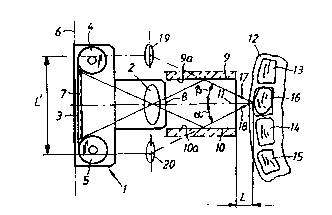
Figure l ~hows, in a top view, the camera in
relation to the lower jaw of a patient,
Figure 2 shows, in a top view, the camera in
relation to objectR or object parts on a model,
Figure 3 ~hows a photograph taken with the camera
according to Figures l and 2, in which photograph there
are two images of two given discrete point~, and
Figure 4 shows, in longit~;n~l ~ection, the
optical ray path in the camera where reflecting surfaces
are used to o~tain virtual lens functions arranged at a
distance from one another for achieving the stereo
imaging effect.
DE~TT~n EMBODIMENT
In Figure 1, a camera housing i~ indicated by l.
Arranged in the camera housing is a len~ ~y~em
21~43~6
W095/16222 PCT/SE94101143
-- 7
comprising a len~ 2 which is of a type known per se. Also
included in the camera housing i8 an arrangement for a
film 3 which is disposed and can be advanced on film
spools 4 and 5. The film is acted on 80 as to be placed
5 with great precision in a plane 6 at the back of the
camera. The action is effected by means of a glass plate
7 which is arranged against the film 80 that the latter
assumes its position in the plane 6 with the precision
mentioned above. Arranged at the opening 8 of the camera
lO housing are first and second reflecting surfaces or
mirrors which extend perpendicular to the plane of the
figure. The mirrors extend parallel to the longit--~;n~l
axis ll of the camera or the main direction of the
camera. The mirroring or reflecting surfaces 9 and lO can
15 themselves have other orientations, although the parallel
orientation shown is preferably used. The camera is aimed
at the mouth cavity of a patient, which mouth cavity is
represented by a jaw bone 12. In the mouth cavity or in
the jaw bone there are teeth 13, 14, 15, an implant 16,
20 dental bridge, etc. The radiation emanating from the
imaged area in the mouth cavity is indicated by the lines
17, 18. This optical radiation is reflected on the inner
surfaces 9a and lOa of the mirrors before it passes
through the lens 2 and reaches the film 3. The
25 arrangement means that two virtual lens functions l9, 20
ari~e, which in turn means that three image fields appear
on the film 3. The first image field is caused by the
real lens 2, and the second and third image fields are
cau~ed by the virtual lens functions l9 and 20. Three
30 images are thus obt~; n~ on the picture and can be
compared, for example in computer equipment for
t determining surfaces of the teeth, the implants, etc. The
lens functions are arranged essentially parallel. In this
connection, reference is made to the Swedish patent
35 application [lacuna], which has the same filing date as
the present Swedish patent application.
The camera is of st~n~d design and is of the
type which has been specified above. The ;n;~ture camera
can be placed at a distance L of about 50 - lO0 mm from
W095/16222 ~ 3 ~ ~ PCT/SE94/01143
the patient' 8 mouth.
Figure 2 shows the camera 1' with the same basic
construction as in Figure 1. In this case a prism 21 is
used which has two parallel inner walls 21a and 21b which
correspond to the reflecting surfaces 9a and lOa
indicated in Figure 1. The prism i8 secured to the camera
housing la. The same applies to the len~ 2', which is
secured to the camera housing by means of gluing or
equivalent. The securing of the prism 21 to the camera
housing la is symbolized by a tubular part 22. The camera
is provided with an ~nn~ flash 23 which is of a type
known per se. The flash arrangement is arranged
concentrically on the said sleeve-shaped m~mher 22. The
radiation 17', 18' enters the free end surface 21c of the
prism 21 and is reflected on the said inner surface 21a,
21b before it passes through the len~ 2' and re~ch~s the
film 3'. The imaging can in this case be carried out on
a patient, a model 24, etc.
Figure 3 shows a photograph 25 which comprises
three sections 25a, 25b and 25c. The section 25a
corresponds to the image, of the actual object or body
part, which is effected by the lens 2'. The sections 25b
and 25c correspond to the image areas which are generated
by the virtual lenses 19 and 20. The said virtual lens~s
are separated by a distance ~' (see Figure 1). The images
in the image fields 25b and 25c thus represent the body
part or the object seen from two different angles, alpha
and beta respectively (see Figure 1). A COrre8rQn~; ng
point is present in the image field 25a, which point does
not, howaver, need to be used (even though it is possible
to do 80) ~n the pre~ent case for determining the
positions of the relevant objects, surface~ or points
thereon.
Figure 4 show~ the relevant part of the optical
ray path in the novel camera. The reflecting surfaces or
the mirrors are shown by 9' and 10', and the virtual lens
functions are ~hown by 19', 20'. The real or actual lens
is indicated by 2'' and the film plane by 3a.
The invention is not limited to the embo~ t
21~ 43 ~ 6
W095tl6222 ^ PCT/SE94/01143
g
shown hereinabove by way of example, but instead can be
modified within the scope of the att~rhe~ patent claims
and the inventive concept.
