Note: Descriptions are shown in the official language in which they were submitted.
WO 95/16394 ~ PCTJUS94J14257
CARDIAC MONITORING AND DIAGNOSTIC SYSTEM
BACKGROUND OF THE INVENTION
This invention relates to cardiac monitors, and
more particularly to a system for simultaneous
arrhythmia monitoring and ECG analysis on a real time
basis.
Electrocardiographs for acquiring and analyzing
multiple leads of conventional ECG are known. For
example, electrocardiographs manufactured by Marquette
Electronics, Inc. of Milwaukee, Wisconsin, include a
twelve lead software program called the 12SL ECG
Analysis Program. Eight of the leads (I, II, V1-V6) are
acquired directly and the remaining four (III, aVR, aVL
and aVF) are derived using Einthoven's Law. Each lead
of ECG can be considered a view of the heart from a
different angle. The program also determines the origin
of the predominate rhythm from major categories such as
electronic artificial pacing, atrial flutter, ectopic
atrial rhythm, sinus rhythm, junction rhythm and atrial
fibrillation. In addition, the program analyzes the
contour of the QRS waveform using conventional criteria
for WollfParkinson-White atrial hypertrophy, QRS
abnormalities, ST abnormalities-QRS related, ST
elevation abnormalities, ST depression abnormalities and
T wave abnormalities. These allow the physician to
determine the existence of rhythm abnormalities,
conduction abnormalities, hypertrophy, infarction and
repolarization. This prior art program acquires ECG
data for non real time or delayed analysis.
Cardiac monitors are also well-known for analyzing
cardiac arrhythmia or beat irregularities. One such
system is the EK-Pro arrhythmia analysis algorithm sold
by Marquette Electronics, Inc. of Milwaukee, Wisconsin.
This system analyzes four ECG leads (I, II, III and V1)
to determine the occurrence of such conditions as
ventricular asystole, ventricular fibrillation,
ventricular tachycardia, VT3-5, R-on-T, ventricular
2~.~~~8~.
WO 95/16394 PCTIUS94/14257
-2-
bradycardia, couplet, bigeminy, accelerated ventricular
rhythm, pause, trigeminy, premature ventricular
complexes, ST deviation, tachycardia, bradycardia, and
irregular beats. ,
Arrhythmia monitors are generally rhythm sensing
systems which can indicate the occurrence of and ,
classify arrhythmias on a real time basis, but do not
indicate their cause. ECG lead analyzers, on the other
hand, can determine the condition of the entire
myocardium. Present practice is to employ an arrhythmia
monitor for triggering an alarm upon the occurrence of
an arrhythmia. The patient will then be connected to an
electrocardiograph in an attempt to determine its cause.
However, in certain situations, such as upon the
occurrence of an acute infarction, the patient is at
highest risk during the initial stages of the infarct
when an area of the myocardium is jeopardized due to
ischemia but before the myocardium is damaged. This
condition directly leads to a high probability of
ventricular arrhythmia during the time when the
patient's heart is already suffering from a lack of
oxygenated blood. However, such an ischemic episode
will not be detected by an arrhythmia monitor prior to
the occurrence of ventricular arrhythmia.
SUMMARY OF THE INVENTION
It is an object of the invention to provide a new
and improved system for heart monitoring and ECG
analysis.
Another object of the invention is to provide a
system for simultaneously detecting the occurrence of
arrhythmias and for indicating their cause. ,
A further object of the invention is to provide a
system for ECG analysis and arrhythmia detection on a
real time basis.
In general terms, the invention comprises a cardiac
monitoring system including means f or acquiring a
plurality of analog cardiac signals through leads
.~~8~
WO 95/16394 PCTIUS94/14257
-3-
connected to predetermined locations on the body of a
patient and for converting said cardiac signals to a
plurality of standardized ECG lead signals, first
analyzing means for analyzing a first group of the ECG
signals for determining the existence of rhythm
abnormalities, second analyzing means for analyzing the
contours of all of the ECG lead signals for conduction,
infarction, hypertrophy and depolarization
abnormalities, and means for simultaneously actuating
the first and second analyzing means for the
simultaneous analysis of the first group of ECG signals
and the plurality of ECG signals whereby the occurrence
of a rhythm abnormality and possible causes can be
determined concurrently on a real time basis.
~ ~. ~ ~ 8 .
WO 95/16394 PCT/US94/14257
-4-
BRIEF DESCRIPTION OF THE DRAWINGS
FIGURE 1 schematically illustrates the cardiac
monitoring and diagnostic system according to the
r
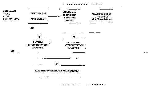
preferred embodiment of the invention;
FIGURE 2 schematically illustrates the arrhythmia
analyzer of the system illustrated in FIGURE 1; and
FIGURE 3 schematically illustrates the ECG wave
contour analyzer of the system illustrated in FIGURE 1.
DETAILED DESCRIPTION OF THE PREFERRED EMBODIMENT
FIGURE 1 schematically illustrates the cardiac
monitor and twelve lead analyzing system according to
the present invention. The system includes a
conventional data acquisition module 12 which is
connected to the patient by ten lead wires RA, LA, LL,
RL, V1, V2, V3, V4, V5 and V6. The acquisition module
12 includes conventional common mode rejection and
filters for removing patient breathing and muscle
artifacts. The acquisition module also converts the
analog lead signals to digital signals and generates the
ECG leads I, II, V1, V2, V3, V4, V5 and V6 which are
acquired directly from the patient leads and leads III,
aVR, aVF, and aVL which are derived. The twelve
digitized ECG signals are provided to a twelve lead
analyzer 14 and a display 16 and the leads I, II, III
and Vl are provided to an arrhythmia analyzer 18. The
outputs from the twelve lead analyzer 14 and the
arrhythmia analyzer 18 are provided to the display 16
which may also provide printed reports and may include
alarms for the arrhythmia analyzer 18. While a twelve
lead analyzer is employed in the preferred embodiment,
those skilled in the art will appreciate that analysis
programs for greater or lesser numbers of leads may also
be employed without deviating from the invention.
' The data acquisition module 14 and the arrhythmia
monitor exist as software in the RAM/ROM of a central
processing unit 19. The arrhythmia software may, for
W~ 95/16394 PCTIUS94/14257
-5-
example comprise the EK-PRO arrhythmia analysis
algorithm and the twelve lead software may comprise the
12SL ECG Analysis Program, both of which are products of
Marquette Electronics, Inc., of Milwaukee, Wisconsin.
The central processing unit also includes a control
a module 20 which permits the operator to operate the
arrhythmia monitor 18 and the twelve lead analyzer
selectively or concurrently. The control 20 may also be
programmed to actuate the twelve lead analyzer 18 upon
the receipt of an arrhythmia call or alarm signal from
the arrhythmia monitor.
The arrhythmia analyzer 18 is shown in Figure 2 to
include a high frequency noise check module 20, a low
frequency noise check 21, a QRS detect module 22, a QRS
correlation module 24 and an arbitrator or module 26.
The high frequency noise check module 20 and the low
frequency noise check module 21 respectively evaluate
each of the leads for high frequency and low frequency
noise, which if present, deactivates the lead so that it
is not further processed. The QRS detection module 22
detects signals falling within the physiologic band and
which it recognizes as valid ECG signals. The QRS
correlation 24 passes the ECG data stream through a list
of active templates, which are incrementally updated so
that they are progressively changed along with the beat
shape.
The arbitrator module 26 processes beats recognized
by the QRS correlator 24 and beats which do not match
any of the existing templates, as determined by the
detection module 22, and a determination is made whether
to create a new template and replace the least useful of
the active templates. These would be templates which
are matched with the least frequency or have not been
recently matched or classified as likely to be
,, 35 artifacts.
A classifier module 28 receives the template
information associated with the beats and takes all the
feature and temporal measurements and arrives at a
determination as to what is represented by that
WO 95/16394 PCT/US94/14257
2~~~~. .
-6-
particular beat, that is a normal QRS, an atrial
artificially paced normal QRS, a premature
supraventricular QRS, ventricular artificially paced
QRS, a ventricular premature QRS, a T wave, a P wave, a
ventricular artificial pacing spike, an atrial
artificial pacing spike or an artifact. The ,.
measurements made to determine individual beat
characteristics are R amplitude, S amplitude, QRS
polarity, T wave polarity, ST segment, noise level, PR
interval, P wave presence, QT interval, QRS duration, RR
interval, RR interval variance, pacemaker signals and
rotation of cardiac vector.
An arrhythmia call logic module 32 employs well-
known criteria to make an arrhythmia call. These
include the duration of usable ECG data, heart rate, the
time between QRS complexes, the occurrence of a
ventricular complex within a repolarization period, the
occurrence of one or more ventricular beats preceded or
followed by nonventricular beats, ST deviations of a
predetermined magnitude, R-to-R intervals and the
intervals between the QRS complex and a pacemaker spike.
With this information, the arrhythmia call logic 32 can
determine if one of the following has occurred: an
artifact, ventricular asystole, ventricular
fibrillation, ventricular tachycardia, VT3-5, R-on-T,
ventricular bradycardia, couplet, bigeminy, accelerated
ventricular rhythm, pause, trigeminy, isolated premature
ventricular complexes, ST deviation, tachycardia,
bradycardia, irregular heartbeat or electronic pacemaker
nonsensing. If an arrhythmia call is indicated, an
appropriate alarm signal is provided to the display 16
and the control 20.
The twelve lead analyzing section 40 receives the
ECG leads I, II, III, Vl-V6, aVF, aVR, and aVL.
Initially, the beat select section makes a template for
each lead. From this point on, the QRS selector looks
for the same shape. If it finds a match, the program
classifies it as another QRS detection. In addition,
the program slides the wave forms past one another
~~.~~81
WO 95/16394 PCTlUS94/14257
looking for the optimal match. If the output of the
filters in the acquisition modules exceed a preselected
value, but there is no match, it is assumed that a
different beat type has been detected and an additional
set of templates are made for further matching tests.
Thus, the beat selector 42 uses a filter and template
matching techniques to both detect and group by shape
the QRS complexes which occur in the ECG record The
QRS detector also defines the points on the ECG record
that can be used to align and time with maximum
correlation, the respective beats of a beat type.
The program then determines which beat type will be
used for the morphology measurements. The program uses
the RR intervals and the location of any pacer spikes in
order to decide which beat has the highest level of
origin in the conduction system. Identical QRS shapes
can even be subdivided as in the case of a sinus rhythm
with premature beats. The selection is not dependent
upon the number of beats per beat type but rather the
beat type which is the most informative for analysis is
the one sought after and any beat type with three or
more complexes can qualify. The beat type that the
computer considers to be most informative of normal
conduction is often referred to as the primary beat.
After a primary beat has been chosen, each of its
associated beats is used in generating a representative
complex for each lead. This is done using the sample
times generated by the QRS detector. These times not
only indicate the occurrence of a QRS but also indicate
when the QRS for a specific beat type are optimally
matched. The representative complex is then generated
with the median voltages from the aligned group of
beats, that is, it is formed by taking, at each sample
time, the middle voltage of the superimposed beats.
After the median for the primary cycle has been
established for each of the twelve leads, the waves of
each complex are identified. This is done separately
for each lead. The program finds the points at which
WO 95/16394 PCT/US94/14257
_g_
the signal crosses the baseline within each complex. If
the crossing points define a wave that has an area
greater than a predetermined value, the wave is
considered to be significant. If the area is less than
this value, the program considers the wave to be
insignificant and it will not label it as a separate
wave. The measurement matrix contains the amplitudes,
with respect to QRS onset, and durations of all of the
individuals waves, including the amplitude and duration
of the P, P', Q, R and S waves, the amplitude of the T
wave, the PR and QT intervals, the QRS duration and the
STJ, STM and STE amplitudes.
The program then utilizes these measurements in
making an interpretation. This includes a rhythm
analysis and a morphology interpretation. The rhythm
analysis first determines the origins of the predominant
rhythm in the sample and chooses from the major
categories consisting of electronic atrial pacing,
atrial flutter, ectopic atrial rhythm, sinus rhythm,
junction rhythm and atrial fibrillation.
The morphology interpretation will determine the
existence of Wolff-Parkinson-White, atrial hypertrophy,
QRS abnormalities such as low voltage QRS, pulmonary
disease pattern, QRS axis, conduction abnormalities,
ventricular hypertrophy, infarction, ST + T abnormality
with ventricular hypertrophy, dating infarcts,
epicardial injury, pericarditis, early repolarization,
nonspecific ST elevation, subendocardial injury,
nonspecific ST depression, digitalis effect, functional
ST depression, ischemia, QRS-T angle and QT interval.
The twelve lead system 40 can be operated
independently of the arrhythmia analyzer 18 or it can be
operated on a real time basis along with the arrhythmia
analyzer for analyzing the same ECG signals or its
operation can be triggered automatically by an
arrhythmia alarm signal from the arrhythmia call logic
module 32.
WO 95/Y6394 PCT/US94/14257
_g_
The arrhythmia analyzer 18 indicates the occurrence
of and classifies arrhythmias, but does not indicate
their cause. The twelve lead analyzer 40, on the other
hand, can determine the condition of the entire
myocardium. If the two are operated concurrently on a
real time basis, for simultaneously analyzing the same
ECG signals, it may be possible to determine the cause
of any arrhythmia detected by the arrhythmia monitor 18.
For example, episodes such as ischemia may be detected
at an early stage before the myocardium is damaged.
Also, because each of the twelve ECG leads is a
view of the cardium along a different axis, the location
of an infarct is indicated by wave abnormalities in
specific leads. For example, septal myocardial
infarction is indicated in leads V1 and V2; anterior
myocardial infarction in leads V2, V3 and V4, lateral
myocardial infarction in leads I, V5, V6 and aVL,
inferior myocardial infarction produces an ST elevation
in leads II, III and aVP. With the twelve lead analysis
and the simultaneous detection and classification of
arrhythmias, the cardiologist is able not only to detect
the occurrence of an arrhythmia but to simultaneously
determine its cause because both systems are running
simultaneously and are analyzing the same ECG lead data
on a real time basis.
While only a single embodiment of the invention as
been illustrated and described, it is not intended to be
limited thereby but only by the scope of the appended
claims.
