Note: Descriptions are shown in the official language in which they were submitted.
W095/1634821 S ~ 7 ~1 PCT~S94/14227
P E C I F I C A T I O N
TITLE
METHOD AND APPARAT~S FOR TREATING A BODY FL~ID
BACKGROUND OF THE INVENTION
5The present invention relates generally to the
collection and therapeutic use of body fluids. More
specifically, the present invention relates to methods
and devices for attempting to substantially reduce or
eliminate potential viral contaminants and other
pathogens in body fluids, such as blood.
Of course, in a wide variety of therapies, such as
transfusions and transplants, body fluids, especially
blood components, such as red blood cells, platelets,
plasma, and bone marrow, are infused from one or more
individuals into a patient. Although such therapies
provide treatments, many of which are life saving, and
cannot otherwise be provided, due to the transmission of
infectious diseases, there may be potential risks to such
therapies.
For example, it is known that blood can carry
infectious agents, such as hepatitis virus, human immune
deficiency virus (an etiological agent for AIDS),
cytomegalovirus, Epstein Barr virus, and herpes virus.
Although screening methods exist to identify blood that
may include such viruses, current screening methods do
not assure that every blood unit that contains such a
virus is identified.
For example, in this regard, one of the difficulties
in testing blood components for viral contamination, such
as HIV, is that many current diagnostic tests are based
on an identification of antibodies. Therefore, a
contaminated blood component will only exhibit a positive
test if it includes antibodies for the virus, e.g., anti-
PCT~S94/14227
Wos~/l6348
2~L54761
;}~ 2
HIV. With respect to intracellular viral infections, an
individual may not generate antibodies immediately upon
infection. Rather, there is a window period that extends
from the initial infection of the patient with a virus
to the generation of antibodies. When an individual is
in this window period, diagnostic tests that are based
on antibodies will not identify the individual, or the
blood unit, as being infected. But, even though the
antibodies are not present, the blood unit can still
transmit an infection.
With respect to HIV infection, it is believed that
this window period can extend from approximately six
weeks up to 48 months. During this time period, an
individual who has been infected with HIV and
accordingly, whose blood will transmit same, will
register a n~gative antibody response. Currently used
screening methods will not identify as contaminated a
blood unit from an individual who is infected with HIV,
but who has not generated anti-HIV.
In order to address the limitations of current
diagnostic techniques and also to deal with the concern
of transmission of viral contaminants and other pathogens
to a patient receiving a transfusion, recent attention
has focussed on the development of viral inactivating
agents. It is envisioned that these viral inactivation
agents would be added to the body fluid prior to the body
fluid being administered to the patient.
For example, a number of photoactive agents that
have antiviral action have been explored. These
photoactive agents are generally agents that upon
activation with light will inactivate or destroy
pathogens, e.g., a virus that may be present. Such
photoactive agents include: psoralens; porphyrins;
21~4761
W095/16348 PCT~S94/14227
phthalocyanines; and dyes, such as methylene blue. See,
for example, U.S. Patent Nos.: 4,748,120; 4,878,891;
5,120,649; and German Patent Application No. DE 39 30 510
Al (Mohr).
Although such agents provide promise for the
treatment of body fluids to eliminate the concern of
viral contamination, there may be regulatory, as well as
possible other concerns with respect to such agents. of
course, the resultant body fluid to which the anti-viral
agent is added will be infused into a patient.
Therefore, it is imperative that the agent does not
create toxicity issues or other in vivo concerns.
With respect to photoactive agents, a still further
issue is that upon activation~ of the agent and
interaction of the agent with a virus, other products may
be generated. For example, methylene blue is a
photoactive agent that has been shown to have efficacy
in inactivating viral contamination in plasma. Although
methylene blue has been, through exhaustive testing,
shown to have no toxicity concerns, upon photoactivation
of methylene blue, photoproducts are generated.
Specifically, Azure A and B are generated upon
photoactivation of methylene blue. The in vivo effect
of these products has not been as well studied as
methylene blue in patients and therefore they rais~
regulatory issues and in vivo concerns.
There therefore is a need for an improved method and
system for treating a body fluid to substantially reduce,
if not eliminate, viral contaminants that may be present
therein.
SUMMARY OF THE INVENTION
The present invention provides a method of treating
a body fluid to substantially inactivate viral
W095/16348~ PCT~S94/14227 -
215~761 j ~
contaminants that may be present therein. Pursuant to
the method, to a body fluid is added a viral inactivation
agent. The resultant product is then passed through a
container, e.g., column including a material having an
affinity for the viral inactivating agent. This allows
the column to remove excess viral inactivating agent.
Additionally, other products, e.g. photoproducts, that
may be generated upon addition of the viral inactivation
agent or any activation thereof are also eliminated. The
body fluid can then be infused into a patient without
regulatory or toxicity concerns.
To this end, in an embodiment, the present invention
provides a method for trea~ing a body fluid to at least
substantially inactivate viral contaminants that may be
present comprising the steps of: providing a body fluid;
adding to the body fluid a viral inactivating agent to
create a resultant product; and passing the resultant
product through a column including material having an
affinity for the viral inactivating agent.
In an embodiment, the material includes charcoal.
In an embodiment, the column is an ion exchange
column.
In an embodiment, the material includes neural
macroporous polymeric beads with a high surface area for
absorbing organics from aqueous solutions.
In an embodiment, the viral inactivating agent is
a light activated agent.
In an embodiment, the viral inactivating agent is
chosen from the group consisting of: porphyrins;
psoralens; phthalocyanines; and dyes.
The present invention also provides a method for
treating a blood product comprising the steps of:
providing a blood product; adding to the blood product
WO95/16348 21 S 4 7 6 1 PCT~S94114227
a light activated viral inactivating agent to create a
resultant product; irradiating the resultant product with
light of a sufficient wavelength to activate the viral
inactivating agent to create a further product; passing
the further product through a column having an affinity
for the viral inactivating agent; and collecting a
product that passes through the column.
In an embodiment, the blood product includes
platelets and the viral inactivating agent is a psoralen.
In an embodiment, the blood product includes plasma
and the viral inactivating agent includes methylene blue.
In an embodiment, the column also has an affinity
for photoproducts generated by irradiating the resultant
product.
The present invention also provides a method for
providing a blood product to a patient comprising the
steps of: collecting a blood product from a donor;
adding to the blood product a light activated viral
inactivation agent; irradiating the blood product and
light activated viral inactivation agent with light of
a sufficient wavelength to activate the viral
inactivation agent to create a resultant product; passing
the resultant product through a column having an affinity
for the viral inactivation agent; collecting a resultant
blood product that passes through the column; and
administering the resultant blood product to a patient.
An advantage of the present invention is that it
provides an improved method for treating a body fluid to
at least substantially inactivate viral contaminants that
may be therein.
Another advantage of the present invention is that
it provides a method for inactivating or eliminating
Wo95/16348 PCT~S94/14227 ~
2154761
,. ~ ....
L ~ '
pathogens from blood or its components before they are
infused into a patient.
Furthermore, an advantage of the present invention
is that it provides a system that allows viral
inactivating agents to be added to a body fluid before
the fluid is infused into a patient and eliminate
toxicity or regulatory concerns.
Still further, an advantage of the present invention
is that it provides a method for eliminating
photoproducts from a system that adds a photoactive agent
to a body fluid.
Moreover, an advantage of the present invention is
that it prevents any post thaw photoactivation of excess
photoactivated agents.
Another advantage of the present invention is that
it allows normal plasma color for treated plasma.
Additional features and advantages of the present
invention are descri~ed in, and will be apparent from,
the detailed description of the presently preferred
embodiments and from the drawings.
BRIEF DESCRIPTION OF THE DRAWINGS
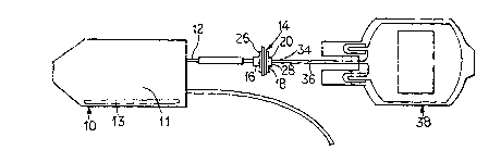
Figure 1 illustrates, schematically, an embodiment
of the system of the present invention.
Figure 2 illustrates graphically results of Example
No. 3 and specifically, the content of fibrinogen in
reference plasma, post-thaw, pretreatment, post
treatment, and post removal.
Figure 3 illustrates graphically results of Example
No. 3 and specifically, the content of Factor V in
reference plasma, post-thaw, pretreatment, post
treatment, and post removal.
Figure 4 illustrates graphically results of Example
No. 3 and specifically, the content of Factor VII in
~ WO95/16348 215 ~ 7 61 PCT~S94/14227
reference plasma, post-thaw, pretreatment, post
treatment, and post removal.
Figure 5 illustrates graphically results of Example
No. 3 and specifically, the content of Factor VIII:C in
reference plasma, post-thaw, pretreatment, post
treatment, and post removal.
Figure 6 illustrates graphically results of Example
No. 3 and specifically, the content of Factor IX in
reference plasma, post-thaw, pretreatment, post
treatment, and post removal.
Figure 7 illustrates graphically results of Example
No 3 and specifically, the content of Factor XI in
reference plasma, post-thaw, pretreatment, post
treatment, and post removal.
Figure 8 illustrates graphically results of Example
No. 3 and specifically, the content of prothrombin in
reference plasma, post-thaw, pretreatment, post
treatment, and post removal.
Figure 9 illustrates graphically results of Example
No. 3 and specifically, the content of activated partial
thromoplastin time in reference plasma, post-thaw,
pretreatment, post treatment, and post removal.
DETAILED DESCRIPTION
OF THE PRESENTLY PREFERRED EMBODIMENTS
The present invention provides a method and
apparatus for use in treating a body fluid, such as
blood, to reduce or eliminate viral contaminations that
may be present therein. It is believed that the present
invention can be used with a variety of viral
inactivating agents. Such agents include, without
limitation, photoinactivation agents, such as psoralens,
porphyrins, dyes, such as methylene blue,
W095/16348 PCT~S94/14227 ~
215~761
8 -
.
phthalocyanines, phenothia~ines, hypericin, and other
compounds that are activated by light.
As has been suggested in the art, a photoactive
viral inactivating agent would be added to a body fluid,
such as blood prior to the blood being infused into a
patient. The resultant blood product including the
photoactive agent then would be activated by light of a
suitable wavelength. Of course, if desired, other viral
inactivating agents that are not based on activation
through light can be utilized in the present invention.
Pursuant to the present invention, as illustrated
in Figure 1, a container 10 will be provided including,
for example, a blood component 11. The blood component
have added thereto a viral inactivation agent 13.
For example, it is known to collect whole blood in
a blood pack. Typically, whole blood is then separated
into its component parts. After the blood is separated
into the respective components, using a system such as
the Optipress~ system marketed by affiliates of Baxter
International, the blood component can be added to the
container 10 including the viral inactivation agent. For
example, methylene blue can be added to the plasma
component. Of course, if desired, whole blood can be
treated with a viral inactivation process. Likewise, if
desired, a separate container is not required and the
viral inactivation agent can be added to the container
in whcih the component is stored.
The-container 10 will include a fluid line 12 that
will be coupled to a column 14. As used herein, column
refers broadly to a chamber or device that includes
material that will remove specific compounds or entities.
Accordingly, column includes cartridges, containers, and
other means for housing such material.
~ WO95/16348 215 4 7 61 PCT~S94/14227
g
Pursuant to the present invention, the column 14
will include materials having an affinity for the viral
inactivation agent and photoproducts generated thereby.
The column will include an inlet 16 allowing product to
flow into an interior 18 defined by the housing 20. In
an embodiment, porous plates (not illustrated) are
located at each end 26 and 28, respectively, of the
interior 18 of the housing 20. The porous plates allow
the body fluid to flow through an affinity matrix located
therein. The resultant product then flows out of the
cartridge 14 through the outlet 34.
In use, after the container containing the blood
product and viral inactivation agent is activated by
light of an appropriate wavelength, the resultant product
flows through fluid line 12 and into the affinity column
14. The affinity column 14 will remove excess viral
inactivation agents, as well as photoproducts. For
example, with respect to methylene blue, excess methylene
blue will be removed, as well as Azure A and B. The
resultant blood product will then flow through fluid line
36 to a container 38. The blood can be stored in the
container 38 and then infused into a patient.
To allow selective flow through the fluid line 12,
breakable cannulas, as are known in the art, can be
provided. Of course, other means for allowing selective
flow through the fluid line 12 can be provided.
It should be noted that although in the illustrated
embodiment the cartridge 14 is a separate and distinct
component from the container 10, a unitary structure can
be provided. In this regard, the column can be integral
with the container or coupled thereto as an outlet port
of the container.
WO9~/16348 ~.~ PCT~S94/14227
~5 1~61
-- 10 --
The material used for the matrix in the affinity
column 14 can comprise a variety of different materials.
For example, charcoal, an ion exchange resin, or biobeads
can be used. As used herein, the term "biobeads" refers
to neural macroporous polymeric beads with a high surface
area for adsorbing organics from aqueous solutions.
Biobeads can vary in their hydrophilic and hydrophobic
polarities. The range of believed useful properties of
biobeads for the present invention is as follows:
polarity (non-polar to intermediate polarity); Dipole
Moment (0.1 to 3.0); bead size (30 to 2000 ~m); average
pore diameter (45 to 300 angstroms); bead surface area
(150 to 1,600 sq. meters/gram dry bead). It has been
found that biobeads available from Biorad Laboratories,
2000 Alfred Nobel Drive, Hercules, CA 94547 under the
name Macro-Prep~ t-butyl HIC function satisfactorily to
remove methylene blue and methylene blue photoproducts
Azure A and B.
By way of example, and not limitation, examples of
the present invention will now be given:
EXAMPLE NO. 1
Removal studies were performed on 4'-aminomethyl-
4,5',8-trimethyl psoralen (AMT). Specifically, three
studies were performed, two using activated charcoal
columns and one using an ion exchange column. The
charcoal columns each consisted of 5.3 grams of activated
charcoal obtained from a commercial water purification
device. The ion exchange column consisted of less than
8.2 grams of Biorad AG 50W-X8 cation exchange resin.
One unit (80 mL) of plasmalyte platelets containing
40 ug/mL of AMT was passed through the first charcoal
filter at a rate of a~out 30 mL/min. This column removed
86~ of the AMT as measured by HPLC. Platelet loss going
~ WO9~/16348 215 4 7 61 PCT~S94/14227
-- 11 --
through the column was 6%. Total protein was reduced by
33%. The platelet morphology score dropped from 355 to
315.
A second charcoal column was tested at a flow rate
of about 5 mL/min. This column removed "100~" of the AMT
as measured by HPLC. Platelet loss was 14~. Total
protein increased by 14%. The platelet morphology score
was unchanged by the column (200).
It is clear from these data that the activated
charcoal can remove significant amounts of the AMT drug.
The removal is inversely proportional to flow rate. The
charcoal also appears to remove about one third of the
plasma protein and 6-7~ of the platelets. At the reduced
flow rate (higher drug removal) the platelet morphology
score dropped appreciably. We did not see any "fines"
coming off the charcoal column.
The ion exchange column clearly removed significant
amounts of AMT at low flow rate, but not as much as the
charcoal. This column did not appear to remove any
plasma protein and platelet loss was higher than with the
charcoal. The platelets did not appear to be effected
by the ion exchange resin.
EXAMPLE N0. 2
In this example, two more AMT removal studies were
performed using biobeads, one with 5.5 grams of 100-200
mesh biobeads and the other with 7.5 grams of 20-50 mesh
biobeads.
One unit (50-60 mL) of platelets (in lactated
ringers) containing about 20 ug/mL of AMT was pumped
through each column at a rate of 7 mL/min. Both columns
removed all measurable AMT, but the 20-50 mesh column
material yielded a "cleaner" HPLC output. The platelet
loss for ~he 100-200 mesh column was 40~ and for the 20-
,
WO95/16348 PCT~S94/14227 ~
21~7~1
- 12 -
50 mesh was 28%. Total protein was~reduced by 13~ in the
100-200 mesh column and by 32% in the 20-50 mesh. The
platelet morphology for the 100-200 mesh column was
unchanged passing through the column at 355, and for the
20-50 mesh column the morphology changed from 130 to 115.
It should be noted that the unit of platelets used for
the 20-50 mesh biobead column had low platelet counts,
bad platelet morphology and low protein content. The
columns did not appear to shed any "fines", nor did the
beads swell.
The biobeads removed AMT as well as the activated
charcoal tested in Example No. 1.
EXAMPLE N0. 3
The following method was performed on ten units of
fresh frozen plasma, which had been thawed using an
Instacool plasma thawer. An approximately 12 ml sample
was collected from each unit as an untreated control
sample and aliquoted into tubes for testing. The tubes
were labeled with the protocol number, the sample letter
and untreated. These units were stored frozen (-
80C+10C) until analyzed.
TREATED SAMPLES
The following procedure was performed on ten units
of fresh frozen plasma, which had been thawed using the
Instacool plasma thawer. An approximately 12 ml
untreated sample was removed from each unit, aliquoted
and stored frozen (-80C+10C) until testing. Each unit
was sterile connected and added to a container containing
- methylene blue. These units were labeled K-T. Each
methylene blue treatment bag (PL732) was wrapped with
aluminum foil and placed on a rotator (Scientific
Products Multipurpose rotator Model 151) at room
temperature and mix end-over-end at 40-60 rpm for 60
WO95/16348 215 ~ 7 6 1 PCT~S94/14227
- 13 -
minutes. After mixing, the units were kept in aluminum
foil and at room temperature prior to irradiation.
An approximately 16 ml pretreatment sample was
removed from each unit and divided into a 4 ml aliquot
for methylene blue testing. Prior to irradiation of the
plasma units, the light output delivered by the
irradiation box was measured. The light output was
recorded. The methylene blue plasma mixture was
irradiated with the LED light source. The LED light
source was placed on top of a horizontal rotator at a
speed of 60 rpm. All units were irradiated with red
light for 8 J/cm2 exposure. The start and stop times
were noted.
An approximately 16 ml post treatment sample was
removed from each unit and divided into a 4 ml aliquot
for methylene blue testing. The remaining plasma in each
plasma unit was passed through a methylene blue removal
cartridge in the removal set of Figure l with a plasma
expressor. The removal cartridge included biobeads
obtained from Biorad Laboratories and sold under the name
Macro-Prep~ t-butyl HIC. An approximately 16 ml post
removal sample was aseptically removed from each unit and
divided into a 4 ml aliquot for methylene blue testing.
The following data was generated. Figures 2-9
graphically illustrate the data.
WO 95/16348 PCT/US94/14227
2~547~1
' ~ -- 1 4 --
Methylene Blue (MB) (ug/ml)
1 uM = .374 ug/ml
Test Sample untreated pretreatment post - post
treatment removal
MB K NT 0.308 0.39 NRQ
MB L NT 0.316 0.365 NRQ
MB M NT 0.409 0.363 NRQ
MB N NT 0.368 0.329 NRQ
MB O NT 0.419 0.353 NRQ
MB P NT 0.401 0.348 NRQ
1 0 MB Q NT 0.422 0.306 NRQ
MB R NT 0.426 0.409 NRQ
MB S NT 0.43 0.344 NRQ
MB T NT 0.292 0.384 NRQ
NRQ = No r~3coverable q~antity
Wo 95/16348 215 4 7 61 PCT/US94/14227
-- 15 --
.cll.ro,.,bin Time
Test Sample untreated p~el~dl~"ent post post
treatment removal
PT K 12 12 12.2 13.8
PT L 11.9 11.8 12.4 11.9
PT M 12.2 12.5 13.7 13.7
PT N 11.5 11.5 11.8 11.4
PT O 12.2 12.1 15.1 12.1
PT P 13.6 12.5 13.4 12.5
PT Q 11.7 12.1 13.8 12.1
PT R 11.6 13.2 15.8 13.7
PT S 11.8 12.1 13.8 13.2
PT T 11.7 14.2 13.1 11.8
AVG 12.02 12.4 13.51 12.62
SD 0.603 0.782 1.249 0.900
Activated Partial T~romboplastin ime
Test Sampie untreated plelredLIllent post post
treatment removal
APTT K 37.1 34.2 35.9 39
APTT L 26 26.6 26.9 27.3
APTT M 28.6 30.4 36.7 28.4
2 0 APTT N 31.7 31.1 32.3 29.7
APTT O 31.2 32.5 38.9 31.3
APTT P 38.9 31.8 36.3 33.6
APTT Q 29.4 30.4 35.9 32.2
APTT R 29.9 34.1 45.3 36.1
2 5 APTT S 29.6 30.7 31.7 39.5
APTT T 31.9 41.2 31.1 31.8
AVG 31.43 32.3 35 32.89
SD 3.882 3.801 4.990 4.185
Wo 95/16348 PCTrUS94/14227
21~761
- 16 -
Factor IX
Test Sample untreaté~ pr~LI ~dll I ,ent post post
treatment removal
Factor IX K 117 92 94 96
Factor IX L 100 87 76 87
Factor IX M 61 59 60 54
Factor IX N 79 75 81 69
Factor IX O 68 75 72 65
Factor IX P 70 63 45 51
Factor IX Q 71 67 64 55
Factor IX R 72 59 70 68
Factor IX S 88 88 87 71
Factor IX T 84 82 76 66
AVG 81 74.7 72.5 68.2
SD 16.964 12.338 13.986 14.227
Factor Xl
Test Sampie untreated pretreatment post post
treatment removal
Factor Xl K 132 118 122 80
Factor Xl L 121 105 96 76
Factor Xl M 118 94 67 44
Factor Xl N 109 104 96 78
Factor Xl O 132 128 126 68
Factor Xl P 77 69 65 43
Factor Xl Q 102 103 91 54
Factor Xl R 99 78 88 44
Factor Xl S 106 91 95 36
Factor Xl T 87 78 75 39
AVG 108.3 96.8 92.1 56.2
SD 18.087 18.564 20.431 17.492
WO 95/16348 2 1 5 4 7 6 1 PCrlUS94114227
Factor Vll
Test Sample ~ wLed pretreatment post post
treatment removal
Factor Vll K 103 95 92 104
Factor Vll L 129 119 119 116
Factor Vll M 63 ~7 57 61
Factor Vll N 87 84 82 93
Factor Vll O 89 85 89 gO
Factor Vll P 59 63 50 60
Factor Vll Q 86 70 72 80
Factor Vll R 63 55 55 61
Factor Vll S 74 66 64 99
Factor Vll T 92 90 87 95
AVG 84.5 78.4 76.7 85.9
SD 21.324 20.001 21.250 - 19.723
Facto~ C
Test Sample ~l ILr~L~d pr~lr~dLI~ ~enL post post
treatment removal
Factor Vlll:C K 47 41 41 44
Factor Vlll:C L 89 88 83 87
FactorVlll:C M 93 81 81 71
2 0 Factor Vlll:C N 56 51 46 46
Factor Vlll:C O 104 101 88 85
Factor Vlll:C P 46 36 39 36
Factor Vlll:C Q 76 70 54 54
Factor Vlll:C R 53 43 46 44
Factor Vlll:C S 63 55 56 54
Factor Vlll:C T 92 75 62 66
AVG 71.9 64.1 59.6 58.7
SD 21.522 22.098 18.265 17.795
PCT/US94/14227
WO 95/16348
2~54761
-- 18 --
Fibrinogen
Test Sample untreated pr~tr~L~I~ent post post
treatment removal
Fibrinogen K 341 290 276 281
Fibrinogen L 308 285 261 248
Fibrinogen M 250 235 204 185
Fibrinogen N - 377 345 318 329
Fibrinogen O 285 273 248 252
Fibrinogen P 200 189 149 137
Fibrinogen Q 308 284 212 214
1 0 Fibrinogen R 273 248 223 235
Fibrinogen S 241 244 196 191
Fibrinogen T 299 266 257 255
AVG 288.2 265.9 234.4 232.7
SD 50.861 41.162 47.664 53.994
Factor V
Test Sample untreated p,~lr~l",ent post post
treatment removal
Factor V K 69 65 58 53
Factor V L 84 80 74 77
Factor V M 77 73 69 62
2 0 Factor V N 84 79 74 71
FactorV O 63 58 60 54
FactorV P 74 67 _ 62 53
Factor V Q 71 63 52 49
Factor V R 77 58 65 63
2 ~ Factor V S 73 62 59 60
Factor V T 53 47 48 43
AVG 72.5 65.2 62.1 58.5
SD 9.384 10.130 8.634 10.244
WO 95/16348 21 5 4 7 61 PCTIUS94/14227
-- 19 --
CONTROL SAMPLES
Sample Fibrinogen Factor V Factor Vll
Rerarence 200400 50-150% 65-135~
range mg/dl
A untreated 281 82 95
B untreated 249 114 144
C untreated 202 79 103
D untreated 302 62 76
E untreated 399 87 71
F untreated 233 96 82
G u~ d 279 93 87
H untreated 263 58 113
I untreated 299 60 80
J untreated 240 111 98
1 5 AVG 274.7 84.2 94.9
SD 53.614 20.077 21.584
Sample Factor Vlll:C Factor IX Factor Xl
Reference 50-150% 60-140% 65-135%
2 0 range
A untreated 69 83 101
B untreated 83 115 107
C untreated 31 86 118
D untreated 62 79 101
E untreated - 55 90 118
F untreated 96 97 80
G untreated 62 118 142
H untreated 99 85 130
I untreated 150 94 97
WO95/16348 PCT~S94/14227 -
2~ 5476~
- 20 -
",.. .
J untreated 83 ." . 98 103
AVG 79 94.5 109.7
SD 32.180 13.109 17.764
Sample Prothrombin APTT
Time (sec) (sec)
Reference
range
A untreated 12.3 30.7
B untreated 11.6 30.8
C untreated 12 31.
D untreated 12.1 . 31
E untreated 11.7 28.4
F untreated 12.4 27.1
G untreated 11.7 25.1
H untreated 12.5 28
I ~Intreated 12.9 28.2
J untreated 11.5 26.5
AVG 12.07 28.7
2 0 SD 0.45~ 2.179
After flowing through the cartridge less than 4
nanograms/ml of methylene blue and photoproducts were
present in the blood. It should be noted that prior to
removal, the blood unit contained 400 nanograms/ml
methylene blue. By way of example for a 70 Kg man
receiving 2 liters of methylene blue treated fresh frozen
plasma he would receive, after removal pursuant to the
present invention, 114 ng/Kg of methylene blue. This
amounts to 1/44,000 that of normal intravenous clinical
W095/16348 2 1 5 ~ 7 6 1 PCT~S94/14227
- 21 -
dose. This reduced level effectively eliminates any
toxicity concerns.
As illustrated in Figures 2-9, except for with
respect to Factor XI, the removal step does not remove
components from the plasma. Figures 2-9 illustrate,
graphically content of specific components in: reference
plasma; post-thaw; pretreatment; post treatment; and post
removal. Specifically, Figures 2-9 graphically
illustrate, content of: Fibrinogen; Factor V; Factor
VII; ~actor VII:C; Factor IX; Factor XI; prothrombin; and
activated partial thromboplastin time, respectively. As
illustrated, the method of the present invention can be
used without destroying the therapeutic benefits of the
blood to be transfused.
It should be understood that various changes and
modifications to the presently preferred embodiments
described herein will be apparent to those skilled in the
art. Such changes and modifications can be made without
departing from the spirit and scope of the present
invention and without diminishing its attendant
advantages. It is therefore intended that such changes
and modifications be covered by the appended claims.