Note: Descriptions are shown in the official language in which they were submitted.
WO 94/25485 PCT/US94/01439
-1-
OXYTOCIN ANTAGONIST
FIELD OF INVENTION
The present invention relates to a novel compound
which is highly active as an oxytocin antagonist and
which exhibits slight antagonism for vasopressin.
BACKGROUND OF INVENTION
Preterm labor is the major cause of prenatal
morbidity and mortality in the United States. Current
methods of inhibiting preterm labor are not always
successful and are often associated with significant
side effects. Since the uterus is a target organ for
oxytocin, and assuming that oxytocin is an important
contributing factor to preterm labor, the development
of a potent oxytocin antagonist would result in
successful inhibition of preterm labor with few
associated side effects.
Structurally, oxytocin (OT) and antidiuretic
hormone (ADH), also called vasopressin, are similar.
Their comparative structures are illustrated below.
1 2 3 4 5 6 7 8 9
Cys-Tyr-Ile-Gln-Asn-Cys-Pro-Leu-Gly-NHZ
S S
WO 94/25485 PCT/US94/01439
10
_2_
OXYTOCIN (OT)
1 2 3 4 5 6 7 8 9
Cys-Tyr-Phe-Gln-Asn-Cys-Pro-Arg-Gly-NHZ
S S
VASOPRESSIN (ADH)
Various investigations in the literature have
reported the synthesis of antagonists to ADH for the
treatment of hypertension and the synthesis of
antagonists to oxytocin. In 1960, Law, H.D. and V.
DuVigneaud, J. Am. Chem. Soc., 82:4579, reported the
first synthesis of an oxytocin antagonist (2-0-
methyltyrosine-OT). In 1967, Chan, Fear and
DuVigneaud, Endocrinolocry, 81:1267, reported the
synthesis of 1-L-Penicillamine-oxytocin and 1-deamino-
penicillamine-oxytocin. This was the first study to
show an in vivo inhibitory effect of an oxytocin
antagonist on uterine contractions and response to
oxytocin in the anesthetized rat.
In 1980, Sawyer, et al., Endocrinology, 106:81,
reported the synthesis of an oxytocin antagonist that
combined the two important features of the antagonist
of Law and DuVigneaud and of the antagonist of Chan,
et al.. The new antagonist was (1-deamino-
penicillamine, 2-0-methyltyrosine) oxytocin. The new
antagonist had a pA2 of 7.8 as determined by the
oxytocic bioassay. The pAz is the negative logarithm
of the molar concentration of the antagonist that
reduces the response to the antagonist by 1/2. It is
defined by Schild, British J. Pharmacology, 2:189
(1947).
In 1983, Manning, et al., J. Med. Chem., 26:1607-
WO 94/25485 r'
PCT/US94/01439
_ -3-
161 reported the synthesis of a number of antagonists
to ADH. One of these antagonists proved to have
potential anti-oxytocic activity [f~,~3-pentamethylene-
~-mercaptopropionic acidl,D-Phe2,Ile'] arginine
vasopressin with a pA2 of 8.2, or in other words, 2.5
times more potent than the antagonist reported by
Sawyer, et al. in 1980 (see page 1610, Table I,
compound no. 1). This oxytocin antagonist can be
called [Pmpl, D-Phe2, Phe3, Ile',Arge] oxytocin. A related
oxytocin antagonist, [Pmpl,D-Trp2,Phe3,Ile',Arge]
oxytocin was disclosed by Wilson and Flouret, Abstract
for Society for the Study of Reproduction Meeting July
14-17, 1986.
In 1981, Melin, et al., EndocrinoloQV, 88:173,
developed an oxytocin antagonist for inhibiting
preterm labor. They synthesized 1-deamino,
ethyloxytocin which had a pA2 of 7.2. They also
showed that this compound inhibited uterine
contractions in rats in vivo and in humans in vitro
and in vivo (Akerland, et al., Obstet. and Gynecol.,
62:309, 1983). In 1985, Akerland, et al., Obstet. and
Gynecol. Scand., 64:499, reported the synthesis of 1-
deamino[D-Tyr(OEt)2,Thr', OrnB] vasopressin with a pAz
of 8.3. They have tested this compound in vitro on
human uterine tissue and have shown it to inhibit
uterine contractions.
United States Patent 4,597,901 discloses the
class of vasopressin antagonists in which cysteine-1
is present in both oxytocin and vasopressin and
substituted with p,Q-cylopentamethylene-p-
mercaptopropionic acid.
Other amino acids of vasopressin are substituted.
The resulting class of compounds is said to be
vasopressin antagonists the biological activity being
WO 94125485 ~'~~ ~ PCT/US94101439
-4 -
manifested as water diuresis.
SUMMARY OF INVENTION
The present invention comprises an oxytocin
antagonist which is an analog of oxytocin. In the
compound of this invention, cysteine-1 of oxytocin is
substituted with Q,p-(3-thiapentamethylene)-f3-
mercaptopropionic acid. In addition, L-tyrosine-2 is
substituted with D-tryptophan, and penicillamine is
substituted for 1-cysteine in the 6 position and L-
arginine is substituted in the 8 position for L-
leucine. The resulting compound [(S)Pmpl,D-
Trp2,Pen6,Arg8] oxytocin is believed to be novel and
has been found to have remarkable properties. It is
highly active as an oxytocin antagonist. At the same
time, and although it is structurally similar to
vasopressin and vasopressin antagonists described in
the literature, the new compound exhibits minimal ADH
antagonism. When these two antagonisms are expressed
as a ratio, the compound of this invention has a very
high anti-oxytocin/anti-ADH activity ratio. This
combination of properties is highly advantageous for
therapeutic use. Effective anti-oxytocin action can
be obtained with minimal anti-ADH side effects. The
compound of this invention is therefore adapted for
inhibiting contraction of the uterine muscle in
response to bodily oxytocin, and can be used to
suppress preterm labor.
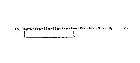
DESCRIPTION OF THE INVENTION
The oxytocin antagonist of this invention is
represented by the formula:
CA 02155872 2000-03-O1
-5-
1 2 3 4 5 6 7 8 9
(S)Pmp-D-Trp-Ile-Gln-Asn-Pen-Pro-Arg-Gly-NHZ
S S
wherein Pmp is p,p-(3-thiapentamethylene)-Q-
mercaptopropionic acid, D-Trp is the D form of
tryptophan, and Ile, Gln, Asn, Pen (Pen =
penicillamine), Pro, Arg, are the L forms of
isoleucine, glutamine, asparagine, penicillamine,
proline and arginine, respectively.
The remarkable properties of the novel compound
of this invention are shown by bioassays, which will
now be described.
ox~rtocin Bioassay
The protocol used for the oxytocin bioassay
procedure is derived from procedures described in a
paper by Sawyer, et al., Endocrinoloctv, 106:81 (1980),
which in turn was based on reports of Munsick, Brit.
J. Pharmacol., 3_:328 (1960), and Holton, Brit. J.
Pharmacol., 3:328 (1948). The assay calculations for
the pA2 estimates are described by Schild, British
J.Pharmacoloav, x,:189 (1947). The major difference in
the present procedure from those reported by others in
the field is that the area under the contraction is
integrated where most other techniques calculate the
amplitude. Integration provides much more consistent
and reliable results although the pA2 estimates are
approximately an order of magnitude lower than those
reported using amplitude of the contraction as the
endpoint.
Method:
1. Animals - a 1.5 cm piece of uterus from a
virgin rat (Holtzman) in natural estrus is used for
WO 94125485 PCT/US94/01439
-ti-
the assay.
2. Buffer/Assay Bath - The buffer used is
Munsicks. This buffer contains 0.5 mM Mg++ which
reduces the pA2 estimates, but the results are
reported to correlate better with in vivo data
(Sawyer, et al., 1980). The buffer is gassed
continuously with 950 oxygen; 5% carbon dioxide giving
a pH of 7.4. The temperature of the assay bath is 37
°C. A 10 ml assay bath is used that contains a water
l0 jacket for maintaining the temperature and inlet and
outlet spikets for adding and removing buffer.
3. Polygraph/transducer - The piece of uterine
tissue used for the assay is anchored at one end and
connected to a Statham Strain Gauge Force Transducer
at the other end which in turn is attached to a Grass
Polygraph Model 79 for monitoring the contractions.
4. Assay Protocol. (a) The tissue is
equilibrated in the assay bath for one hour with
washing with new buffer every fifteen minutes. One
gram of tension is kept on the tissue at all times.
(b) The tissue is stimulated initially with
oxytocin at 10 nM to "acclimate" the tissue and with 4
mM KC1 to determine the maximum contractile response.
(c) A cumulative dose response curve is then
done with oxytocin and a concentration of oxytocin
equivalent to approximately 80% of the maximum is used
for estimating the pA2 of the antagonist.
(d) The tissue is exposed to oxytocin
(Calbiochemical, San Diego, California) for one minute
and washed out. There is a three minute interval
before addition of the next dose of the agonist or
antagonist. When the antagonist is tested, it is
given five minutes before the agonist. The agonist is
given for one minute. All responses are integrated
WO 94/25485 ~ PCT/US94/01439
_7_
using a 7P10 Grass Integrator. This is the major
difference between the present protocol and others in
the literature which usually measure amplitude of the
contractions as the response. A single concentration
of oxytocin, equal to 80% of the maximum response, is
used to test the antagonist. Three different
concentrations of antagonists are used, two that will
reduce the response to the agonist by less than 50%
and one that will reduce the response greater than 50%
(ideally this relation would be 25%, 50% and 75%).
This is repeated three times for each dose of
antagonist for a three point assay.
(e) Calculations for pAz: The dose-
response (DR) ratios are calculated for antagonist and
a Schild's Plot is performed by plotting the Log (DR-
1) vs. Log of antagonist concentration. The line
plotted is calculated by least squares regression
analysis. The pA2 is the concentration of antagonist
at the point where the regression line crosses the 0
point of the Log (DR-1) ordinate. The pA2 is the
negative Log of the concentration of antagonist that
will reduce the response to the agonist by one-half.
As an analog of oxytocin, the novel compound of
this invention may be designated as [(S)Pmpl,D-
Trp2, Pen6,Arg8 ] oxytocin. When this compound was
tested by the above-described assay for competitive
antagonism with oxytocin, in an average of ten assays,
the pA2 value was found to be greater than 8.86.
ADH-Bioassay
The above compound was also tested for antagonism
to vasopressin. Anti-ADH activity can be determined
by measuring the alteration in urine output due to ADH
in the presence and absence of the antagonist. A
WO 94125485 PCTIUS94/01439
_g_
suitable ADH-assay is described in Sawyer, et al.,
Endocrinology, 63:694 (1958). When tested by this
method, it was found that the compound [(S)Pmpl, D-
Trp2, Penb, ArgB] oxytocin exhibited very low activity
as a vasopressin antagonist. The ratio of oxytocin
antagonism to ADH antagonism was very high, viz. over
1,866, as compared with 200 for the compositions
disclosed in the parent applications.
By virtue of its oxytocin antagonist activity
with minimal vasopressin antagonism, the compound of
this invention will be useful in treating symptoms
requiring an oxytocin antagonist in humans and
animals. It can be used to inhibit uterine
contractions and milk letdown as well as to inhibit
preterm labor. Although the structure of the compound
resembles both oxytocin and vasopressin, it exhibits
not only increased anti-oxytocin activity but also
greatly decreased anti-ADH activity. This compound
might also be useful for inhibiting dysmenorrhea or
serving as an antidote for over stimulation of uterine
contraction during labor induction with oxytocin or
for treating hypertension.
The compound of this invention can be
administered to women by various known routes of
administration. For hospital use, intravenous
infusion will usually be the administration route of
choice. However, the compound may also be
administered intraperitoneal, subcutaneously, or
intramuscularly. Oral administration may also be
feasible. If required, tablets or capsules for oral
use may be provided with an enteric coating protecting
the compound from destruction in the stomach while
permitting its release in the intestinal tract. The
sublingual administration by providing suitable doses
CA 02155872 2000-03-O1
_g-
of this compound in tablet triturates placed under the
tongue may also be practical. This is the way the
hormone oxytocin is given to induce milk let-down from
the breasts of the lactating mother.
An effective but nontoxic quantity of the
compound is employed in this treatment. The dosage
regimen for preventing or treating symptoms by the
compound of this invention is selected in accordance
with a variety of factors including the type, age,
l0 weight, sex and medical condition of the woman, the
severity of the symptoms and the route of
administration of the compound. An ordinary~medical
practitioner can determine and prescribe the effective
amount based on the route of administration of the
oxytocin antagonist to prevent or arrest the progress
of the condition to be inhibited. For example, an
effective dose range may range from 0.01 to 100
milligrams per kilogram of body weight per day using
administration by the intravenous route, such as in
sterile normal saline.
The compound of this invention may be prepared by
a novel method. The substitution of tryptophan in
peptides may have been avoided in the past because
Trp-peptides are acid sensitive. Bodanszky, et al.,
J. Med. Chem., x:1258-1261 (1980) and Sawyer, et al.,
Endocrinology, 106:81 (1980) made [Trp-e] oxytocin by
more difficult indirect methods, in order to avoid
acid treatment of the Trp-peptide.
Example I-Synthesis of [ lS) Pmpl D-Trpz Penb Arcrel
oxYtocin
Synthesis of p-mercaptopropionic acid
WO 94/25485 PCT/US94101439
-lo-
derivatives. Tetrahydrothiopyran-4-one, is reacted
with triethylphosphonoacetate by the method of
Wadsworth and Emmons (Wadsworth, W.S., Jr. Emmons,
W.D. (1973) in Organic Synthesis (Baumgarten, H.ed.)
Col. Vol. V, pp. 547-549, John Wiley & Sons, NY),
yielding ethyl 4-tetrahydrothiopyranylidene
(TEP)acetate. Michael addition of 4-methylbenzyl
mercaptan by the method of Yim and Huffman (Yim,
N.C.F. & Huffman, W.F. (1983) Int. J. Pept. Prot Res
21, 568-570) and saponification yields
tetrahydrothiopyranyl-4-(4-methyl-benzylthio)-4-acetic
acid, or (S)PmP(S-Meb). See Fig. 1.
The abbreviations used comply with
recommendations of the IUPAC-IUB Commission on
Biochemical Nomenclature (J. Bio. Chem. 264, 688-673,
(1989)). Where not indicated, amino acids are of the
L-configuration. Other abbreviations used are: OT,
oxytocin; Pmp, p,Q-pentamethylene-p-
mercaptopropionic acid; (S)Pmp, ,0,~-(3-
thiapentamethylene)-Q-mercaptopropionic acid; Boc,
tert-butyloxycarbonyl; Meb, 4-methylbenzyl; Tos, p-
toluenesulfonyl; ONp, 4-nitrophenyl ester; DCM,
dichloromethane; TFA, trifuoroacetic acid; EtOH,
ethanol; DIEA, diisopropylethylamine; DMF,
dimethylformamide; DCC, dicyclohexylcarbodiimide;
HOBt, 1-hydroxy Benzotriazole; MeOH, methanol; CHL,
chloroform; Ac20, acetic anhydride; TEA,
triethylamine; MeCN, acetonitrile; BuOH, n-butanol;
AcOH, acetic acid; Pyr, pyridine; Et20, ethyl ether;
HPLC, high performance liquid chromatography; TLC,
thin layer chromatography; PITC, phenylisothiocyanate;
PTC, phenylthiocarbamyl; UV, ultraviolet; OR, optical
rotation.
Peptide synthesis. All protected peptides
WO 94/25485 PCT/LTS94/01439
-11-
precursors of the antagonists were synthesized
manually by the solid phase (SP) method (Merrifield,
R.B. (1963) J. Am. Chem. Soc. 85, 2149-54). The Boc-
amino acids (Stewart, J.M. & Young, J.D. (1984) in
Solid Phase Peptide Synthesis pp. 1-176, Pierce
Chemical Co., Rockford, IL) strategy of synthesis was
followed. All position l analogs o~f Pmp had the thiol
group protected with the 4-methylbenzyl group.
Completion of coupling was monitored by means of the
ninhydrin test (Kaiser, E., Colescot, R.L., Bossinger,
C.D. & Cook, P.I. (1970) Anal. Biochem. 34, 595-598).
Protected peptides were removed from the resins by
ammonolysis (Manning, M., (1968) J. Am. Chem. Soc. 90,
1348-1349). Protected peptides were freed from
blocking groups on side chain functionalities by
reduction with Na/liquid ammonia (du Vigneaud, V.,
Ressler, C., Swan, J.M., Roberts, C.W., Katsoyannis,
P.G. & Gordon, S. (1953) J. Am. Chem. Soc. 75, 4879-
4880) or liq HF-anisole (Sakakibara, S. & Shimonishi,
Y. (1965) Bull. Chem. Soc. Jpn. 38, 1412-1413) and the
disulfhydryl peptides were cyclized in very dilute
solution (Manning, M., Lammek, B. & Kolodziejczyk,
A.M. (1981) J. Med. Chem. 24, 701-706) to the cyclic
disulfide~by oxidation with potassium ferricyanide
(Hope, D.B., Murti, V.V.S. & du Vigneaud, V. (1962) J.
Biol. Chem. 237, 1563-1566). The free peptides were
freed from small by-products and salts by gel
filtration (Porath, J. & Flodin, P. (1959) Nature
(London) 183, 1657-1659) on Sephadex G-15 (Manning,
M., Wuu, T.C. & Baxter, J.W.M. (1968) J. Chromatoqr.
38, 396-398) and by preparative high performance
liquid chromatography (HPLC) (Flouret, G., Brieher,
W., Mahan, K., and Wilson, L., Jr. (1991) J. Med.
Chem. 34, 642-646). Peptide purity was monitored by
TLC, HPLC, and amino acid analysis (Bidlingmeier,
CA 02155872 2000-03-O1
-12-
B.A., Cohen, S.A. & Tarvin, T.L. (1984) J. Chromatoar.
336, 93-104).
The peptide sequence of each analog was assembled
manually by the SP method using a mechanical shaker
and a special vessel. Where suitable some peptides
were deprotected with liquid HF, using an all-Teflon'
apparatus (Protein Research Foundation, Osaka, Japan).
Boc-amino acids were supplied by Sachem, and synthetic
or ionic resins were supplied by BioRad. all other
reagents were supplied by Aldrich Chemical Co.,
Pierce, or Chemical Dynamics. The purity of peptides
was checked by analytical HPLC with a Millipore
apparatus previously described (Flouret, G., Brieher,
W., Mahan, K., and Wilson, L., Jr. 01991) J. Med.
Chem. 34, 642-646) and an analytical uBondapak'~ Cls
column (30 x 0.39 cm). For preparative HPLC we used-a
Gilson auto-preparative HPLC System 71 as previously
described (Flouret, G., Brieher, W., Mahan, K., and
Wilson, L., Jr. (1991) J. Med. Chem. 34, 642-646) and
a preparative column module 21.4 x 25 cm, with a guard
module, 5 cm, both modules packed with Dynamax-60AT", 8
Vim, C18 (Rainin). The solvents used for
chromatography or synthesis were HPLC grade (Fisher
Scientific). The solvent systems used both for
analytical or preparative HPLC were: (a) 0.05% TFA:
(b) 60% MeCN-40% solvent A. The purity of peptides
was also monitored by thin-layer chromatography (TLC)
on silica gel G pre-coated Uniplates (0.25 mm,
Analtech). The solvent systems used (ratios given by
volume) were: (A) n-BuOH-AcOH-H20 (4:1:1); (B) n-
BuOH-AcOH:H20 (4:1:5, upper phase); (C) n-BuOH-
AcOH:H20 (5:1:1); (D) n-BuOH-AcOH:H20:Pyr (5:1:1:1).
Peptides were visualized with Ehrlich reagent or
chlorine-tolidine (Stewart, J.M. & Young, J.D. (1984)
CA 02155872 2000-03-O1
-13-
in Solid Phase Peptide Synthesis pp. 1-176; Pierce
Chemical Co., Rockford, IL). For amino acid analysis
analogs were hydrolyzed with 6N HC1 for 24 hr. at
110°C and the resulting amino acid components were
derivatized with Phenylsiothiocyanate and analyzed by
the Waters Associates Picotag method (using a Waters
Picotag~ set up as previously described (Flouret, G.,
Brieher, W., Mahan, K., and Wilson, L., Jr. (1991) J.
Med. Chem 34, 642-646). The optical rotations of
peptides were measured with a Rudolph Polarimeter
(precision ~ 0.01°).
Solid-Phase Synthesis of Protected Peptides.
Boc-amino acids were used for the synthesis, and for
protection of side chain functionalities, Boc-
Arg(Tos), Boc-Pen(Meb), and (S)Pmp(Meb). We used Boc-
Gly-Resin (0.7 mmol of Boc-Gly/g) which was prepared
on a 200-400 mesh cloromethylated resin (BioRad), 1%
cross-linked with divinylbenzene, by esterification
with the cesium salt of respective Boc-amino acid
(Gisin, B.F. (19?3) Helv. Chim. Acta 65, 1476-1482).
The Boc-Gly-Resin (0.5-0.7 mmol/g) was taken manually
through the required number of coupling cycles by the
SP method of synthesis as previously modified
(Flouret, G., Brieher, W., Mahan, K., and Wilson, L.,
Jr. (1991) J. Med. Chem. 34, 642-646). In each cycle
the Boc group was removed with 30% trifluoroacetic
acid in DCM and, after neutralization of the resin
with 10% DIEA in DCM, coupling was performed with a
three-fold excess of Boc-amino acid and DCC. Six
molar excess of Boc-Asn-ONp or Boc-Gln-ONp in DMF was
used at the appropriate steps, and the excess reagent
was recovered by precipitation with water. Completion
of the coupling step was monitored by means of the
ninhydrin test which usually gave a negative response.
If the test was positive, the coupling step was
WO 94125485 ~ PCT/US94/01439
-14-
repeated, but if only faintly positive, the peptide
was capped by acetylation with Ac20:DIEA:DCM (1:1:8).
Unprotected Boc-D=Trp was introduced at position 2.
The Boc-group was then removed with 30% TFA in DCM
containing 1% mercaptoethanol and 10% anisole and
(S)Pmp(S-Meb) was incorporated in 3 mole excess in DMF
solution by activation with DCC and HOBt. The final
assembled peptide was removed from the resin by
ammonolysis with MeOH (25 ml) saturated with ammonia.
After 3 days, the resin was removed by filtration, and
extracted three times with hot DMF. The methanolic
filtrate and the DMF extracts were pooled and
evaporated to dryness. The residue was dissolved in
DMF (2-3 ml) and the protected peptide amide was
precipitated from the pooled DMF extracts by treatment
with water or EtOH:Et20, yielding 400-600 mg of
protected peptide. TLC analysis of protected peptides
obtained after ammonolysis usually showed one major
component with minor impurities, hence, they were used
directly for deprotection and preparation of the free
analogs.
Ethyl-4-tetrahydrothiopyranylidene acetate: This
ester was prepared as described for the preparation of
ethyl-4-tetrahydropyranylidene acetate (Wadsworth,
W.S., Jr. Emmons, W.D. (1973) in Organic Synthesis
(Baumgarten, H. ed.) Coll. Vol. V, pp. 547-549, John
Wiley & Sons, New York) and vacuum distillation, oil
(73% yield).
Tetradrothiopyranyl-4-(4-methyl-benzylthio)-4-
acetic acid, or (S)Pmp(4-S-Meb). This protected acid
was prepared from the preceding ester, by the method
of Yim and Huffman as described for Pmp (Int. J. Pept.
Prot. Res. 21, 568-570, 1983), mp. 113-1158 (50-70%
yield).
CA 02155872 2000-03-O1
-15-
[ ( S ) Pmpl, D ~ Trp2, Penb, ArgB) OT, or Ant~g I II .
(S)Pmp(S-Meb)-D-Trp-Ile-Gln-Asn-Pen(Meb)-Pro-Arg(Tos)-
Gly-NHZ (600 mg), assembled by the SP method (starting
with 0.5 mmole of amino acid-resin) as described
above, was dissolved in liquid ammonia (200 ml)
freshly distilled from sodium and treated under
anhydrous conditions with a sodium stick until a pale
blue color lasted for about 15-30 sec. After
evaporation of ammonia in a vacuum, the solid residue
was dissolved in 20 ml of 50% AcOH. This dissolved
peptide was added to deaerated water (2 L)_(this large
volume can be sharply reduced by a modified procedure)
the pH was adjusted to 7.0 by the addition of
concentrated ammonium hydroxide and cyclization to the
peptide disulfide was brought about by titration of
the disulfhydryl peptide with O.O1N potassium
ferricyanide until a permanent yellow color resulted
and then adding 20% excess of potassium ferricyanide
solution. After 20 min, the ferrocyanide and
ferricyanide salts were removed by stirring for 10 min
with AG1 X-2T"' (C1-) ion exchange resin (15 g. ) and then
by passing the suspension through a column containing
additional ion exchange resin (15 g.), using
additional 0.2N AcOH (100 ml) for washings. The
combined filtrate and washings were lyophilized.
Analysis of the solid obtained containing the peptide
was accomplished on an analytical uBondapakT" Cla column
(30 x 0.39 cm), monitoring at 220 nm, and eluting
isocratically with 55% solvent B (solvent A, 0.05%
TFA; solvent B 60% MeCN-40% of 0.05% TFA), at a rate
of 1.8 ml/min. Under these conditions there was good
resolution of impurities. The residue was dissolved
in the smallest possible volume of 50% acetic acid and
was applied to a Sephadex'1'r' G-15 column ( 115 x 2 . 7 cm)
WO 94125485 ,~ ~ PCT/US94/01439
-16-
and eluted with the same solvent at a rate of about
50-60 ml/hr (6). The eluate was monitored in a UV
spectrophotometer at 254 nm. The fractions
corresponding to the major peak were monitored by
analytical HPLC, with an analytical ~.Bondapak C18
column (30 x 0.39 cm), eluting with 57% solvent B, and
detecting peptides at 220 nm. The pure fractions by
HPLC criteria were pooled and lyophilized. The
residue was dissolved in 0.2 N AcOH (20 ml) and was
applied to a preparative Dynamax-60A, 8 Vim, C18
(Rainin) column, 21.4 x 25 cm, with a 5 cm guard
module. A gradient was run from 0 to 45% B over 45
minutes, eluting at a rate of 5 ml/min, monitoring the
eluent at 280 nm. Center portions of the main
component eluted after approximately 3.5 hr. The
purer fractions determined by analytical HPLC, were
pooled and lyophilized, yielding Antag III (240 mg,
42% from initial resin). Analogue purity was
established by thin layer chromatography (TLC) in four
separate solvent systems, by analytical HPLC, and by
amino acid analysis. The analogue gave the expected
amino acid analysis ratios ~ 10%. D-Tryptophan in
peptides, was estimated by UV spectrophotometry at 280
nm (13). The lower value found for tryptophan, 0.96,
suggests that the peptide lyophilisate may have TFA,
and/or HZO.
TABLE 1
[ alpha] DZ'~-39 ° ( 1N, AcOH)
TLC: (A) n-BuOH-AcOH-H20 (4:1:1) Rf 0.27
(B) n-BuOH-AcOH:HzO (4:1:5, upper phase) 0.42
(C) n-BuOH-AcOH:HzO (5:1:1) 0.19
(D) n-BuOH-AcOH:H20:Pyr (5:1:1:1) 0.56
, CA 02155872 2000-03-O1
-17-
EXAMPLE II-Comparative testinct of Comeounds
For comparison with [ ( S ) Pmpl, D-Trp2, Penb , Arge ]
oxytocin (antagonist D in Table 2), three related
compounds were synthesized. One of these was the
compound described by Manning, et al., J. Med. Chem.,
26:1607-1613 (1983). This compound can be called
[Pmpl, D-Phe2, Phe3, Ile', Arge] oxytocin. This compound
is called antagonist A in Table 2. The other compound
was [Pmpl, D-Trp2, Phe3, Ile',ArgB] oxytocin is referred
to as antagonist B in Table 2. The third comparative
compound [Pmpl, D-Trp2, Arge] oxytocin is referred to as
antagonist C in Table 2. The four compounds were
comparatively studied in bioassays.
oxytocic Bioassay-
The protocol used for the oxytocin bioassay
procedure is derived from procedures described in a
paper by Sawyer, et al., Endocrinolocxv, 106:81 (1980),
which in turn was based on reports of Munsick, Brit.
J. Pharmacol.. 3:328 (1960), and Holton, Brit. J.
Pharmacol., 3_:328 (1948). The assay calculations for
the pA2 estimates are described by Schild, Brit. J.
Pharmacol. (1947). The major difference in procedure
from those previously reported was the integration of
the area under the contraction instead~of merely
calculating the amplitude. Integration provides more
consistent and reliable results, although the pA2
estimates are about an order of magnitude lower than
those reported using amplitude of the contraction as
the endpoint.
WO 94/25485 r,~ PCT/US94/01439
-18-
Method: Animals. A 1.5 cm piece of uterus from
a virgin rat (Holtzman) in natural estrus is used for
the assay.
Buffer/Assay Bath. The buffer used is Munsick's.
This buffer contains 0.5 mM Mg++ which reduces the pAZ
estimates, but the results are reported to correlate
better with the in vivo data (Sawyer, et al., 1980).
The buffer is gassed continuously with 95% oxygen:5%
carbon dioxide giving a pH of 7.4. The temperature of
the assay bath is 37°C. A 10 ml assay bath is used
that contains a water jacket for maintaining the
temperature and inlet and outlet spikets for adding
and removing buffer.
Polyctraph/Transducer. The piece of uterine
tissue used for the assay is connected to a Statham
Strain Gauge Force Transducer which in turn is
attached to a Grass Polygraph Model 79 for monitoring
the contractions.
Assay Protocol. (a) The tissue is equilibrated
in the assay bath for one hour with washing with new
buffer every 15 minutes. One gram of tension is kept
on the tissue at all times.
(b) The tissue is stimulated initially with
oxytocin at 10 nM to "acclimate" the tissue and with 4
mM KC1 to determine the maximum contractile response.
(c) A cumulative dose response curve is then
determined with oxytocin and a concentration of
oxytocin equivalent to approximately 80% of the
maximum used for estimating the pA2 of the antagonist.
(d) The tissue is exposed to oxytocin
(Calbiochemical) for one minute and washed out. There
is a three minute interval before addition of the
next dose of the agonist or antagonist. When the
antagonist is tested, it is given five minutes before
WO 94125485 ~ : PCT/US94101439
_ -19-
the agonist. The agonist is given for one minute.
All responses are integrated using a 7P10 Grass
integrator. This is the major difference between our
protocol and others in the literature who usually
measure amplitude of the contractions as the response.
A single concentration of oxytocin, equal to 80% of
the maximum response, is used to test the antagonist.
Three different concentrations of antagonists are
used, two that will reduce the response greater than
50% (ideally this relation would be 25%, 50% and 75%).
This is repeated three times for each dose of
antagonist for a three point assay.
The anti-ADH activity is measured by the
alteration in urine antagonist by ADH in the presence
and absence of the antagonist to determine the
specificity of the antagonist. The anti-ADH assay is
described in Sawyer, et al., EndocrinoloQV, 63:694
(1958).
Additional studies were performed to determine if
the results of the rat bioassays reflected the binding
affinity to the uterine OT receptors in the rat and
human. The relative binding affinities of 5 different
oxytocin antagonists were compared.
Oxvtocin Receptor Assays.
Method. Rats. Uterine tissue was removed on day
21 of pregnancy ( del ivery = Days 211,2 to 2 21,Z ) from
Holtzman rats. The tissue was emptied of its
contents, rinsed in ice cold buffer, cut into small
pieces and frozen at -70°C until homogenization.
Humans. Human myometrial tissue was
collected from patients at the time of cesarian
section after informed consent. The tissue was rinsed
in cold buffer, cut into small pieces and frozen at -
70°C until homogenization.
WO 94125485 PCT/US94101439
j~'~ '
-20-
Isolation of oxytocin receptors Oxytocin
receptors (OTrs) reside on the cell membrane and are
present at high concentrations at the end of pregnancy
in uterine tissue. Frozen tissue is homogenized in
Tris buffer, the homogenate filtered, and the filtrate
centrifuged at 1000 g for 15 minutes at 4°C. The
supernatant is centrifuged at 40,000 g for 30 minutes
and the pellet containing the cell membranes
resuspended in 10% sucrose. Density gradient
ultracentrifugation is then performed by placing the
10 % sucrose suspension on top of 35% sucrose and
centrifuging for 30 minutes in a swing bucket rotor at
105,000 g. The membranes at the interface of the
10%/35% sucrose are removed and resuspended in Tris
buffer containing EDTA for 30 minutes. This procedure
removes divalent cation and results in dissociation of
any endogenously bound OT to the receptor. This
mixture is then centrifuged for 15 minutes at 100,000
g and the pellet containing the membrane OTrs
resuspended in Tris, PMSF, Mg++, buffer by sonication.
OT receptor assay The binding assay consist of
0.1 ml of 20,000 cmp of tritium labeled OT (New
England Nuclear, 37.1 Ci/nmol), 0.1 ml of OT
antagonist added at increasing concentration, 0.25 ml
of buffer and 0.05 ml of membrane (70-150 ug protein).
A nonspecific tube has 100X of cold OTA added to it.
The incubation is for 30 minutes at 30.°C. The
membrane is pelleted by centrifugation in ultraclear
mini tubes (5 x 41 mm) for 30 minutes at 105,000 g.
The resulting pellet containing the bound 3H-OT is
dissolved in 0.1 N NaOH at 45 °C for 30 minutes and
this mixture is then placed in liquid scintillation
counting fluid and counted for dpms in a scintillation
counter.
WO 94125485 ~ PCT/US94/01439
-21-
The data is analyzed by nonlinear curve fitting
methods using McPherson's EBDA (J Pharmacol Methods
14:213-228, 1985) and Munson and Rodbard's LIGAND
(Anal Biochem 107:220-239, 1980) program for
saturation and competition analysis for determining
Kds and Kis.
Results of the comparative bioassay and receptor
studies are shown in Tables 2-4.
TABLE 2
Oxytocic Bioassay ADH Bioassay
Relative Relative Ratio
Anti-Oxytocic Anti-ADH Anti-OT
OTA* pA2 Activity pA2 Activity AntiADH
A 7.35 0.7 7.66 1.000 0.70
B 7.51 1.0 7.40 0.550 1.82
C 7~77 1.7 5.51 0.007 242.86
D 8.86 22.4 < 5 75 0 012 >1866 7
*Compound A is [Pmpl, D-Phez, Phe3, Ile',ArgB] oxytocin, as
described by Manning, et al., J. Med. Chem., 26:1607-
1613 (1983).
Compound B is [Pmpl, D-Trp2, Phe3, Ile',Arge] oxytocin =
ANTAG I
Compound C is [Pmpl,D-Trp2,Arg8] oxytocin = ANTAG II.
Compound D is [ (S) Pmpl, D-Trp2, Pen6,Arg8] oxytocin =
ANTAG III.
In Table 2, Compound D, comprising the novel
compound of this invention, demonstrated a higher
anti-oxytocic activity than of the other three
compounds. Further, it had a much lower anti-ADH
WO 94/25485 ~ ~ PCT/US94/01439
-22-
activity. The ratio of anti-OT/anti-ADH for Compound
D is greater than 1,866.70, while this ratio for
Compound A was 0.7, Compound B 1.8 and Compound C
242.9. These data therefore indicate that Compound D
can be expected to produce less anti-ADH side effects
when administered at an effective oxytocin antagonist
dose than either Compounds A, B or C.
Table 3 illustrates the comparison of the binding
affinities (Kas) estimated from the rat uterine
receptor assay (Kas) versus the bioassay. Correlation
of the Loglo Ka with Loglo EDSO was highly significant
(r = 0.92; p<0.01). Comparison of the relative
activity of ANTAG III (compound D) to ANTAG I
(compound B) by the rat uterine receptor assay and
bioassay is shown in Table 5. By both assays ANTAG
III is approximately 20X more potent than ANTAG I.
Table 4 shows the binding affinity of the
different OTAs to the human uterine OTr compared to
the rat bioassay. Correlation of the Loglo Ka to the
2o Loglo EDSO was highly significant (r = 0.95; p<0.01).
Comparison of the relative binding activity of ANTAG
III versus ANTAG I to the human OTr (hOTr) is shown in
Table 5. By this estimate ANTAG III (compound D) is
about 80X more potent than ANTAG I. Therefore, it
appears that the rat assays might be under estimating
the relative potency of ANTAG III in the human. This
possibility is further supported by the in vivo
studies performed in the pregnant baboon described
below.
Example III testing of Compounds in Pregnant Baboons
The purpose of this study was to ascertain the
relative in vivo activity of four oxytocin antagonist
using the tethered pregnant baboon model and compare
these results to previous activity estimates using rat
WO 94/25485 ~ PCT/US94/01439
-23-
assays and human OTr assays. The baboon is an
excellent animal model because of its physiologic and
anatomic similarity to humans (see articles by our
laboratory: Am J Obstet Gynecol. 163:1815-1882, 1990;
Am J Obstet Gynecol 165:456-560, 1991; Am J Obstet
Gynecol 165:1487-1498, 1991: articles by other
laboratories: Endocrine Reviews 11:124-150, 1990;
11:151-176, 1990). Pregnant tethered baboons were
studied between 130 to 145 days of pregnancy Delivery
- day 184). The oxytocin antagonists were
administered as a single bolus injection of 1 mg
intra-arterially followed 1 minute later by the
infusion of oxytocin. Oxytocin was infused
continuously beginning at 10 mU and doubling the dose
every 20 minutes up to 400 mU/minute or until the
contractile force (CF=(freq x mean amplitude)/10
minutes] response was significant (ie CF>50). If
there was no significant response the oxytocin
challenge test was repeated 24 hours later. The
antagonists-response interval (ARI) was determined by
multiplying the time to the first significant response
in minutes by the contraction to pulse ratio (CF/OT
concentration). Results: The results are shown in
Table 5. The ARI was highly correlated with the rat
and human OTr estimates of binding affinity (Ka) (r-
0.98; p<.O1) with 4/5 oxytocin antagonists. One
oxytocin antagonist (antagonist F in Table 5) showed
no inhibitory activity at the dose tested although it
had moderately good binding affinity in the rat and
human OTr assays and rat bioassay. This oxytocin
antagonist was not produced in our laboratory and is
not an oxytocin analog. One mg of the best oxytocin
antagonist tested (Compound D, the novel compound of
this invention) blocked the response to oxytocin
antagonist for greater than 24 hours. Comparison of
WO 94/25485 ~ PCT/US94/01439
-24-
the relative activities of the different OTAs to
compound B (ANTAG I) suggests that compound D,
[ ( S ) PMP1, D-Trp2, Penb , ArgB ] oxytoc in ( ie ANTAG II I ) is
about 130 times more potent than compound B. In
summary, the relative activity ratios (see Table 5) of
ANTAG III (D) to ANTAG I (B) by rat bioassay, rat
receptor assay, human receptor assay and in vivo
baboon bioassay were 22, 20, 82 and 133,
respectively. These data indicate that the rat assays
might be underestimating the potency of ANTAG III in
primates.
TABLE 3. COMPARISON OF RELATIVE UTERINE RECEPTOR
BINDING (Ka) AND BIOASSAY INHIBITORY ACTIVITY (ED50)
FOR OXYTOCIN ANTAGONISTS
RECEPTOR ASSAY OXYTOCIC BIOASSAY
ED50
Oxytocin Antaqonist Ka l10+8M-11 i"M~
A 0.39 44.76
B 1.16 30.90
C 2.03 16.98
C2 11.50 1.91
D 24.40 1.38
E 0.51 102.33
F 5.89 19.93
RECEPTOR ASSAY - PREGNANT RAT UTERUS (P-21)
BIOASSAY - ESTROUS RAT UTERUS
A - DESCRIBED BY MANNING ET AL. J. MED. CHEM. 26:1607,
1983
B - [Pmpl, D-Trp2, Phe3, ILe', Arge] OXYTOCIN = ANTAG I
C - [Pmpl, D-Trp2, ArgB] oxytocin = ANTAG II
C2 - [Pmpl, D-Trpz, Penb, ArgB] oxytocin = ANTAG II-2
D - [ ( S ) PMP1, D-Trp2 , Penb , ArgB ] oxytoc in = ANTAG I I I
E - [Mpal, D-Tyr (Et) Z, Thr', OrnB] oxytocin ( ie.ATOSIBAN)
F - L-366,948 FROM MERCK PHARMACEUTICAL
WO 94125485 ~ ~ PCT/US94/01439
-25-
TABLE 4. COMPARISON OF UTERINE RECEPTOR BINDING (Ka)
IN HUMAN MYOMETRIAL TISSUE TO RAT UTERINE BIOASSAY
INHIBITORY ACTIVITY (ED50) FOR OXYTOCIN ANTAGONISTS
RECEPTOR ASSAY BIOASSAY
OTA Ka ( 10+BM-1 ) E D5 0 l nM l
B 0.51 30.9
C 1.82 ~ 17.0
D 41.7 1.38
E 0.53 102.3
F 2.49 20.0
TT/1TTT/1T1 1
1\L~rJrl: iV1\ L~L7Jlli - m1iv11iL1icltfL 11JJUL~ mriri~,w rrcvrl wvr21J1v
A'1'
TERM BY C-SECTION
BIOASSAY - ESTROUS RAT UTERUS
COMPOUNDS B-F ARE DESCRIBED IN TABLE 3
TABLE 5. COMPARISON OF THE RATIOS OF BIOLOGIC
ACTIVITY (ARI) OF 5 OXYTOCIN ANTAGONISTS (OTAs) IN THE
PREGNANT BABOON TO THE RATIOS OF OXYTOCIN RECEPTOR
(OTr) BINDING AFFINITY AND BIOASSAY ACTIVITY IN THE
RAT AND HUMAN
OTA AR1 ARI rOTr hOTR B10ASSAY
(OTA/ANTI)(OTA/ANTI)(OTA/ANTI)(ANTI/OTA)
B 59 1 1.0 1.0 1.0
2 5 C 547 9 1.8 3.6 1.8
D 7856 133 20.0 81.8 22.1
- - 0.4 1.0 0.3
F 0 0 5.1 4.9 1.5
71 T T
.~i~i n~v1C~VVt~iViv7-i~irJrVi~JG 11V1LW CVtIL 11V 111 L' Yt'CL~LT(VAIV~1~
BABOON (SEE TEXT FOR EXPLANATION OF CALCULATION)
rOTr - RAT OXYTOCIN RECEPTOR
hOTr - HUMAN OXYTOCIN RECEPTOR
BIOASSAY - RAT OXYTOCIC BIOASSAY
ANTI-ANTAG I
COMPOUNDS B-F ARE DESCRIBED IN TABLE 3.
Although the invention has been described
primarily in connection with special and preferred
embodiments, it will be understood that it is capable
of modification without departing from the scope of
WO 94/25485 PCT/US94/01439
-26-
the invention. The following claims are intended to
cover all variations, uses, or adaptations of the
invention, following in general, the principles
thereof and including such departures from the present
disclosure as come within known or customary practice
in the field to which the invention pertains, or as
are obvious to persons skilled in the field.