Note: Descriptions are shown in the official language in which they were submitted.
WO 94/20843 ~ ~ ~ ~ PCT/CA94/00120
METHOD AND DEVICE FOR ISOELECTRIC
FOCUSING WITHOUT CARRIER AMPHOLYTES
BACKGROUND OF THE INVENTION
FIELD OF THE INVENTION
This invention relates to a device and method
for isoelectric focusing of ampholytes contained in a
buffer. The focusing facilitates fractionation of
ampho'ytic components.
DESCRIPTION OF THE PRIOR ART
Isoelectric focusing is an electrophoretic
method that has been used previously to separate
ampholyte analytes, such as proteins, based on
differences in their isoelectric points. Analytes are
placed in an electrostatic field produced in a medium
such as agarose gel with a well-defined pH gradient.
Analytes are initially protonated and deprotonated
depending on the pH of the buffer in which they are
located and they migrate in the electrostatic field
towards their respective isoelectric points where the
net charge of the analytes is zero and therefore their
mobility is nil. Ampholyte analytes can be
concentrated and focused in narrow zones frequently
giving resolution between analyte bands better than
0.01 pH units. Isoelectric focusing using capillaries
has advantages over the gel format because of superior
speed and because the capillary can have an inside
diameter as small as 5 m which a~lows analysis of very
small samples. When a capillary is used, a pH gradient
is created using carrier ampholytes, which are
polyaminopolycarboxylic acids. These carrier
ampholytes are expensive and introduce complexity in
' purifying the proteins. In addition, they interfere
with ultraviolet detection. It is also known to create
' a pH gradient, which results from a temperature
gradient, by using a system of two circulating baths at
different temperatures attached to each end of the
WO 94/20843 PCT/CA94/00120
r.. ,..
- 2 -
separation vessel. Unfortunately, the temperatures are
not stable due to Joulean heating and this procedure is
very inconvenient. In all of the prior art methods
that do not use carrier ampholytes, the pH gradient is
created separately from an electric current that is
used for the actual separation or fractionation of the
ampholytes.
SUMMARY OF THE INVENTION
It is an object of the present invention to
provide a device and method of isoelectric focusing and
fractionation where an electric current is used to
create a temperature gradient along a separation vessel
and that same electric current is used to create an
electric field gradient for isoelectric focusing and
fractionation. It is a further object of the present
invention to provide a device and method for
isoelectric focusing and fractionation where a
temperature gradient is created due to the physical
characteristics of a separation vessel using a power
source that generates a constant current.
A device used for isoelectric focusing of
ampholytes contained in a buffer has an elongated
separation vessel with two ends. The vessel contains
the buffer and the vessel has physical characteristics
such that a temperature gradient can be created within
contents of the vessel using a power source that
generates a constant current. The power source has one
terminal connected at one end of the vessel and another
terminal connected at the other end of the vessel. The
power source is connected to create a temperature
gradient along the buffer in said vessel with a ,
detection system to monitor progress of said focusing.
A method of isoelectric focusing of
ampholytes contained in a buffer uses an elongated
separation vessel with two ends. The vessel has
physical characteristics such that a temperature
WO 94/20843 PCT/CA94/00120
A ~ ~~~~~~~
- 3 -
gradient can be created within contents of the vessel
using a constant current from a power source. The
power source and an imaging detection system are
arranged to monitor progress of said focusing. The
method comprises the steps of filling the vessel with
a
buffer containing ampholytes, connecting the power
source to create a temperature gradient along said
buffer in said vessel and monitoring the progress of
said focusing using said detection system.
A method of fractionating ampholytic
components of biological material contained in a buffer
uses an elongated separation vessel with two ends. The
vessel has physical characteristics such that a
temperature gradient can be created within contents of
the vessel using a constant current generated by a
power source having one terminal connected at one end
of said vessel and the other terminal connected at the
other end. There is a reservoir for the terminals at
each end of the vessel. One of the reservoirs is a
cathodic reservoir and the other reservoir is an anodic
reservoir. There are several separate anodic
reservoirs. The method comprises the steps of
activating the current until all components of the
buffer having a pI, which is low enough, pass through
the vessel into a first anodic reservoir, replacing the
first anodic reservoir with a second anodic reservoir
and activating the system with a slightly lower current
than that which was used with the first reservoir,
thereby causing part of the ampholytes located by
focusing-at one end of the vessel and having a low
enough pI to be charged negatively and to migrate into
the second anodic container, replacing the second
anodic container with a third anodic container and
repeating the process with an even lower current to
cause another part of the ampholytes located by
focusing at one end of the vessel with a low enough pI
PCT/CA94/00120~
WO 94/20843
- 4 -
to migrate into the third anodic container, continuing
to repeat the process with successive anodic containers
and successive reductions in current until sufficient
fractions of ampholytic components are obtained.
BRIEF DESCRIPTION OF THE DRAWINGS
In the drawings:
Figure 1 is a schematic perspective view of
part of an isoelectric focusing system;
Figure 2 is a schematic side view of an
isoelectric focusing system;
Figure 3a is a schematic side view of a cone-
shaped capillary with ampholytes separated into sample
zones;
Figure 3b is a graph of a temperature
gradient along the capillary of Figure 3a;
Figure 3c is a graph of the pH gradient along
a length of the capillary;
Figure 4 is a side view of a cone-shaped
capillary with a reservoir at each end;
Figure 5 is a sectional side view of a
capillary having a constant inside diameter and a
conductor of varying thickness;
Figure 6 is a schematic perspective view of a
continuous f low separation vessel having a V-shaped
cross-section;
Figure 7 shows tie magnitude of successive
absorption signals along the length of a capillary with
a coated interior wall; and
Figure 8 shows the magnitude of successive
absorption signals along the length of a capillary with
an uncoated interior wall.
DESCRIPTION OF A PREFERRED EMBODIMENT
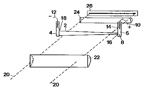
In Figure 1, an elongated separation vessel 2
has two ends with a reservoir 4, 6 at each end. The
vessel and the reservoirs contain a buffer 8. The
buffer is either a mixture of a weak acid and
WO 94/20843 ~ PCT/CA94/00120
i ~ ~~
- 5 -
conjugated base or a weak base and conjugated acid
(usually in water). A power source (not shown) that
generates a constant current has one terminal 10
connected at one end of said vessel 2 and another
terminal 12 connected at another end of said vessel 2.
The power source is connected to create a temperature
gradient along the buffer 8 in the vessel 2.
Simultaneously, the electric current.created by the
power source will produce an electric field gradient
required for isoelectric focusing on ampholytic
components. A membrane 14 is an ultrafiltration
membrane that surrounds the electrodes to prevent
access of proteins or other substances in the buffer to
the surface of the electrodes so that absorption,
oxidation, reduction or degradation will not occur.
The vessel 2 has physical characteristics so that a
temperature gradient will be created within the buffer
8 when the power source is connected to pass a constant
electric current through the vessel. The vessel 2 is a
cone-shaped capillary having a narrow end 16 and a wide
end 18. A positive terminal is connected into the
reservoir 6 at the narrow end 16 and a negative
terminal 12 is connected into the reservoir 4 at the
wide end 18. The tapered shape of the capillary causes
a temperature gradient to be created within the buffer
from one end of the capillary to the other. The
temperature gradient in turn causes a pH gradient to be
created.
When the electric current is passed through
the buffer, the narrow end of the capillary gets hotter
than the wide end 18, creating a temperature gradient
along the buffer. The electric field gradient created
. by the presence of the current focuses or fractionates
the ampholytes with ampholytes having the same
isoelectric point moving to the same zone of the
capillary.
WO 94/20843 PCT/CA94/00120
. i
- 6 -
A detection system monitors progress of the
focusing. The detection system consists of a light
source (not shown in Figure 1) which generates a light
beam 20. The light beam 20 passes through a
cylindrical lens 22 through the capillary, which is
transparent, and through a lens 24 to a sensor 26. The
sensor 26 determines the differences in an absorption
signal of the light beam as it passes through the
capillary.
In Figure 2, a more detailed schematic
drawing of the capillary or separation vessel 2 and the
detection system is shown with the same components that
are shown in Figure 1 being designated by the same
reference numeral. In Figure 2, a light source 28 has
a reflector 30 on one side and produces the light beam
which is directed through a filter 32. After the
filter 32, the beam 20 passes through a focusing lens
34, which focuses the beam on a pinhole 36 in a
partition 38. After passing through the pinhole, the
20 beam 20 passes through a collecting lens 40 and then
through the cylindrical lens 22 which focuses the beam
on the capillary 2. After passing through the
capillary 2 and the buffer 8 contained within the
capillary 2, the beam passes through the lens 24 and is
directed onto the sensor 26 where the magnitude of the
absorption signal is measured continuously.
Preferably, the power supply is a DC power
supply having a high voltage of approximately 1 kV.
Preferably, the voltage ranges from 100 volts per cm of
length of the separation vessel to 1 kV per cm of
length of the separation vessel.
From Figures 3a, 3b and 3c, it can be seen
that the capillary gets much hotter at the narrow end
than at the wide end and that pH is much lower at the
narrow end than at the wide end.
O 94/20843 PCT/CA94/00120
The amount of heat generated per unit length
of the separation vessel filled with the electrolyte
buffer at a dimension x is dQ(x)/dx and is proportional
to the electrophoretic current, I, and the magnitude of
the electric field along the separation vessel at this
dimension fE(x)~. In other words:
d~ (x) - E(x)I
dx
The value of the electrophoretic current I is
constant along the capillary axis (see Figure 3a) and
can be calculated from:
I = KA(x) E(x)
where A(x) is a cross-section area of the separation
channel at given dimension x and K is the electrolyte
conductivity and therefore:
dg (x) - I2
dx KA (x)
For cone-shaped capillary geometry of the separation
vessel the relationship is:
2
dg (x) - I
dx K~rR~( x j
where R(x) is the capillary radius at given dimension.
x.
The above equation clearly indicates that by
reducing the capillary diameter along the capillary
axis, the amount of heat generated in the system is
increasing rapidly which results in the temperature
gradient in the capillary as shown in Figure 3b. The
exact temperature profile is produced in the cone-
shaped capillaries can be calculated by solving the
differential equation describing heat transfer in the
system and considering appropriate electrolyte flows.
Since the dissociation constant of a weak
acid or base, which determines the pH of the buffer, is
r
a thermodynamic property the thermal gradient generated
inside the cone-shaped capillary produces a
corresponding pH gradient as shown in Figure 3c.
CA 02157373 2003-06-04 ---
In Figure 4, there is shown a device which
can be used to rapidly concentrate and purify small
amounts of biological material prior to analysis. In a
first step, an upper reservoir 4 is filled with
ampholytes contained in a buffer 8. The reservoir 6 at
the lower end of the vessel 2 has a positive terminal
from the power source (not shown) connected therein.
The reservoir 4 has a negative or ground terminal from
the same power source connected therein. After
application of separation voltage, target proteins
within the buffer 8 are concentrated and focused inside
the vessel 2 which is uncoated. Then,
that part of the sample containing the positively
charged ampholytes (the pI of which is higher than the
buffer pH) and neutral species is removed from the
reservoir 4. Then, the content of the capillary is
emptied for collection. The electroosmotic flow, which
flows from the lower to upper reservoir prevents the
neutral species from entering the capillary, but it
also slows down the focusing process. A positive
pressure can be applied to the upper reservoir 4 or a
vacuum to the reservoir 6 to increase
hydrodynamic flow in the capillary. The device shown
in Figure 4 can also be used to fractionate ampholytic
components of biological material when using several
anodic containers (only one of which is shown in Figure
4). Initially, all sample components, for which pI is
low enough to allow passage through the capillary, will
be collected in the reservoir 6 and will constitute
a first fraction. Then, the reservoir 6
will be replaced with a second anodic container (not
shown) and a tip 42 of the vessel 2 will be placed
in the second container and the current in the system
will be slightly lowered, thereby lowering the
temperature in the tip 42. The current is lowered by
lowering the potential. This will cause an increase in
WO 94/20843 PCT/CA94100120
~~
3
_ g _
pH in the buffer in the tip. Proteins within the
capillary, which have a low enough pI and are located
at the very tip of the capillary, will be charged
negatively and will migrate to the second anodic
container. This process can be repeated several times
by successively lowering the current still further to
fractionate biological material focused and
concentrated inside the cone-shaped capillary to create
further fractions as desired. The process can be
performed in a continuous and automated fashion and can
be used for concentration fractionation and separation.
In Fiaure 5, a separation vessel 44 has a
constant inside diameter but a conductive outer wall 46
of varying thickness. When electrodes are connected to
the wall 46 with a positive electrode connected at a
narrow end 48 and a negative or ground electrode
connected at a wide end 50 and a constant current is
passed through the wall 46, a temperature gradient is
created in the same manner as the temperature gradient
for the separation vessel 2 shown in Figure 1 except
that the electrodes are connected to the conducting
wall 46 as well as being connected into the buffer
within the vessel. A temperature gradient is created
within the wall 46 and the heat from the wall 46 is
conducted into the buffer within the vessel 44 to
create a similar temperature gradient within the
buffer. The positive electrode (not shown) is
connected to the narrow end of the wall 46 and the
negative electrode (not shown) is connected to the wide
end of the wall 46. While a single power source is
preferred, separate power sources could be used, one
for the wall 46 and one for isoelectric focusing.
In Figure 6, there is shown a continuous flow
separation vessel 52 having two non-parallel walls 54,
56. The walls converge with one another so that the
vessel has a V-shaped cross-section with an inlet ~8
WO 94/20843 PCT/CA94/0012
- 10 -
and an outlet 60 so that a buffer can flow into and out
of said vessel continuously in a direction normal to
said cross-section. Collectors 62 at the outlet 60 -
collect part of the buffer that contains focused
ampholytes of interest in the location along the length
of the vessel where the collectors are lacated. While
the electrodes are not shown in Figure 6, the positive
electrode would be connected into the buffer at a
narrow end 64 and the negative electrode would be
connected into the buffer at the wide end 66. The end
walls themselves could be membrane protected
electrodes.
In Figure 7, there is shown the magnitude of
the absorption signal along the length of the capillary
at 0 minutes, 27 minutes, 32 minutes and 37 minutes of
isoelectric focusing. These results were obtained with
an inside wall of the capillary coated with a non-
cross-linked poly (acrylamide) to eliminate
electroosmotic flow. In addition, agarose gel plugs
were used at both ends of the capillary to reduce
potential hydrodynamic flew. Initially, the cone-
shaped capillary and two reservoirs (as shown in Figure
1) were filled with a pH = 7.3 TIRS buffer. Then, 1 kV
of potential was supplied across the capillary for 10
minutes to achieve the uniform temperature and
corresponding pH gradients. Then, a few drops of 0.1
mg/mL sample solution consisting of two forms of
hemoglobin (methemoglobin, pl = 7.20 and oxyhemoglobin
pl = 7.00) was introduced into the reservoir 4 at the
wide end of the capillary containing the grounded
electrode (cathode). Since the sample components have
a slightly lower pl (by a fraction of a pH unit)
compared to the chosen buffer pH, they are initially
negatively charged and begin migrating through the
capillary towards the positive electrode 10 until they
reach their isoelectric point and the migration ceases.
WO 94/20843 ~ a PCT/CA94/00120
- 11 -
The analytes are trapped inside the capillary and, as
expected, they form two narrow zones corresponding to
methemoglobin 68 and oxyhemoglobin 70. It can be seen
that the magnitude of the absorption signal
progressively increases with time which confirms that
the accumulation of analyte in the capillary occurs.
In other words, the peaks increase in size with time.
The width of the isoelectric focusing (IEF) bands can
be estimated from the distance between the two forms of
hemoglobin present in the sample (0.2 pH units) and
corresponds to about 0.04 pH units. In Figure 3, the
estimated change in pH between the two ends of the
cone-shaped capillary is about one pH unit. This
corresponds to a temperature difference of about 400
between the two ends. To widen the pH range, buffers
which have a larger temperature co-efficient of pH can
be used. Also, the temperature differential can be
increased by narrowing the smaller end of the capillary
or by increasing the electrophoretic current by
increasing the electrical potential. This approach
might require cooling down the buffer reservoirs to
prevent denaturing of proteins at high temperatures
generated inside the capillary. Steady state
temperature conditions were not reached in Figure 3
even after 30 minutes since the bands were still
changing position along the capillary between 32
minutes and 37 minutes. However, the drift that
occurred between 32 minutes and 37 minutes was smaller
than the drift that occurred between 27 minutes and 32
minutes and therefore the system was very close to
reaching steady state temperature conditions.
In Figure 8, the magnitude of the absorption
. signal across the length of the capillary in successive
times is shown. The cone-shaped capillary used to
produce the results of Figure 8 had an uncoated
interior wall. The concentration ef analyte in the
CA 02157373 2003-06-04
- 12 -
buffer was about ten times higher than the
concentration that produced the results for Figure 7.
The sample therefore accumulated much faster in the
system of Figure 8 than it did for the system in Figure
7. The bands of analyte in Figure 8 are not located in
the appropriate isoelectric point, but rather at a
somewhat higher pH at which electrophoretic velocity of
the proteins are equal to the flow generated by
electroosmosis. The electrolyte flow in the capillary
cools the system and results in the pH values being
shifted towards the narrow end of the capillary. The
presence of the flow also produces more rapid thermal
equilibration as indicated by the very stable analyte
band position in the capillary. For example, it can be seen
that analyte band 72 lies at approximately the 19.5 mm
location along the capillary after 3 minutes, after 4
minutes and after 7 minutes. The resolution in Figure
8 decreased about SOo compared to the resolution in '
Figure 7 and is most likely caused by the flow in the
system.
The lowermost results in Figure 8 were
obtained after the separation voltage was increased
from approximately 1 kV to approximately 1.5 kV.
Analyte bands 72, 74 move towards the wider end of the
capillary since the amount of heat generated in the
capillary is increased and the pH values shift towards
the cooler end. The intensity of the band is decreased
because of the increase in the band's width and
increase of the capillary diameter at the new position.
From Figure 7, it can be seen that the
isoelectric focusing technique of°the present invention
can be used not only for analytical and preparative
separations but also for preconcentration and
purification of biological samples prior to analysis.
From Figure 8, it can be seen that the isoelectric
focusing system can be used for trapping and
WO 94/20843 ~~~ PCTICA94100120
~~~
J~
- 13 -
concentrating target ampholytes (for example, proteins,
peptides, amino acids or any other substances having an
isoelectric point) in the capillary followed by
mobilization of analytes towards the detection or
collection point by increasing the electrical
potential.
EXAMPLE #1
Cone-shaped capillaries of 4 cm in length
were used as the separation concentration channel and
were drawn from 5 mm inside diameter glass tubes. The
i. d. of one end of the capillary was 0.2 mm and
another end was 1 mm. The capillary was mounted on a
cartridge and its two ends were connected to buffer
reservoirs as shown in Figure 1. In some experiments,
the inner wall of the capillary was coated with non-
cross-linked poly (acrylamide) to eliminate
electrcosmosis by the reported way. Cross-linked poly
(acrylamide) could also be used. The separation was
driven by a high-voltage DC power supply (Spellman,
Plainview, NY), and the separation voltage was about 1
kV. The anode was inserted into the buffer reservoir
at the narrower end of the capillary, and the another
end of the capillary was connected to ground.
The protein sample used in the experiment was
human hemoglobin (Sigma, St. Louis, MO) which contained
two major isoforms; methemoglobin (75~) a:~d
oxyhemoglobin (25~). All chemicals were reagent grade,
and solutions were prepared using deionized water. The
buffer as 0.05 M TRIS bu~fer at pH 7.3. This buffer
has a large temperature co-efficient of pH (dpH/dT is
-0.028K-1 at 25oC) (10). Protein solutions were
prepared in the TRIS buffer. The solutions were
. filtered using 0.2-um pore size cellulose acetate
filters (Sartorius, Gottingen, Germany).
A UV-vis absorption imaging detector was
employed for the monitoring of the protein zones
WO 94/20843 PCT/CA94/0012
- 14 -
focused inside the capillary. As shown in Figure 2,
the light source of the detector was a halogen lamp.
The sensor was a 1024 pixel CCD (Type 53903-1024Q,
Hammamatsu, Hammamatsu City, Japan). A bandpass
coloured filter (400 nm - 600 nm) was used to cut near
IR and ultraviolet radiations of the lamp. The light
beam was first collimated as shown in Figure 2, and
then focused into the capillary by three cylindrical
lenses. The image of the capillary was projected into
the CCD sensor as shown in Figure 2.
Two sample introduction methods were used in
the experiment. In the first method, the coated
capillary was filled with the buffer, and plugs of 1$
agarose gel (prepared in the buffer) were placed in
both reservoirs to avoid hydrodynamic flow in the
system, and then the voltage was applied. After 10
minutes, a few drops of 0.1 mg/mL sample solution was
added to the top of the reservoir at the cathodic end
of the capillary. In the second method, the reservoirs
and the uncoated capillary were filled with the protein
solution and the voltage was applied. In all
experiments, the current passing through the capillary
was kept at about 0.8 mA by adjusting the applied
voltage to about 1 kV. All experiments were done in
triplicate to ensure reproducibility.
In using the isoelectric focusing system of
the present invention, the reservoir 6 at the narrow
end of the capillary (anodic end) can be kept at a
predetermined temperature to produce a low enough pH of
the buffer to positively charge target proteins present
in the reservoir (i.e. the sample that was added to the
anodic reservoir). These proteins will then migrate
through the capillary toward the cathode and will be
trapped in the capillary at their isoelectric points or
will be collected at the reservoir 4 connected to the
wide end of the capillary. The system can be used to
WO 94/20843 ~ PCT/CA94/00120
- 15 -
purify the biological material by fractionating it with
respect to the isoelectric point. The cathodic end
reservoir 4 will contain proteins which have a lower pI
than the buffer in the reservoir 4 which is kept at
room temperature. The anodic reservoir will contain
proteins at a pI that is higher than the pH of the
buffer in that reservoir while the capillary will
contain proteins which have intermediate pI. The
anodic reservoir 6 will be heated up in the process and
that will speed up the concentration and focusing
process when the sample is introduced to both the
cathodic and anodic reservoirs.
Numerous variations, within the scope of the
attached claims, will be readily apparent to those
skilled in the art.
~: ll~F ~ ~, ~t ~~~4.~~+~s~,V